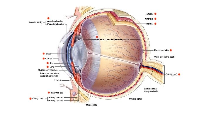
RETINAL IMAGING AND FLUORESCEIN ANGIOGRAPHY RETINAL ANATOMY Choroid


")


")


")


")


")


")


- Slides: 82
RETINAL IMAGING AND FLUORESCEIN ANGIOGRAPHY
RETINAL ANATOMY *Choroid *Retina *Optic nerve
Retinal Vasculature Central retinal artery Branch retinal arteries Arterioles Capillaries Venules Branch retinal veins Central retinal vein
Reasons for physician wanting FA Macular degeneration White Dot Syndromes To determine if exudative and what is the best treatment protocol APMPPE (Acute posterior multifocal placoid pigment epitheliopathy Histoplasmosis MEWDS multiple evanescent white dot syndrome To assess choroidal neovascularization CME To assess leakage, typical flower petal pattern PIC punctate inner choroidopathy Panuveitis Diffuse subretinal fibrosis
Reason for physician ordering FA Vascular Diabetes CRAO Nonproliferative diabetic retinopathy BRAO Diabetic macular edema CRVO Ischemia BRVO Proliferative diabetic retinopathy HTN
Setting up FA *Schedule if possible *Eat prior to procedure (reduces chance of nausea and vomiting) *Should be well hydrated (optimizes vein access) *Optimal dilation with 1% Tropicamide and 2. 5% phenylephrine (x 2 sometimes) *Informed consent
Color Photos F 1 and F 2
Mosaic
Seven Standard Fields
Red Free Photos Green filter
Set up 5 cc Fluorescein Sodium 10% or 2 cc Fluorescein Sodium 25% use filter needle if in glass ampule IV kit Tourniquet Alcohol wipes Gauze Tape Bandage 23 or 25 G butterfly needle Gloves
Starting Angiogram Filter in place (exciter only on our Topcon 50 DX)
Position patient
Start timer and injection
Start photographing One photo taken as soon as dye is completely injected to let physician know injection time. Take one photo every second for approx 40 -45 seconds. Photograph fellow eye. Photograph both eyes at around one minute. (End of early phase).
Angiogram continued After one minute pictures, patient gets break Sit back Remove needle Many times this is about when adverse effects occur Mid-phase pictures at 3 minutes Late phase pictures at 5 -15 minutes, depends on pathology, will need to adjust flash.
Early phase: Choroidal/Arterial Choroidal Flush ~10 seconds
Early phase: arterial Artery fills 1 -2 seconds after Average arm to eye 12 seconds Delayed arm to eye can mean: -carotid disease -heart disease -PVD(peripheral vascular dx)
Early phase: arteriovenous phase Complete filling of retinal capillary bed Veins begin to fill First fill along vein wall (laminar flow)
Laminar flow
Early Phase: venous phase Complete filling of veins Best time to view perifoveal capillaries
Mid phase 2 -4 minutes after injection Veins and arteries equal Diminished brightness Dye removed from bloodstream
Late phase: Five to fifteen minutes post injection Elimination of dye from retina And choroidal vasculature Disc staining Other areas of hyperfluorescence
Risks of Fluorescein injection Extravasation of dye into tissues Small butterfly needles helpful due to blood being injected first. If dyes gets into tissues stop ASAP If happens, use ice and beware of necrosis and phlebitis. Educate patient
Flushing Nausea Vomiting Usually occurs at one minute mark Dependent on amount of dye, speed of injection and possibly concentration 25% Advise patient to eat and be hydrated prior to procedure If happens, advise deep breaths and reassure that it will pass quickly Have basket available “just in case” Phenergan can be used if they have had in past and physician determines FA essential to diagnosis and treatment
Vasovagal response Happens usually due to anxiety Be ready for them to pass out Frequently happens in younger patients
Hives Liquid Benadryl Make sure patient knows that they need to let you know of this or any other reaction so it can be documented in medical record and taken into account if they need another FA in future.
Bronchospasm Laryngeal edema Liquid Benadryl Epipen Document in medical record
Anaphylaxis Epipen Crash cart Physician in area whenever FA is done
Hypotension Syncope Seizures MI/cardiac arrest CVA Need for physician and emergency medical equipment/crash cart available Call for code Call 911 Epinephrine Corticosteroids
Abnormalities of Angiogram Hypofluorescence *Reduction or absence of normal fluorescence due to blockage such as blood or abnormalities in choroidal or retinal perfusion. (occlusion or ischemia)
Abnormalities of Angiogram Hyperfluorescence Increased transmission or abnormal presence of dye. Autofluorescence hyperfluorescence in absence of dye (optic nerve head drusen) Pseudofluorescence usually found in old filters that need replacement Transmission defect absence of pigment allowing choroidal fluorescence to be seen (window defect)
Transmission defect
Hyperfluorescence Leakage due to extravasation of dye due to DME, CSR. Occurs with neovascularization from PDR and AMD
Staining Late hyperfluorescence from dye accumulation. Occurs with drusen, chorioretinal scar, optic nerve. Visible where there is reduction/absence of RPE.
Pooling Accumulation within distinct space such as CSR or serous detachment
Improving Images 1. 2. 3. 4. Focus ocular eyepiece. Place white paper in front of lens to focus reticle. Turn eyepiece to high plus power. Relax eyes by focusing on distance for few seconds to decrease accommodation. 5. Focus with both eyes open to prevent accommodation. 6. Turn toward plano and stop when reticle is just in focus. 7. Repeat several times. 8. Check it every time you use camera, especially if sharing camera with other staff members. 9. Position patient properly with chin and forehead placed correctly 10. Pull focusing knob toward you, slowly turn away until image just in focus.
Artifacts
Iris
Blink
Dust
Pathology
Cotton wool spots
Exudate Blot hemes
Microaneurysms
Intraretinal microvascular abnormalities (IRMA)
IRMA
IRMA
Retinal neovascularization
Neovascularization
Neovascularization
Rubeosis/neovascularization of iris (NVI)
Preretinal hemorrhage from PDR
Ischemia
Crossing changes/AV nicking
Crossing changes
Venous Beading in diabetic retinopathy Also enlarged foveal avascular zone
Venous beading
Cystoid Macular Edema
Retinitis pigmentosa
White dot syndrome: Acute posterior multifocal placoid pigment epitheliopathy (APMPPE)
APMPPE Red Free
CRAO with Cilioretinal artery
CRAO with Cilioretinal artery
CRAO with PDR (1 minute post injection)
Choroidal folds
Choroidal folds red free
Choroidal folds FA late
Kissing choroidals (Choroidal hemorrhage)
Kissing Choroidals
Kissing choroidals
Diabetic papillopathy
Diabetic papillopathy red free
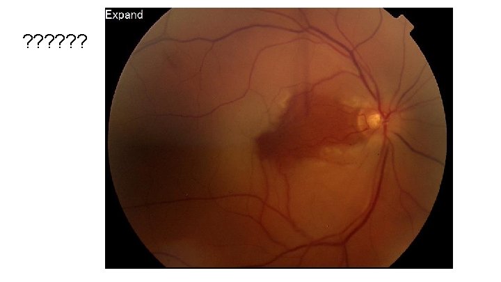
Central Serous Retinopathy
CNVM with histoplasmosis
CNVM Histo FA