Retinal Detachment Presentation and Principle of Management By
 By Dr. Suhail Mushtaq Boobak MBBS, DOMS,")
Retinal Detachment (Presentation and Principle of Management) By Dr. Suhail Mushtaq Boobak MBBS, DOMS, MCPS, ICO(UK) FCPS, FRCS (UK) Associate Professor Ophthalmology Sargodha Medical College, University of Sargodha

Retinal Detachment Separation of neurosensory retina from the retinal pigment epithelium.

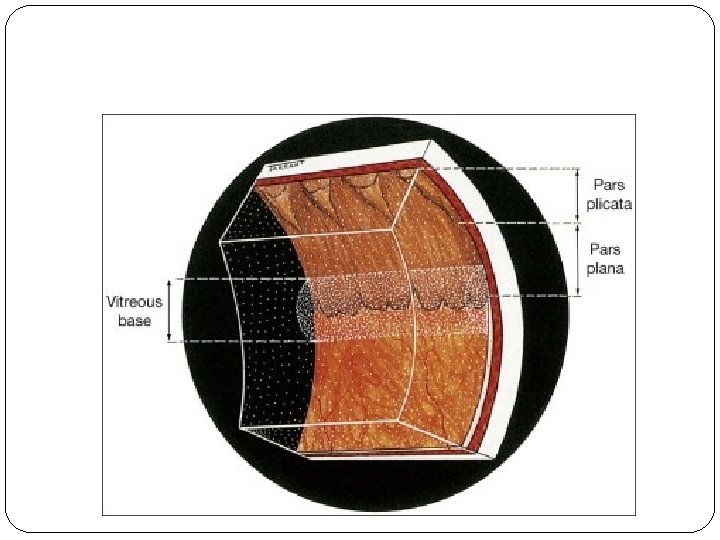
1. Definitions and classifications • Retinal breaks • Retinal 2. detachment Anatomy • Anatomical landmarks • Variants of ora 3. Examination serrata • • Vitreous base Indirect techniques ophthalmology • Scleral indentation

Indirect ophthalmology Condensing lenses Technique • Keep lens parallel to patient’s iris pla The higher the power, the less the magnification, the shorter the working • Avoid tendency to move towards pati distance but the greater the field of view • Ask the patient to move eyes and he into optimal positions for examination •
 2 - Tractional 3 -")
Classification 1 - Rhegmatogenous ( Simple , Primary ) 2 - Tractional 3 - Exudative ( Serous, Secondary ) 4 - Combined tractionalrhegmatogenous.

Separation of sensory retina from RPE by subretinal f Rhegmatogenous - caused by a Non-rhegmatogenous retinal break exudative

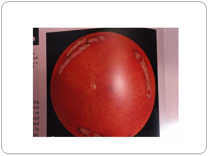
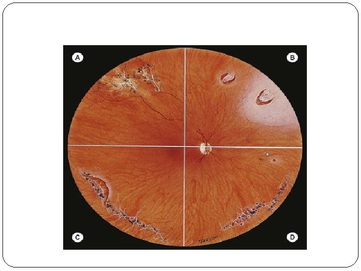
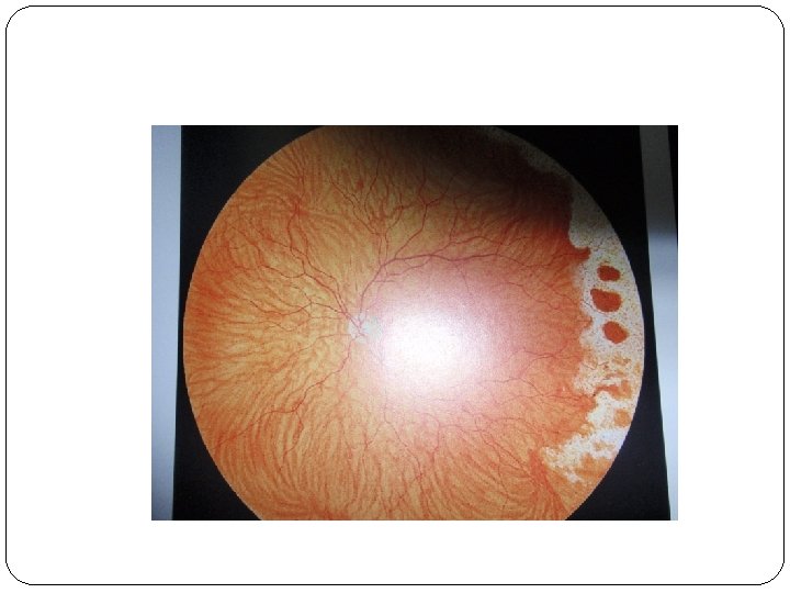
Pathogenesis of rhegmatogenous retinal detachment a- Vitreoretinal traction b-Predisposing peripheral retinal degeneration c-Myopia d-Trauma

Pathogenesis of tractional retinal detachment a- Diabetic tractional retinal detachment b- Traumatic tractional retinal detachment Pathogenesis of exudative retinal detachment a- Vascular diseases b- Inflammatory diseases c- Neoplastic diseases.


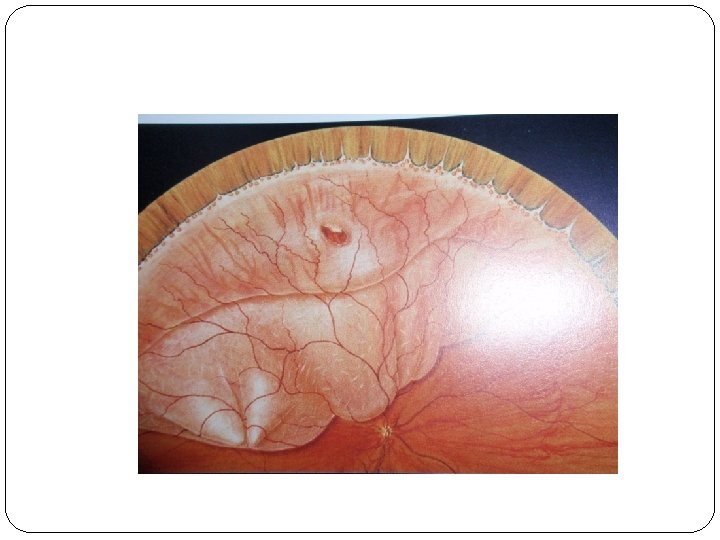
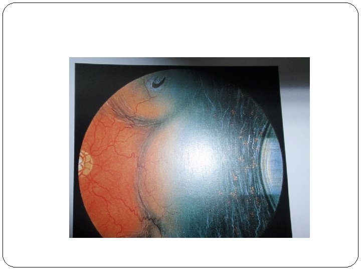
Diagnosis of Rhegmatogenous retinal detachment Symptoms 1 - Photopsia 2 - Floaters 3 - Visual Field Defects Signs 1 - Marcus Gunn pupil 2 - Low IOP 3 -Mild iritis 3 - Tobacco dust 4 - Retinal break 5 Convex configration & Subretinal fluid(Fresh RD) 6 -Retinal thinning, 2 ndary intraretinal

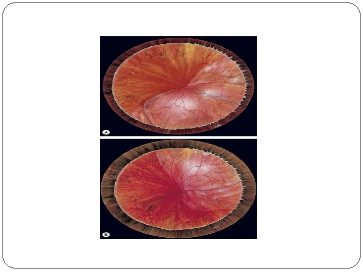
Diagnosis of tractional retinal detachment �Symptoms Visual field defects Signs a - Retinal detachment –Concave configuration b - Retinal mobility is severely reduced.


Diagnosis of exudative retinal detachment �Symptoms Floaters may be present Signs a - Retinal detachment –Convex configuration b - Retinal mobility is present
 2 -Scleral")
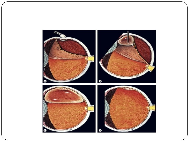
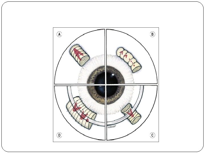
�Treatment 1 -Pneumatic retinopexy(SF 6 -sulphur hexafluoride, C 3 F 8 -perfluoropropane) 2 -Scleral buckling 3 -Drainage of SRF 4 -Pars Plana Vitrectomy
 4 -Reopening")
�Causes of failure 1 -Missed breaks 2 -Buckle failure 3 -PVR(Proferative vitreoretinopathy) 4 -Reopening of retinal break

�Postoperative complications 1 -Raised itnraocular pressure 2 -Cataract 3 -Band keratopathy

Thanks
- Slides: 26