
Retina Imaging Conference Tala Kassm DO April 14

- Slides: 29
Retina Imaging Conference Tala Kassm DO April 14, 2016 University of Louisville Department of Ophthalmology and Visual Sciences
Subjective n CC: “I can’t see from my left eye over 2 -3 months. ” n HPI: 47 year old white male presents with progressive vision loss in his left eye over 2 -3 months. He initially thought it was something wrong with his glasses which he only uses to read. Denied eye pain, flashes or floaters.
History POH: None PMH: Gastroesophageal reflux disease, anxiety, no hypertension Family Hx: Noncontributory Meds: Clonazepam, Fluoxetine, Omeprazole Allergies: NKDA Social history: denies alcohol, tobacco or illicit drug use Review of Systems: denies recent trauma, joint pain,
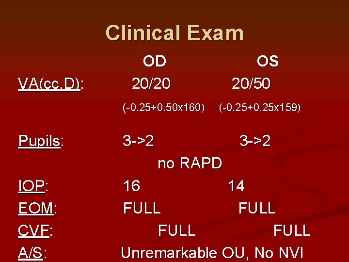
Clinical Exam VA(cc, D): OD 20/20 (-0. 25+0. 50 x 160) Pupils: OS 20/50 (-0. 25+0. 25 x 159) 3 ->2 no RAPD IOP: EOM: CVF: A/S: 16 14 FULL Unremarkable OU, No NVI
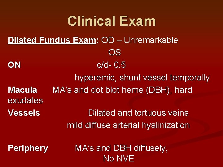
Clinical Exam Dilated Fundus Exam: OD – Unremarkable OS ON c/d- 0. 5 hyperemic, shunt vessel temporally Macula MA’s and dot blot heme (DBH), hard exudates Vessels Dilated and tortuous veins mild diffuse arterial hyalinization Periphery MA’s and DBH diffusely, No NVE
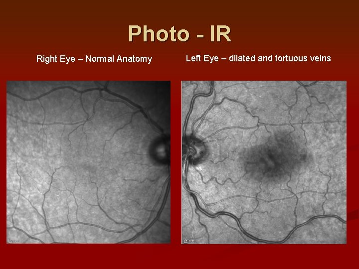
Photo - IR Right Eye – Normal Anatomy Left Eye – dilated and tortuous veins
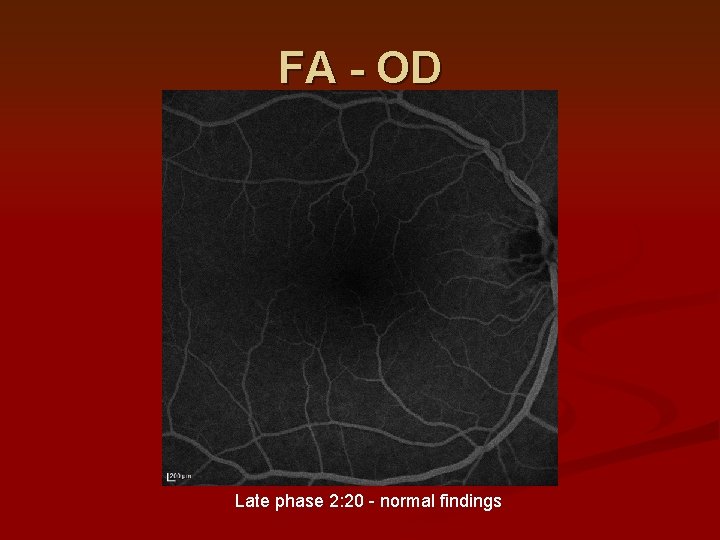
FA - OD Late phase 2: 20 - normal findings
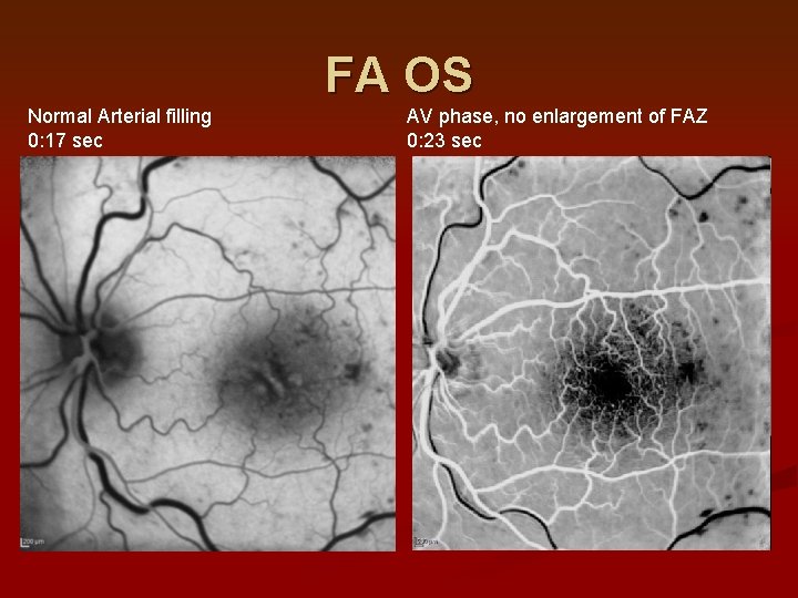
FA OS Normal Arterial filling 0: 17 sec AV phase, no enlargement of FAZ 0: 23 sec
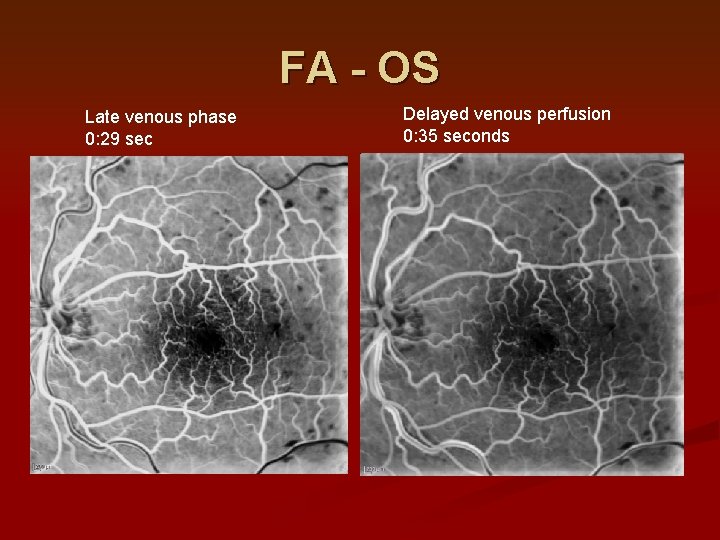
FA - OS Late venous phase 0: 29 sec Delayed venous perfusion 0: 35 seconds
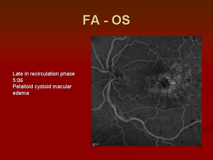
FA - OS Late in recirculation phase 5: 06 Petalloid cystoid macular edema
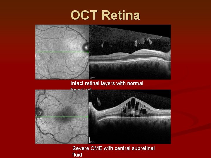
OCT Retina Intact retinal layers with normal foveal pit Severe CME with central subretinal fluid
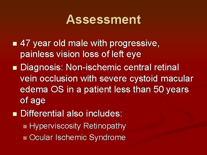
Assessment 47 year old male with progressive, painless vision loss of left eye n Diagnosis: Non-ischemic central retinal vein occlusion with severe cystoid macular edema OS in a patient less than 50 years of age n Differential also includes: n Hyperviscosity Retinopathy n Ocular Ischemic Syndrome n
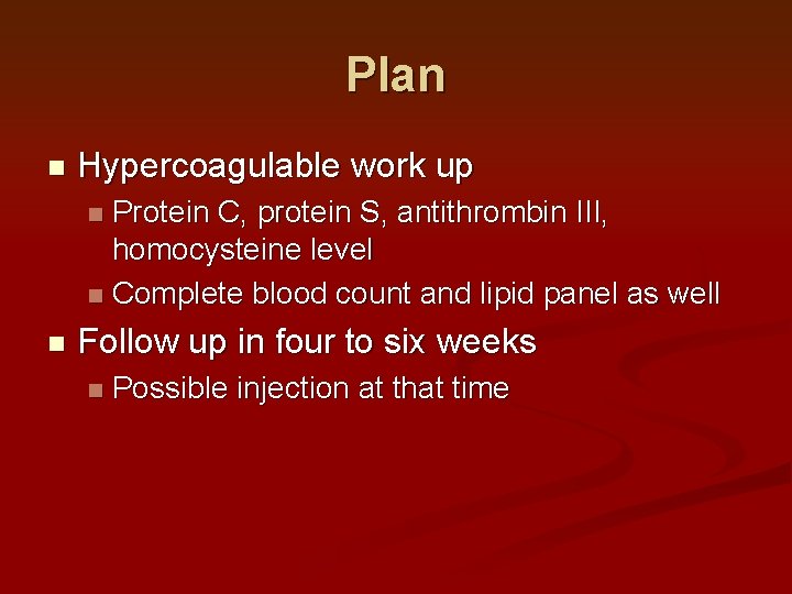
Plan n Hypercoagulable work up Protein C, protein S, antithrombin III, homocysteine level n Complete blood count and lipid panel as well n n Follow up in four to six weeks n Possible injection at that time
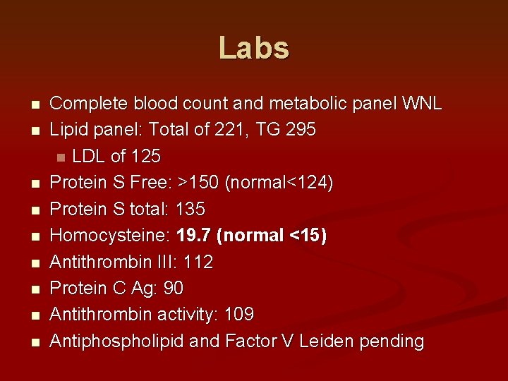
Labs n n n n n Complete blood count and metabolic panel WNL Lipid panel: Total of 221, TG 295 n LDL of 125 Protein S Free: >150 (normal<124) Protein S total: 135 Homocysteine: 19. 7 (normal <15) Antithrombin III: 112 Protein C Ag: 90 Antithrombin activity: 109 Antiphospholipid and Factor V Leiden pending
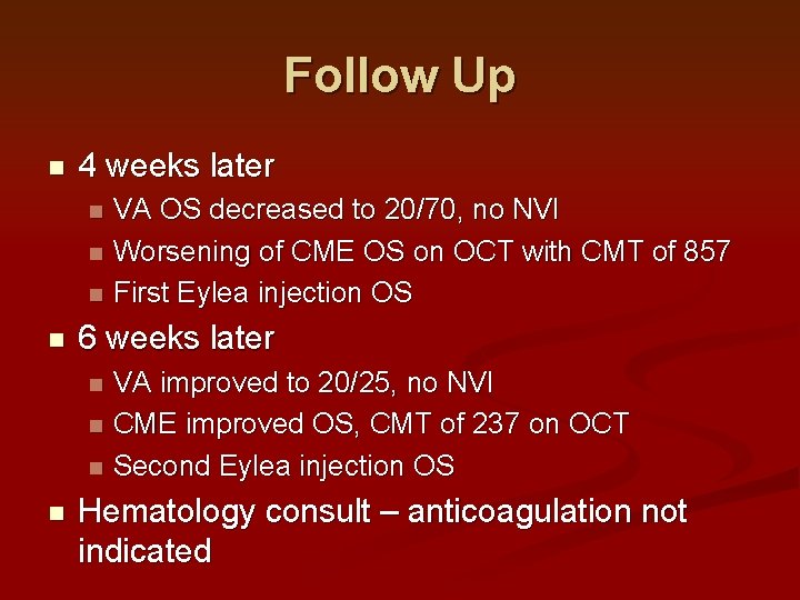
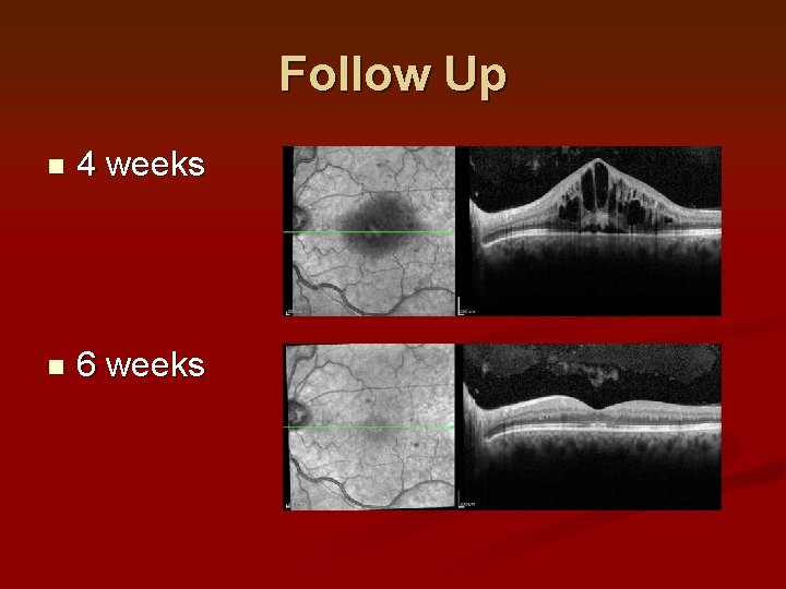
Follow Up n 4 weeks later n n 6 weeks later n n VA OS decreased to 20/70, no NVI Worsening of CME OS on OCT with CMT of 857 First Eylea injection OS VA improved to 20/25, no NVI CME improved OS, CMT of 237 on OCT Second Eylea injection OS Hematology consult – anticoagulation not indicated
Follow Up n 4 weeks n 6 weeks
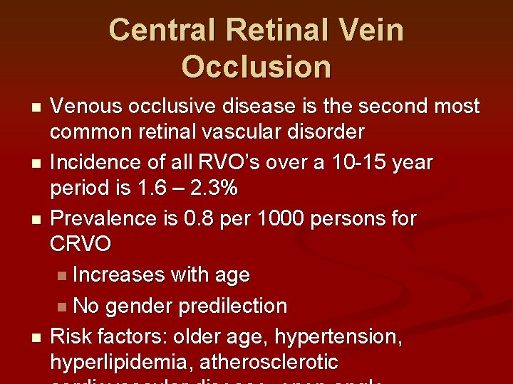
Central Retinal Vein Occlusion n n Venous occlusive disease is the second most common retinal vascular disorder Incidence of all RVO’s over a 10 -15 year period is 1. 6 – 2. 3% Prevalence is 0. 8 per 1000 persons for CRVO n Increases with age n No gender predilection Risk factors: older age, hypertension, hyperlipidemia, atherosclerotic
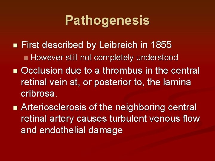
Pathogenesis n First described by Leibreich in 1855 n However still not completely understood Occlusion due to a thrombus in the central retinal vein at, or posterior to, the lamina cribrosa. n Arteriosclerosis of the neighboring central retinal artery causes turbulent venous flow and endothelial damage n
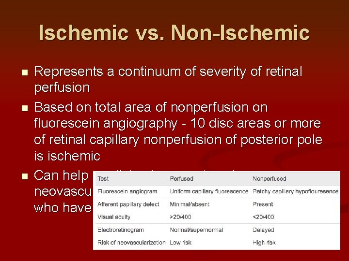
Ischemic vs. Non-Ischemic n n n Represents a continuum of severity of retinal perfusion Based on total area of nonperfusion on fluorescein angiography - 10 disc areas or more of retinal capillary nonperfusion of posterior pole is ischemic Can help predict subsequent ocular neovascularization and identification of patients who have a poorer visual prognosis
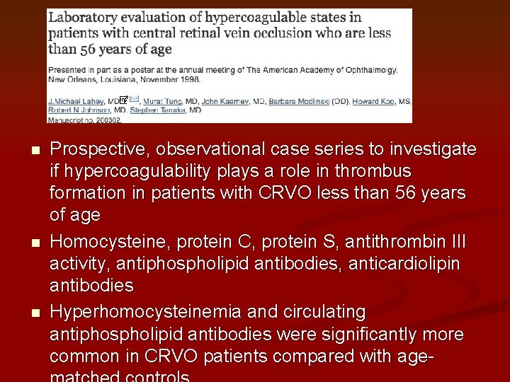
n n n Prospective, observational case series to investigate if hypercoagulability plays a role in thrombus formation in patients with CRVO less than 56 years of age Homocysteine, protein C, protein S, antithrombin III activity, antiphospholipid antibodies, anticardiolipin antibodies Hyperhomocysteinemia and circulating antiphospholipid antibodies were significantly more common in CRVO patients compared with age-
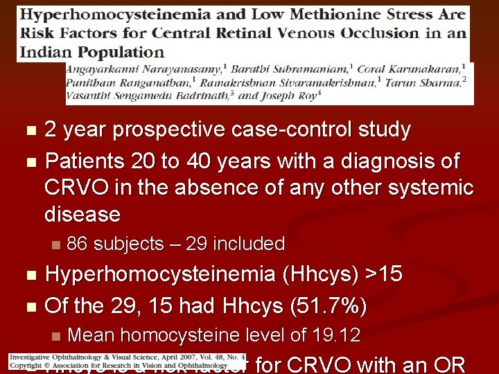
2 year prospective case-control study n Patients 20 to 40 years with a diagnosis of CRVO in the absence of any other systemic disease n n 86 subjects – 29 included Hyperhomocysteinemia (Hhcys) >15 n Of the 29, 15 had Hhcys (51. 7%) n n n Mean homocysteine level of 19. 12 Hhcys is a risk factor for CRVO with an OR
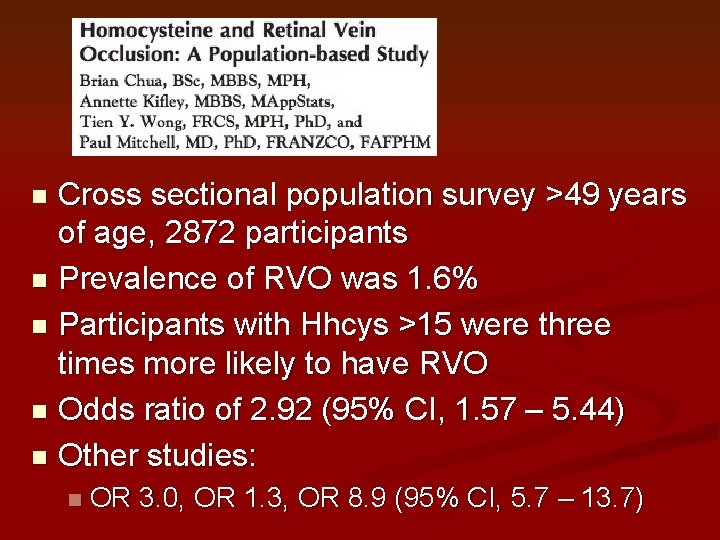
Cross sectional population survey >49 years of age, 2872 participants n Prevalence of RVO was 1. 6% n Participants with Hhcys >15 were three times more likely to have RVO n Odds ratio of 2. 92 (95% CI, 1. 57 – 5. 44) n Other studies: n n OR 3. 0, OR 1. 3, OR 8. 9 (95% CI, 5. 7 – 13. 7)
Complications n Neovascularization of Iris and Angle In nonperfused CRVO: 45 -80% n Greatest predictors of anterior segment neovascularization – visual acuity worse than 20/200 or degree of nonperfusion n Early treatment with PRP – resolution of NVI in 2/3 cases n n Neovascular glaucoma 90 day glaucoma (onset typically within 3 months) n Occurs in 20 to 63% of CRVO’s n
Management of CRVO n n Medical treatment and workup Laser photocoagulation – CVOS n n n Prophylactic PRP decreases the risk of iris neovascularization compared to untreated eyes – not statistically significant PRP more likely to result in prompt regression of NVI in previously untreated group vs. prophylactically treated group Frequent follow-up exams during early months, prompt PRP if NVI develops to reduce risk of NVG
Management of CRVO n Intravitreal corticosteroids n SCORE study – patients treated with 1 mg and 4 mg of triamcinolone n At one year 27% and 26% respectively had improvement of 15 ETDRS letters n Dexamethasone injectable intravitreal implant n Improved period visual acuity outcomes over a 6–month
Management of CRVO n Intravitreal Anti- VEGF Therapy Ranibizumab and aflibercept both FDA approved n Bevacizumab used off-label n Used in practice as monthly dosing, asneeded treatment or treat-and-extend approach for macular edema n May also be used to reduce iris neovascularization and treat neovascular glaucoma in the short term n
Course and Outcome n Prognosis depends on subtype of CRVO n n Of ischemic, 10% achieve vision better than 20/400 Usually predicted based on visual acuity at initial evaluation Presenting VA >20/40, maintained in 65% of people n Presenting VA <20/200, remained at that level in 80% n n Nonischemic CRVOs experience full
References n n n Scott IU, Ip MS, Van Veldhuisen PC, et al. A randomized trial comparing the efficacy and safety of intravitreal triamcinolone with standard of care to treat vision loss associated with macular edema secondary to branch retinal vein occlusion: the Standard Care vs. Corticosteroid for Retinal Vein Occlusion (SCORE) study report 6. Arch Ophthalmol. 2009; 127(9): 1115 -1128. BCSC: Retina and Vitreous pages 116 -125. Baseline and early natural history report. The Central Vein Occlusion Study. Arch Ophthalmol. 1993; 111(8): 1087 -1095. Klein R, Moss SM, Meuer SM, Klein BE. The 15 – year cumulative incidence of retinal vein occlusion: the Beaver Dam Eye Study. Arch Ophthalmol. 2008; 126(4): 513 -518. Stem MS, Talwar N, Comer GM, Stein JD. A longitudinal analysis of risk factors associated with central retinal vein occlusion. Ophthalmology. 2013; 120(2)362 -370. Gunther, J. Scott IU, Ip Michael. Retinal Venous Occlusive Disease. Albert & Jakobiec’s Principles & Practice of Ophthalmology. 2000, 1994(132): 1755 -1773.
Thank you for listening