
Results from a Simultaneous PETMRI Breast Scanner Bosky
Results from a Simultaneous PET-MRI Breast Scanner Bosky Ravindranath 1, Sachin Junnarkar 2, Martin Purschke 2, Sean Stoll 2, Xiaole Hong 3, Pai. Jung Huang 4, Sri Harsha Maramraju 1, Paul Vaska 2, Craig Woody 2, David Schlyer 2 1 Biomedical Engineering, Stony Brook University, Stony Brook, NY, 2 Brookhaven National Laboratory, Upton, NY, 3 Aurora Imaging Technology, Inc. , North Andover, MA, 4 Taipei Medical University Hospital, Taipei, Taiwan. 1
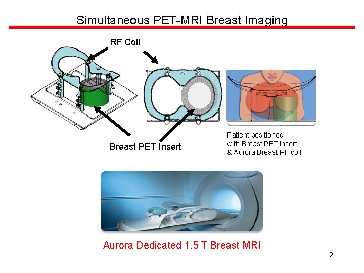
Simultaneous PET-MRI Breast Imaging RF Coil Breast PET Insert Patient positioned with Breast PET insert & Aurora Breast RF coil Aurora Dedicated 1. 5 T Breast MRI 2
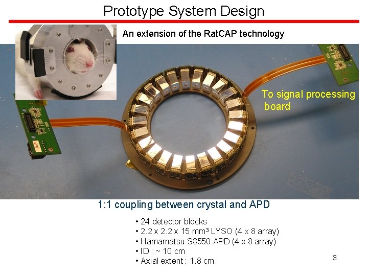
Prototype System Design An extension of the Rat. CAP technology To signal processing board 1: 1 coupling between crystal and APD • 24 detector blocks • 2. 2 x 15 mm 3 LYSO (4 x 8 array) • Hamamatsu S 8550 APD (4 x 8 array) • ID : ~ 10 cm • Axial extent : 1. 8 cm 3
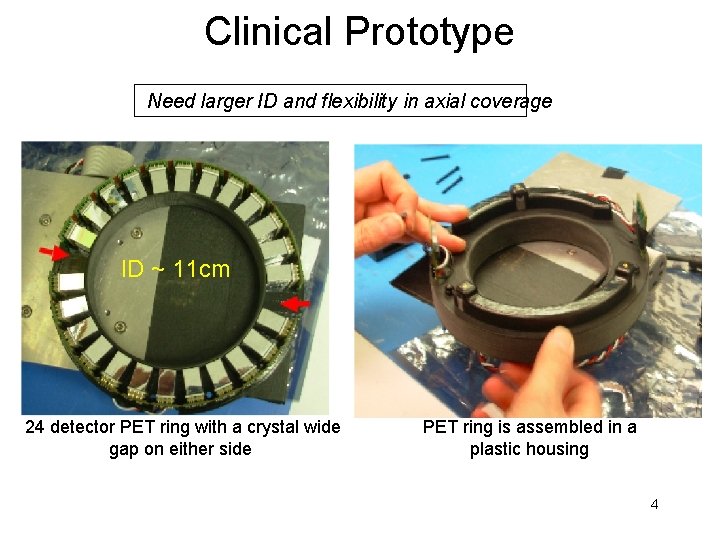
Clinical Prototype Need larger ID and flexibility in axial coverage ID ~ 11 cm 24 detector PET ring with a crystal wide gap on either side PET ring is assembled in a plastic housing 4
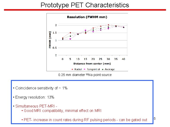
Prototype PET Characteristics 0. 25 mm diameter 22 Na point source • Coincidence sensitivity of ~ 1% • Energy resolution: 13% • Simultaneous PET-MRI • Good MRI compatibility, minimal effect on MRI • PET- increase in count rates during RF pulsing periods - can be gated out 5
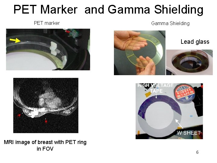
PET Marker and Gamma Shielding PET marker Gamma Shielding Lead glass MRI image of breast with PET ring in FOV 6
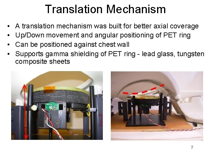
Translation Mechanism • • A translation mechanism was built for better axial coverage Up/Down movement and angular positioning of PET ring Can be positioned against chest wall Supports gamma shielding of PET ring - lead glass, tungsten composite sheets 7
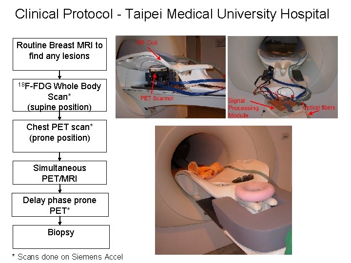
Clinical Protocol - Taipei Medical University Hospital Routine Breast MRI to find any lesions 18 F-FDG Whole Body Scan* (supine position) Chest PET scan* (prone position) Simultaneous PET/MRI Delay phase prone PET* Biopsy * Scans done on Siemens Accel
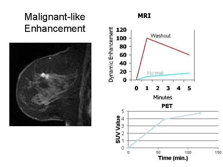
Malignant-like Enhancement Washout SUV Value Normal 5 4 3 2 1 0 PET 0 50 100 Time (min. ) 150
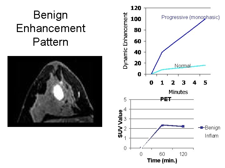
Benign Enhancement Pattern Progressive (monophasic) Normal PET SUV Value 5 4 3 Benign 2 Inflam 1 0 0 60 Time (min. ) 120
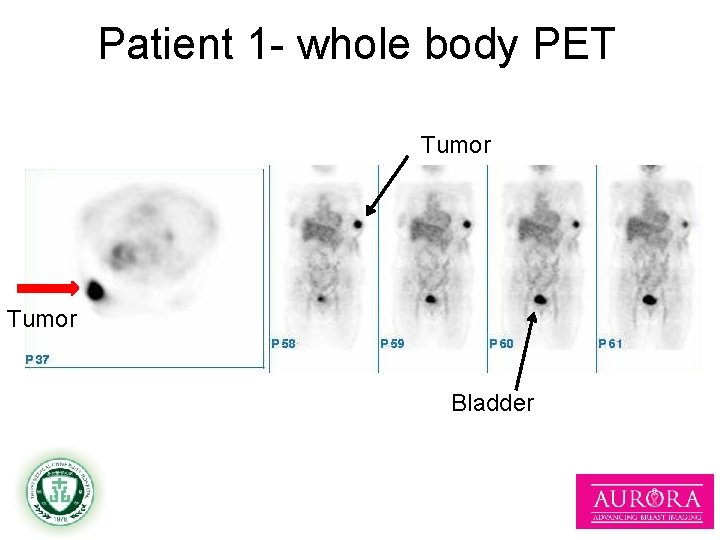
Patient 1 - whole body PET Tumor Bladder 11
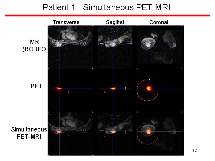
Patient 1 - Simultaneous PET-MRI Transverse Sagittal Coronal MRI (RODEO) PET Simultaneous PET-MRI 12
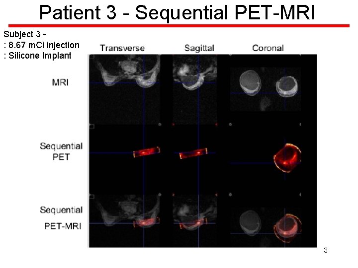
Patient 3 - Sequential PET-MRI Subject 3 : 8. 67 m. Ci injection : Silicone Implant 13
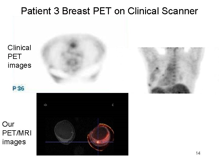
Patient 3 Breast PET on Clinical Scanner Clinical PET images Our PET/MRI images 14
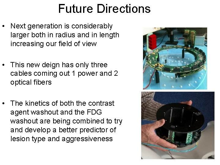
Future Directions • Next generation is considerably larger both in radius and in length increasing our field of view • This new deign has only three cables coming out 1 power and 2 optical fibers • The kinetics of both the contrast agent washout and the FDG washout are being combined to try and develop a better predictor of lesion type and aggressiveness 15
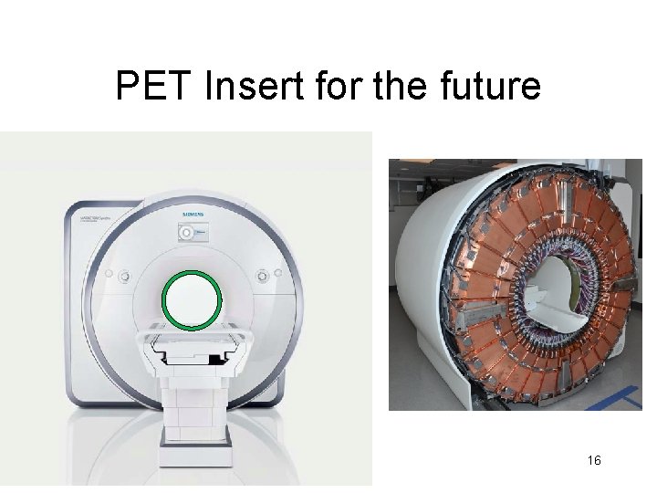
PET Insert for the future 16
Things to Remember • The power of PET and MRI lies in the ability to analyze the images, not in making nice pictures • Think about kinetics as a way to better utilize PET and MRI data • Think about ways to retrofit the 30, 000 MR scanners in the world to make PET/MR available for the clinic 17
Acknowledgements • • • Dr. David Schlyer, BNL § Dr. P. J. Huang, TMUH Dr. Paul Vaska, BNL § Dr. Hsu, TMUH Dr. Craig Woody, BNL § Dr. Xiaole Hong, Aurora Imaging Technology Dr. Sachin Junnarkar § Dr. David Bennett, Aurora Imaging Technology Dr. Sri Harsha Maramraju, SBU § Dr. Ken Cheng, Aurora Imaging Technology Sean Stoll, BNL § Olivia Cheng, Aurora Imaging Technology Dr. Martin Purschke, BNL § The Aurora and TMUH team of technologists and Bill Lenz, BNL nurses who have provided support for this study Richard Ruggiero, BNL Dr. Gene-Jack Wang, BNL Dr. Dardo Tomasi, BNL This work was supported by the U. S. Department of Energy (OBER) and Aurora Imaging Technology under Prime Contract No. DE-AC 0298 CH 10886. 18

Thank You Thanks to the BNL LDRD program, DOE and Aurora Imaging Technologies for Support of this work
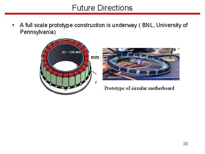
Future Directions • A full scale prototype construction is underway ( BNL, University of Pennsylvania) mm Prototype of circular motherboard 20
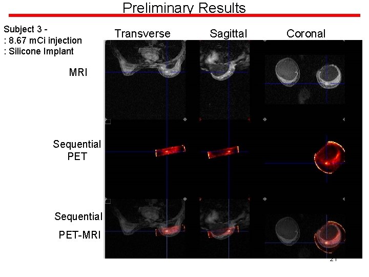
Preliminary Results Subject 3 : 8. 67 m. Ci injection : Silicone Implant Transverse Sagittal Coronal MRI Sequential PET-MRI 21
Points to Remember • A proof of concept simultaneous PET/MRI study was conducted with 4 patients with biopsy proven or highly suspicious breast cancer • This is the first time a study of such kind has been carried out in breast cancer patients • This study lays the groundwork for a better understanding of the challenges of simultaneous PET/MRI within the patient population 22
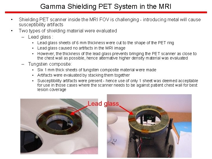
Gamma Shielding PET System in the MRI • • Shielding PET scanner inside the MRI FOV is challenging - introducing metal will cause susceptibility artifacts Two types of shielding material were evaluated – Lead glass : • Lead glass sheets of 6 mm thickness were cut to the shape of the PET ring • Lead glass caused no artifacts in the MRI image • However, the thickness of the lead glass prevents bringing the PET scanner as close to the chest wall as possible, hence alternative higher density material was evaluated – Tungsten composite: • Six 1 mm thick sheets of tungsten composite material were made • Artifacts were evaluated by stacking them together • Susceptibility artifacts were present - hence use of only 1 sheet was deemed acceptable for use in those cases where the scanner needs to be against patient chest wall for best lesion coverage 23
Image Reconstruction and Data Correction – Image reconstruction is carried out using the Maximum Likelihood Expectation Maximization technique – The gaps between the crystals and the missing detector block are accounted for during reconstruction – System matrix estimation using SIMSET involved accurate modeling of the crystal position within the ring, the field of view of the PET scanner is further divided into ~ 1 mm 3 voxels and decays are simulated in each voxel. – Variation in detector efficiency is measured by acquiring uniform phantom data at the operating threshold voltage and gain settings and is fed into the reconstruction algorithm for applying corrections – Randoms fraction is estimated using delayed window technique – Normalization for correcting variation in detector efficiency is applied by measuring system response to a uniform phantom under operating conditions 24
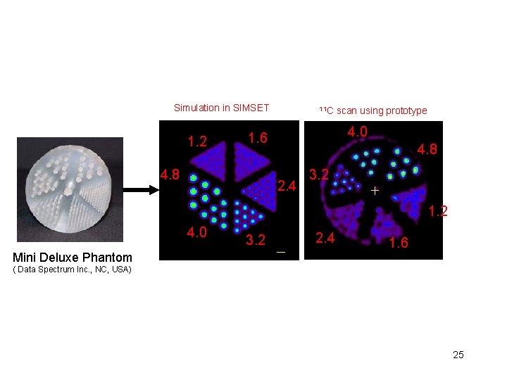
Simulation in SIMSET 1. 2 11 C 4. 0 1. 6 4. 8 scan using prototype 4. 8 2. 4 3. 2 1. 2 4. 0 Mini Deluxe Phantom 3. 2 2. 4 1. 6 ( Data Spectrum Inc. , NC, USA) 25
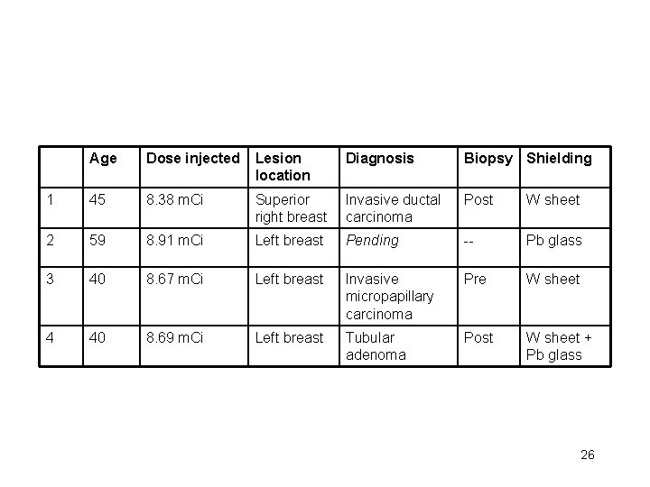
Age Dose injected Lesion location Diagnosis Biopsy Shielding 1 45 8. 38 m. Ci Superior right breast Invasive ductal carcinoma Post W sheet 2 59 8. 91 m. Ci Left breast Pending -- Pb glass 3 40 8. 67 m. Ci Left breast Invasive micropapillary carcinoma Pre W sheet 4 40 8. 69 m. Ci Left breast Tubular adenoma Post W sheet + Pb glass 26
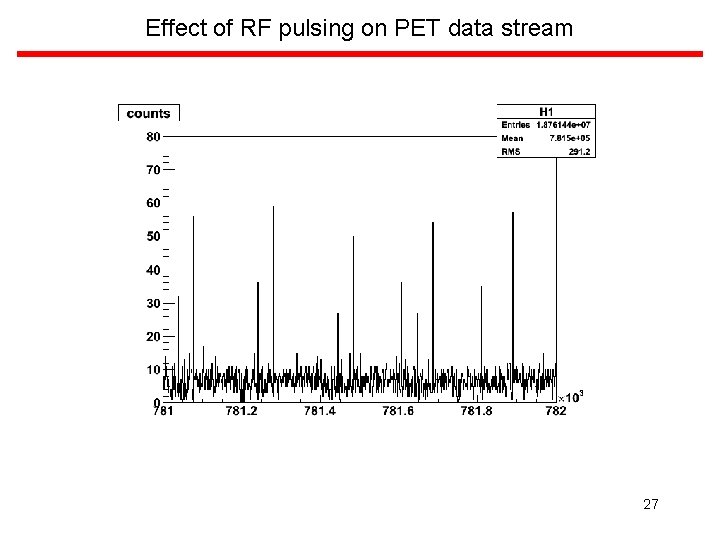
Effect of RF pulsing on PET data stream 27
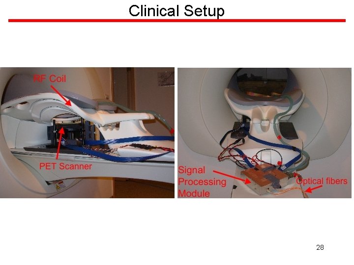
Clinical Setup 28
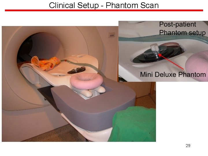
Clinical Setup - Phantom Scan 29
- Slides: 29