RESPONDING TO THE ENVIRONMENT HUMANS HUMAN NERVOUS SYSTEM
 HUMAN NERVOUS SYSTEM PRESENTER: Ms Z. Sanda")


")
- Slides: 22
RESPONDING TO THE ENVIRONMENT (HUMANS) HUMAN NERVOUS SYSTEM PRESENTER: Ms Z. Sanda
EXAMINATION GUIDELINES
INTRODUCTION • All living organisms must be able to respond to the environment in order to survive. • To achieve this, humans have two co-ordinating systems which allow them to adapt to changes within the environment and maintain their internal conditions. • In humans there are two co-ordinating systems, namely, the nervous system and the endocrine system. • The nervous system is made up of nerves and is a fast responding system. • The endocrine system involves hormones which are released into the blood and is a much slower responding system.
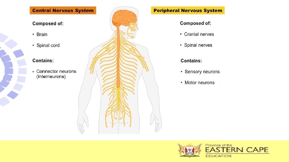
ORGANIZATION OF THE NERVOUS SYSTEM
NERVOUS SYSTEM • All bodily activities, voluntary or involuntary are controlled by the nervous system → it is the body’s control & communication centre • It consists of receptors to detect stimuli from the environment and convert these stimuli into nerve impulses which are transmitted by specialized cells called neurons to the central nervous system ( brain and spinal cord) for interpretation and from there to the effectors such as glands or muscles which respond to stimuli and allow for changes to take place.
Key Terminology • Receptor • Effector − Structure that receives a stimulus and converts it into an impulse e. g. touch receptors in the skin, light receptors in the eye − Structure in the body that responds to a stimulus e. g. muscles and glands • Stimulus (stimuli-pl) − A detectable change in the internal or external environment e. g. internal – body temperature, water, sugar levels; external – pain, environmental temperature, touch, danger • Impulse − An electrochemical message transmitted by nerves • Neurons − Specialized cells in the nervous system that transmit impulses
FUNCTIONS OF NERVOUS SYSTEM • It detects changes in the environment, thus allows the body to react to these changes. • It allows co-ordination of the various activities of the body.
PERIPHERAL NERVOUS SYSTEM • Is all the nervous tissue outside the CNS • Consists of: Ø 12 pairs of cranial nerves. Ø 31 pairs of spinal nerves. • These nerves form a communication network all over the body
ORGANISATION OF THE PERIPHERAL NERVOUS SYSTEM SYMPATHETIC PERIPHERAL AUTONOMIC regulates the internal environment so that homeostasis can be maintained. Carries information from the CNS to the smooth muscles and glands (INVOLUNTARY) SOMATIC carries information from the sense to the CNS and from the CNS to the skeletal muscles (VOLUNTARY) responsible for the fight or flight (action) function in emergency situations (AROUSAL) PARASYMPATHETIC restores the body to a normal state (rest) after an emergency situation. (CALMING) NOTE: The sympathetic and parasympathetic system work antagonistically to each other i. e. have opposite effects
Differences between the Sympathetic and Parasympathetic Divisions of the Autonomic Nervous System Sympathetic nervous system Parasympathetic nervous system • Mostly stimulating in effect • Increases heart rate • Constricts many arteries • Relaxation of the bladder wall • Dilates pupils • Inhibits peristalsis • Widens bronchioles • Mostly inhibitory in effect • Decreases heart rate • Dilates arteries • Contraction of bladder wall • Constricts pupils • Accelerates peristalsis • Constricts bronchioles
FUNCTIONS OF PERIPHERAL NERVOUS SYSTEM • Transmits impulses from the receptors to the central nervous system via the sensory neurons • Transmits impulses from the central nervous system to the effectors via the motor neurons
STRUCTURE AND FUNCTIONING OF THE NERVE • Nerves are made up of nerve fibres that are held together by connective tissue. • The nerve fibre in turn is made of specialised nerve cells called neurons which carry nerve (electrical) impulses through the body • Neurons are therefore regarded as structural units of nervous system. Nerves Nerve fibres neurons
3 MAIN TYPES OF NEURONS Sensory neuron Motor neuron 14
Interneuron/ Connector Neuron 15
TYPES OF NEURONS • Sensory neuron • Connector or interneuron • Motor neuron • carries the impulses from the receptors (nerve ending or sense organ) to the CNS (brain and spinal cord) • carries impulses inside the central the CNS from sensory to motor neuron • carries the impulses from CNS to the effector organs (muscle and glands)
FLOW OF INFORMATION THROUGH THE BODY
STRUCTURE OF A NEURON All neurons are made up of three basic parts namely; cell body, dendrites and an axon • dendrites – transmitting impulses towards the cell body • cellbody (with a nucleus) – controlling the metabolism of the cell • an axon – transmitting impulses away from the cell body • myelin sheath – insulating the axon and speeding up the transmission of impulses; myelin sheath is enclosed by a membrane called the neurilemma which helps to repair damaged neurons Cytoplasm
TRANSMISSION OF NERVE IMPULSES • More than one neuron is involved when neurons carry nerve impulses. • The axon terminals of one neuron lie next to the dendrites of the next neuron. • Neurons are not directly connected to each other • The microscopic gap between axon terminals of one neuron and the dendrite of another neuron is called a synapse • Nerve impulses are carried across a synapse by means of chemical substance called neurotransmitters e. g. Acetylcholine
Revision questions 1. Give the correct biological term for each of the following descriptions. 1. 1 A part of the neuron that conducts impulses towards the cell body DENDRITE 1. 2 The microscopic space between two adjacent neurons is a/an SYNAPSE 1. 3 A change in the internal or external environment that will be detected by a receptor and converted into an impulse STIMULUS 1. 4 Chemical substance which carries impulses across the synapse NEUROTRANSMITTER 1. 5 A part of the neuron that insulates the axon and speed up transmission of impulses MYELIN SHEATH
2.
2. 2 2. 3 2. 4 Identify the type of neuron represented by: (a) B MOTOR NEURON (b) C CONNECTOR NEURON / INTERNEURON (c) E SENSORY NEURON Give the LETTER only of the part that represents the: (a) Receptor F (b) Effector A Give the LETTER and NAME of the: (a) Region where the impulse is transmitted chemically D -SYNAPSE (b) Part that has an insulating function G -MYELIN SHEATH