RESPONDING TO THE ENVIRONMENT CENTRAL NERVOUS SYSTEM MRS


- Slides: 32
RESPONDING TO THE ENVIRONMENT CENTRAL NERVOUS SYSTEM MRS R BISHOP
WHAT THE GUIDELINES SAY…
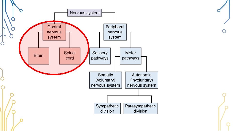
NERVOUS SYSTEM Central Nervous System Peripheral Nervous System
STRUCTURE & FUNCTION OF THE BRAIN Ø It has the following functions: • Receives and interprets all sensations (sight, hearing, smell, taste etc. ) • Controls higher thought processes such as memory, judgement, reasoning etc. • Controls voluntary and involuntary actions • Co-ordinates voluntary actions e. g. walking running • Maintains muscle tone, balance and equilibrium Ø The brain is the enlarged upper part of the spinal cord. Ø It is made up of interneurons
Protection of the Brain CEREBRUM CRANIUM It is well protected by: Ø The bony skull Ø 3 membranes called meninges. Ø The cerebrospinal fluid MENINGES CEREBROSPINAL FLUID
Protection of the Brain • The outer tough dura mater forms the lining for the skull. • The middle arachnoid membrane • The inner delicate pia mater has many blood vessels to supply oxygen & food • The space between the meninges is filled with cerebrospinal fluid which absorbs shock & prevents damage to nervous tissue. It also provides oxygen & food to neurons & removes waste products.
Protection of the Brain • Cerebral Spinal Fluid is also found in ventricles in the brain • And between the meninges that surround the spinal cord ? Meningitis
PARTS OF THE BRAIN Cerebrum Medulla Oblongata
Parts of the Brain Hypothalamus
• The CNS has two kinds of tissue: • grey matter and white matter Grey matter, which has a pinkishgrey color in the living brain, contains the cell bodies, dendrites and axon terminals of neurons White matter is made of axons connecting different parts of grey matter to each other. White matter is made of axons covered by fatty myelin sheath
PARTS OF THE BRAIN: MEDULLA OBLONGATA • The medulla oblongata is the lowest part of the brain and is an extension of the spinal cord • It has white matter on the outside and grey matter on the inside • This is where nerve change over from the left side of the body to the right side of the brain and visa versa Its functions are: • It transmits nerve impulses between the spinal cord and the higher parts of the brain • It controls autonomic functions, including the following: § § rate and depth of breathing heart beat Narrowing and widening of the blood vessels Blinking Corpus callosum
PARTS OF THE BRAIN: THE CEREBELLUM PAR • Situated below the cerebrum and behind the Pons and medulla oblongata • • Functions are: • It controls the tension in the muscles (muscle tone) to maintain balance and posture • Controls muscle activity responsible for speech It co-ordinates and controls the actions of all voluntary muscles to make smooth and precise movement possible e. g. walking, skipping and running Corpus callosum
PARTS OF THE BRAIN: HYPOTHALAMUS* PAR • The thalamus is situated under the corpus callosum and the hypothalamus is situated under the thalamus • It plays a role in the maintenance of homeostasis, by regulating the following: § § § • • Hypothalamus Corpus callosum Blood pressure Heartbeat Body temperature Water balance Hunger, appetite, thirst Sleep It plays a role in the control of emotions such as anxiety, anger and fear It controls the functioning of the pituitary gland, which secretes several vitally important *The hypothalamus is not mentioned here in the guidelines but comes uphormones in other chapters of work
PARTS OF THE BRAIN: THE CEREBRUM • Occupies 2/3 of the cranial space PAR • Divided into right and left hemisphere • The right hemisphere controls actions on the left side of the body and visa versa • The two hemispheres are connected by the corpus callosum • It is highly folded to increase surface area It’s functions are: • Controls voluntary actions • Interpret information from the sense organs. e. g. sight, sound, smell, taste and touch can arise • Seat of higher mental functions such as memory, intelligence etc. Corpus callosum
THE CEREBRUM CAN BE DIVIDED INTO VARIOUS AREAS Frontal lobe – problem solving, emotions, planning, decision making etc. Parietal lobe – touch, spacial orientation, Non-verbal thought Occipital lobe – vision, colour perception Temporal lobe – hearing, speech, memory etc
PARTS OF THE BRAIN: THE PARCORPUS CALLOSUM Corpus callosum • Corpus callosum consists of about 200 million axons that interconnect the left and right hemisphere of the brain • The primary function of the corpus callosum is to allow communication between the left and right side of the brain. .
PARTS OF THE BRAIN: THE CORPUS CALLOSUM PAR Interesting Fact: *At least 1 in 3000 infants is born without a corpus callosum. . Many born without this structure go undiagnosed for years—only neuroimaging can confirm the failed development of this brain area. Instead people are diagnosed with disorders such as autism, depression, or ADHD. It can cause *Physical problems *Mental processing problems *Social problems *Developmental problems This insignificant part of the brain may be a lot more important than we think. Corpus callosum
How much can you Give the nameremember? and function of each of the parts! 1. Cerebrum – controls all voluntary movements and functions - interprets all sensations - responsible for all higher order thinking 2. Corpus callosum – allows communication between left and right side of the brain 3. Cerebellum – co-ordinates and controls voluntary movement - responsible for balance - controls muscular activity responsible for speech 4. Medulla oblongata – controls autonomic functions such as heart beat, breathing and blood vessel dilation - transmits nerve impulses from spinal cord to higher brain
Questions
STRUCTURE & FUNCTION OF THE SPINAL CORD Ø It is an elongated, cylindrical tube Ø It is protected by the vertebral column and the 3 meninges (dura mater, arachnoid, pia mater) Ø 31 pairs of nerves leave the spinal cord
The spinal canal has 3 parts: Ø Inner central canal containing cerebrospinal fluid which supplies neurons with food & oxygen Ø H-shape grey matter which has dorsal and ventral horn. Ø The white matter on the outside forms bundles of nerve fibres running along the spinal cord, to and from the brain. The brain has grey matter on the outside and white matter on the inside. The is opposite to the spinal cord which has white matter on the outside and grey on the inside.
Ø Sensory neurons enter on the dorsal route. Ø The cell bodies of the sensory neuron are outside the spinal cord in a swelling called the dorsal root ganglion. Ø Connector/Inter- neurons are found in the grey matter. Ø Motor neurons leave the spinal cord on the ventral route
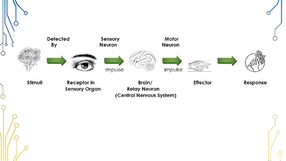
How does coordination take place? STIMULUS RECEPTOR COORDINATOR EFFECTOR RESPONSE Cold outside Change in the internal or external environment Cold receptors in the skin Sense receptors Brain (via spinal cord) Sensory neuron Brain Motor or neuron Spinal Cord Muscles of arms Put on jersey Muscle or Gland The action that takes place
Pathway of an impulse to the brain Ø Sense receptors pick up the stimulus (which is the changes in the environment around us e. g. sight) Ø They convert the stimulus to an impulse. Ø The impulse is sent along a sensory nerve to the CNS (spinal cord or brain). Ø The sensory neurons synapse with interneurons. Ø The interneurons conduct the impulse to the appropriate area of the brain where it is interpreted. Ø The message is sent back along the interneuron which synapses with the motor neuron.
The spinal canal: Pathway of an impulse to the brain CONSIDER THE FOLLOWING SITUATION: Mr and Mrs Abraham were in a serious car accident. When they arrived at the hospital the doctor assessed there condition. Ø Mrs Abraham was in a lot of pain especially in her legs that were crushed but she could not move her legs. Ø Mr Abrahams on the other hand could move all his limbs but although his leg was severely broken and cut he could not feel the pain. The doctor noted that there had been
Mrs Abrahams – motor neuron was damaged. Message could be sent to brain on sensory neuron so therefore she felt pain. But no message travelled from brain to muscle. Therefore motor neuron must be damaged
Mr Abraham – sensory neuron was damaged He could not feel pain so no message was going to the brain. He could move his foot so a message was coming from the brain on the motor neuron.
Functions of the spinal canal: Ø Pathway for impulses to and from the brain and from receptors and effectors Ø Serves as a reflex centre for certain actions