Respiratory System Unit 4 Ventilation Breathing the act

Respiratory System Unit 4
![�Ventilation [Breathing]- the act of moving air in/out of the lungs �Respiration- process where](http://slidetodoc.com/presentation_image_h2/416f05a91c643f567e1509df6c7d3436/image-2.jpg "�Ventilation [Breathing]- the act of moving air in/out of the lungs �Respiration- process where")
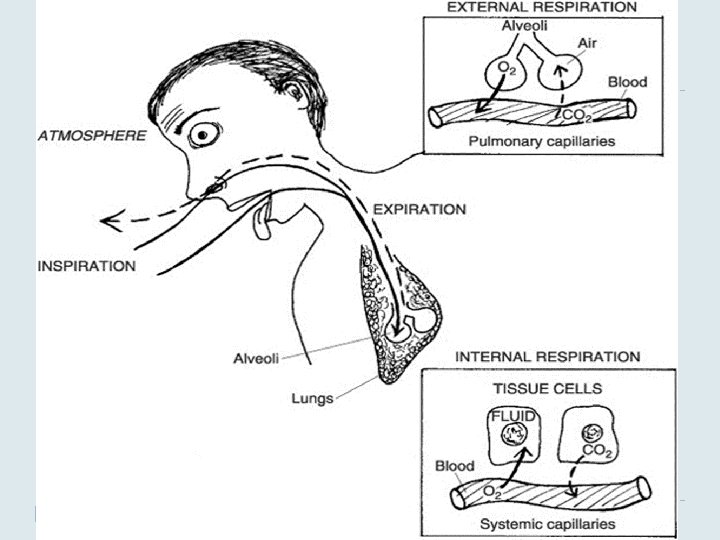
�Ventilation [Breathing]- the act of moving air in/out of the lungs �Respiration- process where ventilation occurs along with the exchange of gases in the lungs and the body tissues

Types of Respiration �External Respiration -exchange of gases between air in lungs and blood [alveoli and pulm. capillaries] �Internal Respiration -exchange of gases between the blood stream and body’s tissues

�Upper Respiratory Tract Organs - Nose, Nasal Cavity, Sinuses, Pharynx �Lower Respiratory Tract Organs - Larynx, Trachea, Bronchi, Lungs, Bronchial Tree, Alveoli
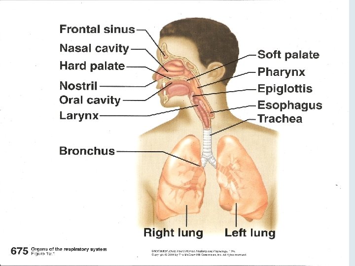

Upper Respiratory Tract Nose - made of bone and cartilage - nares/nostrils-openings guarded by hairs that filter Nasal Cavity -divided sagitally by nasal septum -lined with mucous membrane 1. Warms air 2. Moistens air 3. Traps particles
![� Nasal Conchae [superior, middle, inferior turbinate] -bony, scroll like projections on lateral wall](http://slidetodoc.com/presentation_image_h2/416f05a91c643f567e1509df6c7d3436/image-9.jpg "� Nasal Conchae [superior, middle, inferior turbinate] -bony, scroll like projections on lateral wall")
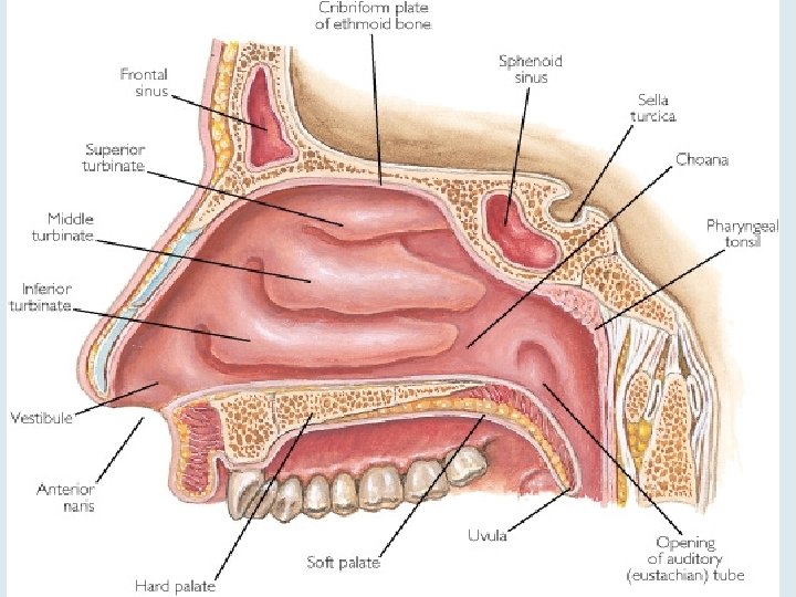
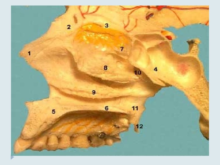
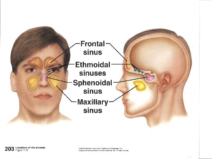
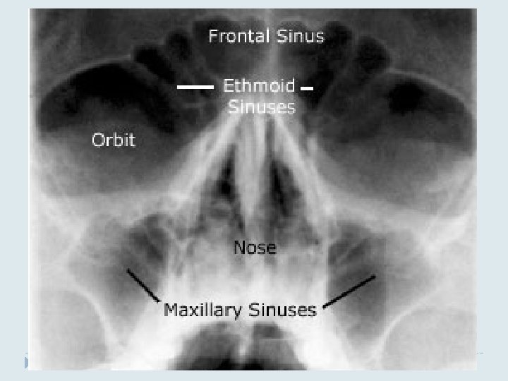
� Nasal Conchae [superior, middle, inferior turbinate] -bony, scroll like projections on lateral wall of each cavity -increase surface area inside [helps mucous membrane] -channel and speed up airflow through nose Sinuses[paranasal] -air filled hollow spaces inside 4 bones of skull [ethmoid, sphenoid, maxillary and frontal bones] - positioned around nose -lined with mucous membrane 1. Drain fluids into nasal cavity 2. Decrease weight of skull 3. Resonate the voice
![� Pharynx [throat] -3 regions: Nasopharynx-posterior to nasal cavity Oropharynx-posterior to oral cavity Laryngopharynx-](http://slidetodoc.com/presentation_image_h2/416f05a91c643f567e1509df6c7d3436/image-14.jpg "� Pharynx [throat] -3 regions: Nasopharynx-posterior to nasal cavity Oropharynx-posterior to oral cavity Laryngopharynx-")
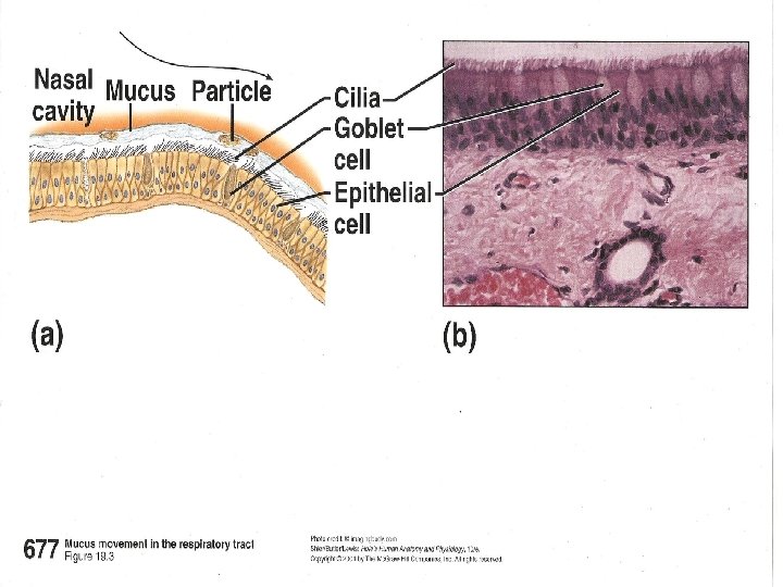
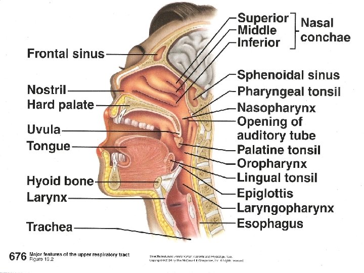
� Pharynx [throat] -3 regions: Nasopharynx-posterior to nasal cavity Oropharynx-posterior to oral cavity Laryngopharynx- posterior to larynx [voicebox] *Pseudostratified ciliated epithelial tissue lines most of tract. Cilia encourages flow of mucous -trapped particles travel down esophagus into stomach
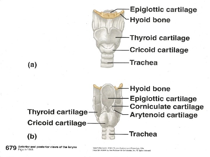
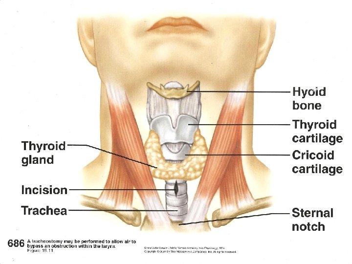
![Lower Respiratory Tract � Larynx [voicebox] -walls made of cartilage -Thyroid cartilage [adam’s apple]](http://slidetodoc.com/presentation_image_h2/416f05a91c643f567e1509df6c7d3436/image-16.jpg "Lower Respiratory Tract � Larynx [voicebox] -walls made of cartilage -Thyroid cartilage [adam’s apple]")
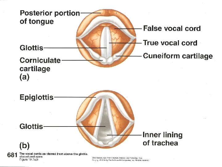
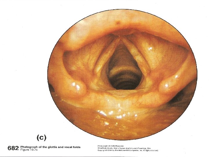
Lower Respiratory Tract � Larynx [voicebox] -walls made of cartilage -Thyroid cartilage [adam’s apple] -Epiglottic cartilage- covered with mucous membrane -epiglottis blocks opening to trachea [glottis] when swallowing False Vocal cords- musc/conn. tissue; same function as epiglottis True Vocal cords- musc/elastic fibers; vibrate with air passage; helps form words
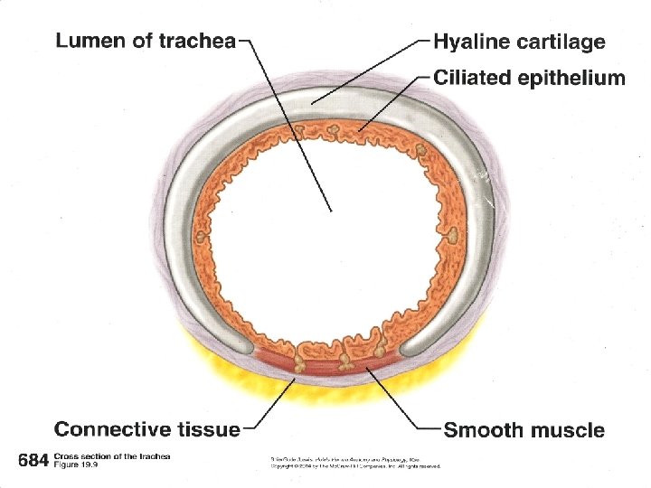
![� Trachea [windpipe] -Anterior to esophagus -4 -5 inches long -Connects larynx to thoracic](http://slidetodoc.com/presentation_image_h2/416f05a91c643f567e1509df6c7d3436/image-20.jpg "� Trachea [windpipe] -Anterior to esophagus -4 -5 inches long -Connects larynx to thoracic")
� Trachea [windpipe] -Anterior to esophagus -4 -5 inches long -Connects larynx to thoracic cavity -Divides inferiorly at an area called the carina, into R and L primary bronchi (each lung) -Lined w/ciliated mucous membrane -Made of “C”-shaped cartilage rings allowing for expansion of esophagus when consuming food -Also smooth muscle
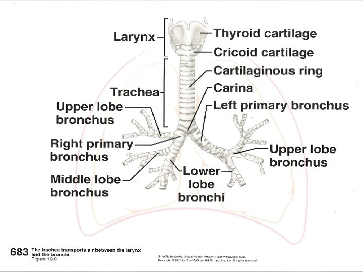

Primary bronchi lead into the lungs and divide to form the bronchial tree Primary bronchi secondary bronchi tertiary bronchioles alveolar ducts alveoli
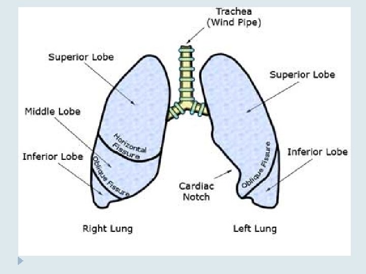
![Lungs [Right and Left] -Right – 3 lobes (upper, middle, lower) – divided by](http://slidetodoc.com/presentation_image_h2/416f05a91c643f567e1509df6c7d3436/image-25.jpg "Lungs [Right and Left] -Right – 3 lobes (upper, middle, lower) – divided by")
Lungs [Right and Left] -Right – 3 lobes (upper, middle, lower) – divided by 2 fissures (horizontal b/w upper and middle lobes; oblique fissure b/w middle and lower) -Left - 2 lobes (upper, lower) – divide by oblique fissure. Lung is slightly smaller to accommodate space for the heart

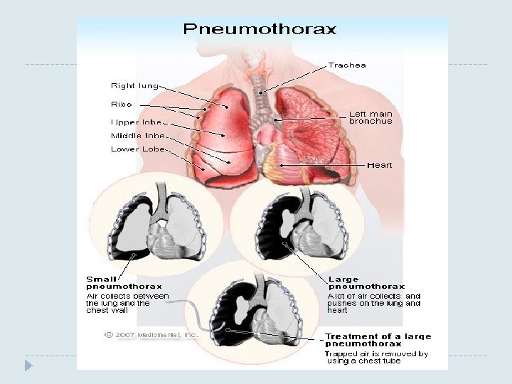
Pleural Cavity -double-layered membrane covering -visceral pleura: inner layer on the lung surface -parietal pleura: outer layer lines the inside of the thoracic cavity -space in b/w the pleural layers filled with serous fluid to reduce friction as lungs inflate and deflate. Pleurisy- infection of the pleural layers. Reduced serous fluid and increased friction when breathing
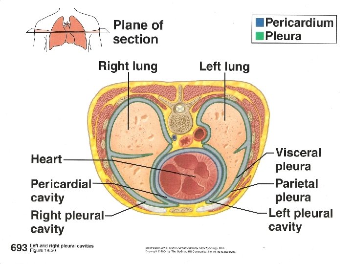

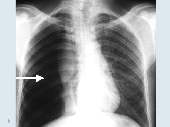
-Serous fluid creates surface tension b/w the pleural layers -keeps them adhered to each other -essential for keeping the lungs inflated. � When the outer pleural membrane (parietal pl. ) moves outward because it is attached to the thoracic wall , the inner pleural layer (visceral pl. ) moves with it. � Pneumothorax: air gets in the pleural space � Atelectasis: too much air gets in the pleural space disrupting surface tension b/w the pleural layers and causes collapsed lung.
- Slides: 32