Respiratory system Regulation of breathing Control of ventilation

Respiratory system

Regulation of breathing

Control of ventilation https: //sleep. sharepoint. com/siteimages/Chapter%203. png

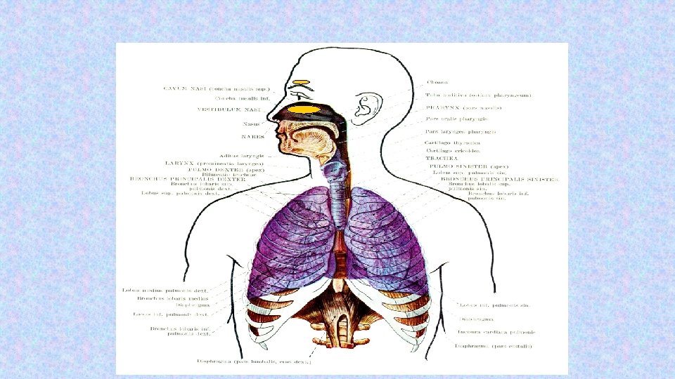
• Breathing is an automatic process that takes place unconsciously. Automaticity of breathing comes from regular (rhythmic) activity of groups of neurons anatomically localized in the medulla and its vicinity.

• They can be divided into three main groups: • dorsal respiratory group – placed bilaterally on the dorsal side of the medulla oblongata, only inspiratory neurons, sending axons to motoneurons of inspiratory muscles (diaphragm, external intercostal muscles; their activation=inspiration, their relaxation=expiration; participates on inspiration at rest and forced inspiration • ventral respiratory group - located on the ventrolateral part of the medulla oblongata, the upper part: neurons whose axons of motor neurons activate the main and auxiliary inspiratory muscles; the lower part: expiratory neurons which innervate expiratory muscles (internal intercostal muscles). Neurons in this group operate only during forced inspiration and forced expiration. • Pontine respiratory group - pneumotaxic center - dorsally placed on top of the pont, contributes to the frequency and depth of breathing; affects the activity of respiratory neurons in the medulla oblongata.

Chemical factors affecting the respiratory center: Central chemoreceptors - on the front side of the medulla - sensitive only to increase of arterial p. CO 2 (by increasing H+ ) - Notice: - central chemoreceptor are stimulated by other types of acidosis (lactate acidosis, ketoacidosis)

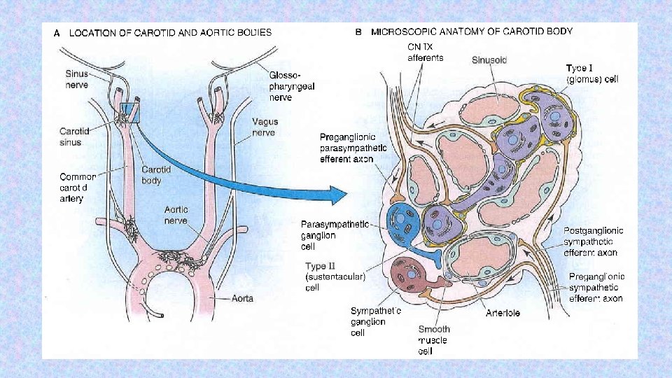
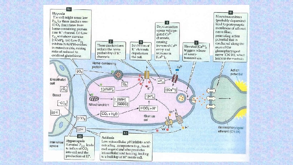
Peripheral chemoreceptors – located in the aortic and carotid bodies -primarily sensitive to decrease in arterial p. O 2, particularly to decrease of O 2 under 10 -13 k. Pa in the arterial blood. They convey their sensory information to the medulla via the vagus nerve and glossopharyngeal nerve. Mechanism of action: Decreased ATP production in mitochondria leads to depolarization of receptors membrane and to excitation of chemoreceptor http: //www. medicine. mcgill. ca/physio/resp-web/sect 8. htm,

Modulation of respiratory output Major parameters for feedback control – classical gases: p. O 2, p. CO 2, p. H In additin to these, the respiratory system receives input from two other major sources: 1. variety of stretch and chemical/irritant receptors that monitor the size of airways and the presence of noxious agentsreceptors in respiratory system 2. Higher CNS centers that modulate respiratory activity for the sake of nonrespiratory activities Irritants receptors on mucose of respiratory system – rapidly adapting Stimulus: agens - chemical substances (histamin, serotonin, prostaglandins, ammonia, cigarette smoke). Respons: increase mucus secretion, constriction of larynx and brochus C-fibre receptors (juxtacapillary=J receptors)– free nerve ending of n. vagus (unmyelinated axon) in intersticium of bronchus and alveolus; Stimulus: Mechanical irritans (pulmonary hypertension, pulmonary oedema)+chemical Response: hypopnoe, rapid shallow breathing, bronchoconstriction, cough Stretch receptors slowly adapting (mechanoreceptors in tracheobronchial tree that detect the changes in lung volume by sensing the stretch receptors of the airway wall), inform to brain about the lung volume to optimize respiratory; its irritants triggered decrese activity of respiratory centre – Hering-Breuer´s reflexes. (protecting the lungs from overinflation/deflation)

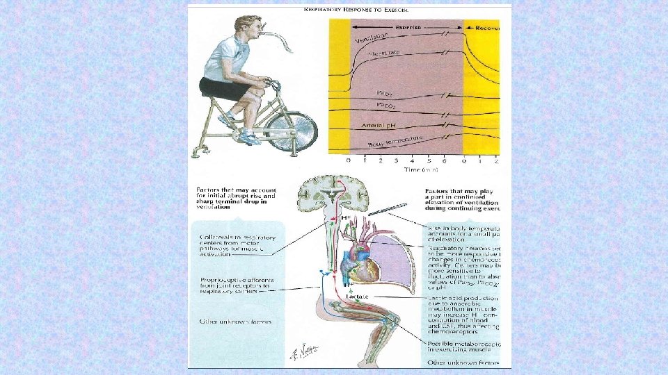
Baroreceptors – suppresses activity of respiratory centre Irritants of proprioreceptors of muscles, tendons during active and pasive movements of limbs Influenced activity of respiratory neurons (increase minute ventilation during work load) Limbic system, hypothalamus – strong pain, emotion Tractus corticospinalis =cortex – activated RC during work load temperature

• Types of anesthaesia in animal experiments Intra-muscular application = i. m. inhalation 13

• Anatomy of neck in laboratory rat ENDOTRACHEAL CANNULA TRACHEA A. CAROTIS N. VAGUS Upraveno dle: Poopesko Peter a kol. (1990) 14

HERING-BREUER „inflation“ REFLEX STOP BREATHING Breathing at rest ARTIFACTS Breathing at rest 15

Changes of breathing after VAGOTOMY inspirium One-side VAGOTOMY expirium Both-side VAGOTOMY 16

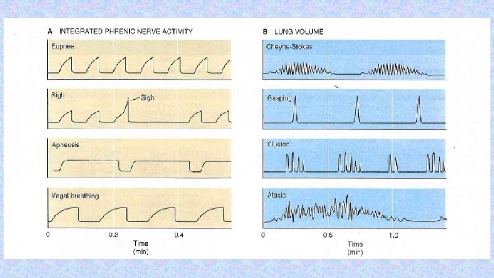
Periodic breathing • It is not regular, rhythmic, but respiration occurs in periods ("a moment to breathe, take a moment to not breathe„) • CHEYNE-STOKES • BIOT‘S • „gasping“ • KUSSMAUL

Hypoxia, hypoxemia • Hypoxia is a general name for a lack of oxygen in the body or individual tissues. • Hypoxemia is lack of oxygen in arterial blood. • Complete lack of oxygen is known as anoxia. The most common types of hypoxia: 1. Hypoxic - physiological: stay at higher altitudes, pathological: hypoventilation during lung or neuromuscular diseases 2. Transport (anemic) - reduced transport capacity of blood for oxygen (anemia, blood loss, CO poisoning) 3. Ischemic (stagnation) - restricted blood flow to tissue (heart failure, shock states, obstruction of an artery) 4. Histotoxic - cells are unable to utilize oxygen (cyanide poisoning - damage to the respiratory chain)

p. O 2 = 1 mm. Hg

Hypercapnia • Hypercapnia - increase of concentration of carbon dioxide in the blood or in • • tissues that is caused by retention of CO 2 in the body possible causes: total alveolar hypoventilation (decreased respiration or extension of dead space) mild hypercapnia (5 -7 k. Pa) causes stimulation of the respiratory center (therapeutic use: pneumoxid = mixture of oxygen + 2 -5% CO 2) hypercapnia around 10 k. Pa - CO 2 narcosis - respiratory depression (preceded by headache, confusion, disorientation, a feeling of breathlessness) hypercapnia over 12 k. Pa - significant respiratory depression - coma and death.

Travelling by aircraft (On board aicraft is pressure as on 2000 m above sea level) High risk for patients with diseases: - concentration of hemoglobin lower than 60 % - severe step of atherosclerosis - cardial insuficiency - respiratory insuficiency - non-treated hypertension (BP ower 200/100 mm. Hg)

Toxicity of oxygen The toxicity seems to be due to the production of the superoxid anion and H 2 O 2 Causes: - lost of possibility binding CO 2 in venous blood - in lungs – pulmonary edoema – decrease CO 2 expenditure Critical values > 40 k. Pa (300 mm. Hg) –dependence on time

Toxicity of oxygen Exposure – 8 hours: - respiratory passages became irritated - Substernal distress - Nasal congestion - Sore throat - Cough - 24 -48 hours: - damage of lungs – decrease production of surfactant

Recommendation: 100 % - give discontinuosly

THANK YOU FOR YOUR ATTENTION
- Slides: 27