RESPIRATORY SYSTEM II EXTRAPULMONARY BRONCHUS primry BRONCHUS Generally
")
RESPIRATORY SYSTEM (II)
 Generally have the same histological appearance as")
ﺧﺎﺭﺝ ﺍﻟﺮﺋﻪ EXTRAPULMONARY BRONCHUS (primry BRONCHUS) Generally have the same histological appearance as the trachea. ﺍﺫﺍ ﺭﺍﺟﻊ ﺍﻟﺪﺭﺱ ﺍﻟﺴﺎﺑﻖ ﻟﻠﺘﻔﺎﺻﻴﻞ
 ﺗﺘﻜﻮﻥ ﻫﻴﺴﺘﻮﻟﻮﺟﻴﺎ ﻣﻦ 1234")
INTRAPULMONARY ﺩﺍﺧﻞ ﺍﻟﺮﺋﻪ BRONCHI (2 ry & 3 ry BRONCHI) ﺗﺘﻜﻮﻥ ﻫﻴﺴﺘﻮﻟﻮﺟﻴﺎ ﻣﻦ 1234 - Mucosa. Muscle coat. Submucosa. Adventitia. ﺗﻔﺼﻞ ﻻﺣﻘﺎ

INTRAPULMONARY BRONCHUS

INTRAPULMONARY BRONCHUS
 Mucosa: It has longitudinal ﻃﻮﻟﻲ mucosal folds. a- Epithelium: Respiratory epith.")
INTRAPULMONARY BRONCHUS (1) Mucosa: It has longitudinal ﻃﻮﻟﻲ mucosal folds. a- Epithelium: Respiratory epith. b- L. P. : (Lamina propria) ﻳﺘﻜﻮﻥ ﻣﻦ Fibroelastic C. T. (loose C. T. rich in elastic fibers). It contains seromucous glands. “ “ lymphoid elements. N. B. No elastic lamina ﺍﻟﺪﻛﺘﻮﺭ ﻳﻘﻮﻝ ﻛﻠﻤﻪ ) ﻧﻮ ( ﻣﻌﻨﺎﻫﺎ ﺳﺆﺎﻝ ﻳﻌﻨﻲ ﺭﻛﺰ ﻋﻠﻴﻬﺎ.
 Muscle coat (complete): Two distinct ﻣﺨﺘﻠﻔﺘﻴﻦ layers of SMF ( smooth")
INTRAPULMONARY BRONCHUS (2) Muscle coat (complete): Two distinct ﻣﺨﺘﻠﻔﺘﻴﻦ layers of SMF ( smooth muscle fibers ) spirally ﺣﻠﺰﻭﻧﻲ arranged in opposite direction (crisscrossing ﻣﺘﻘﺎﻃﻌﻪ bundles of spirally arranged smooth muscle fibers “SMF”). (3) Submucosa: C. T. contains: a- Seromucous glands. b- Lymphoid elements.
 Adventitia: Contents: a- Loose C. T. : Contains radially arranged elastic")
INTRAPULMONARY BRONCHUS (4) Adventitia: Contents: a- Loose C. T. : Contains radially arranged elastic fibers to connect with counterparts of neighboring bronchial tree. b- Irregular plates of hyaline cartilage (complete layer). c- Solitary lymphoid nodules. ﺍﻱ ﺷﻴﺀ ﺍﺣﻤﺮ ﻭﻋﻠﻴﻪ ﺧﻂ ﻫﺬﺍ ﺭﻛﺰ ﻋﻠﻴﻪ ﺍﻟﺪﻛﺘﻮﺭ ﻭﻫﺬﺍ ﻓﻲ ﻛﻞ ﺍﻟﻤﺤﺎﺿﺮﻩ

BRONCHIOLES ﻻ ﻳﻮﺟﺪ ﺑﻬﺎ ﻏﻀﺎﺭﻳﻒ ﻭﻻ ﺑﺎﻟﺘﻲ ﺑﻌﺪﻫﺎ ﻋﺸﺎﻥ ﻛﺬﺍ ﻣﻦ ﻫﻨﺎ ﺍﻧﺴﻰ ﺷﻴﺀ ﺍﺳﻤﻪ ﻏﻀﺎﺭﻳﻒ 1 - Preterminal ( ﻗﺒﻞ ﺍﻟﻨﻬﺎﺋﻴﻪ 1 ry ) Bronchioles (Bronchioles): Are less than 1 mm in diameter. Each bronchiole supplies pulmonary lobule. 2 - Terminal ( ﺍﻟﻨﻬﺎﺋﻴﻪ 2 ry ) Bronchioles. 3 - Respiratory ( 3 ry ) Bronchioles.

BRONCHIOLE
 Mucosa: has longitudinal folds: A- Epithelium: 1 - Simple ciliated columnar")
Preterminal Bronchioles (1) Mucosa: has longitudinal folds: A- Epithelium: 1 - Simple ciliated columnar epith ﻣﻬﻤﻪ ﺟﺪﺍ ﻣﻌﺮﻓﻪ ﻧﻮﻉ ﺍﻻﺑﻴﺜﻴﻠﻮﻳﻢ ﻻﻧﻪ ﻣﺤﻞ ﻣﻘﺎﺭﻧﻪ ﻻﻧﻪ ﺳﻮﻑ ﻳﺘﻐﻴﺮ ﻓﻲ ﺍﻟﺘﺮﺍﻛﻴﺐ ﺍﻟﻘﺎﺩﻣﻪ. with occasional goblet cells (in larger preter. br. ). 2 - ﻣﻬﻤﻪ : ﺗﺮﻛﻴﺐ ﺍﻟﺠﺰﺀ ﺍﻻﺧﻴﺮ ﻣﻨﻬﺎ Simple cuboidal mostly ciliated with occasional Clara cells BUT NO goblet cells (in smaller preter. bronchioles). B- Lamina propria: Fibroelastic C. T. (rich in elastic Fs. ) (2) Smooth muscle: 2 helically arranged SM layers. (3) Adventitia: loose fibroelastic C. T. N. B ﻣﻬﻤﻪ ﺟﺪﺍ. No cartilage, No seromucous glands, No lymph nodu. Les.

TERMINAL BRONCHIOLE

Terminal Bronchioles Similar structure to preterminal. bronchioles, but: Epithelium: Simple cuboidal partially ciliated epithelium With Clara cells. ﻣﺜﻞ ﺗﺮﻛﻴﺐ ﺍﻟﺠﺰﺀ ﺍﻻﺧﻴﺮ ﻣﻦ ﺍﻟﺴﺎﺑﻘﻪ N. B. Are less than 0. 5 mm in diameter. N. B. Each supplies lung acinus.

RESP. BRONCHIOLE & ALV. DUCT

Respiratory Bronchioles Are similar in structure to terminal bronchioles. But: their walls are interrupted by the presence of few pulmonary alveoli. ﻣﻬﻤﻪ ﺟﺪﺍ ﻭﺧﺼﻮﺻﺎ ﺑﺎﻟﻌﻤﻠﻲ
. Dome-shaped apices with microvilli. Numerous apical secretory")
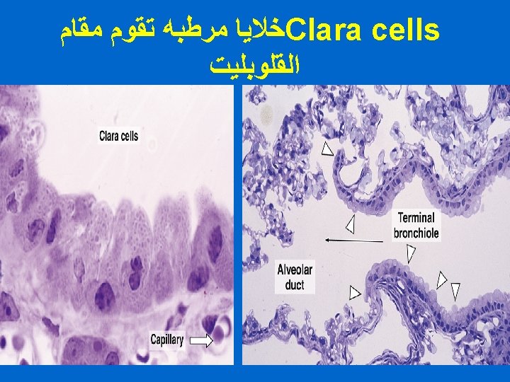
CLARA CELLS Structure: columnar cells (non ciliated). Dome-shaped apices with microvilli. Numerous apical secretory granules (of glycoproteins). Abundant r. ER. Function: ﻣﻬﻤﻪ 1 - Protect the bronchiolar epith. by their glycoproteins secretion. 2 - Degrade toxins in inhaled air. 3 - Divide to regenerate the bronchiolar epith. 4 - Produce surfactant-like material. : ( ﻣﻮﺍﻗﻊ ﺍﻧﺘﺸﺎﺭﻫﺎ ) ﻣﻬﻤﻪ ﺟﺪﺍ 1 - last portion of preterminal bronchioles 2 - all terminal bronchioles 3 - all respiratory portion

ALVEOLAR DUCTS The wall of alveolar ducts consist almost of pulmonary alveoli. Alveolar ducts do NOT have walls of their own; They are merely ﻓﻘﻂ linear arrangement of pulmonary alveoli. N. B. Alveolar duct → ends by: atrium → communicates with: 2 -3 alveolar sacs


PULMONARY ALVEOLI Definition: They are small outpouchings ﺗﺠﺏ ﺧﺎﺭﺟﻲ of respiratory bronchioles, alveolar ducts & alveolar sacs. Topics: ﺍﻟﻌﻨﻮﺍﻧﻴﻦ ﺍﻟﺘﻲ ﺳﺘﺘﻢ ﻣﻨﺎﻗﺸﺘﻬﺎ *Interalveolar septa. *Blood-air barrier ( Blood-gas barrier) *Alveolar epithelium. *Lung macrophages (alveolar macrophages).
")
1 - INTERALVEOLAR SEPTA Definition: The region between 2 adjacent ﻣﺘﺠﺎﻭﻩ alveoli. Components: (A) Alveolar Epithelium: lines both sides of interalveolar septum. (B) Interstitium.

INTERALVEOLAR SEPTA & PULMONARY ALVEOLI
 Type I Pneumocytes (Type I alveolar cells) (Squamous alveolar cells). (2)")
ALVEOLAR EPITHELIUM (1) Type I Pneumocytes (Type I alveolar cells) (Squamous alveolar cells). (2) Type II Pneumocytes (Type II alveolar cells) ( Septal cells) ( Great alveolar cells)

ALVEOLAR EPITHELIUM Type I Pn. Type II Pn.
 Type I Pneumocytes: - line 95% of the alveolar surface. -")
ALVEOLAR EPITHELIUM (1) Type I Pneumocytes: - line 95% of the alveolar surface. - Count: less numerous ﻻﻥ ﺣﺠﻤﻬﺎ ﻛﺒﻴﺮ than type II pneumocytes. - L/M: simple squamous epith. , highly attenuated cells. - E/M: Abundant pinocytotic vesicles, Are connected together and with type II cells by occluding junctions. -Function: Exchange of gases.
")
Type II Pneumocyte (E/M)

Type II Pneumocytes
 Type II Pneumocytes: - Line 5% of the alveolar surfaces. - Are more")
(2) Type II Pneumocytes: - Line 5% of the alveolar surfaces. - Are more numerous than type I pneumocytes. - L/M: Are cuboidal cells ( other textbooks: rounded cells). Usually found in groups of 2 -3 cells. Usually found at sites of union of septa. Foamy ﺭﺍﺋﻎ - ﻣﻨﺘﻔﺦ or vesicular cytoplasm. Nucleus: central, rounded, vesicular. ﻫﺬﺍ ﺍﻟﻨﻮﻉ ﻣﻦ ﺍﻟﺨﻼﻳﺎ ﻫﻮ ﺍﻟﻤﺴﺆﻮﻝ ﻋﻦ ﺍﻟﺘﻌﻮﻳﺾ ﻓﻬﻮ 2 ﻭﺗﺎﻳﺐ 1 ﻳﻌﻮﺽ ﺧﻼﻳﺎ ﺗﺎﻳﺐ

Type II Pneumocytes: - E/M: connected with type I cells by occluding junctions Dome-shaped ﻗﺒﻲ ﺍﻟﺸﻜﻞ apical surface. Short apical microvilli. Abundant mitochondria, RER , Well-developed Golgi. Membrane-bound Lamellar bodies (contain concentric or parallel lamellae limited by a unit membrane) (contain pulmonary surfactant).

Type II Pneumocytes: Function: 1 - Synthesis & secretion of pulmonary surfactant ﺍﻟﺘﻲ ﻭﻇﺎﺋﻔﻪ : a- It reduces effort to inflate pulm. Alveoli. b- It has bactericidal effect. 2 - Phagocytosis of pulmonary surfactant. 3 -Renewal of alveolar epithelial cells: Type II cells can divide to regenerate both type I & type II pneumocytes.
 Continuous Pulmonary Capillaries: -The richest capillary network in the")
Interstitium of interalveolar septa (1) Continuous Pulmonary Capillaries: -The richest capillary network in the body - Continuous blood capillaries - Endothelium shows numerous pinocytotic vesicles. (2) Interstitial C. T. : a- C. T. Fibers: elastic fibers & type III collagen (reticular fibers). b- C. T. Cells: Fibroblasts, Macrophages, Mast cells, Lymphocytes.

Blood-air barrier
 Definition: It is the region of the interalveolar septum that")
BLOOD-GAS BARRIER (BLOOD-AIR BARRIER) Definition: It is the region of the interalveolar septum that is traversed by O 2 & CO 2 Components: 1 - Thin layer of surfactant. 2 - Type I pneumocyte. 3 - Fused basal laminae of type I pneumocytes & endothelial cells of the pulmonary capillary. 4 - Endothelial cells of the pulmonary capillary.
 Sites: (1) In lumen of pulmonary alveoli. (2) In pulmonary")
Alveolar Macrophages (Dust Cells) Sites: (1) In lumen of pulmonary alveoli. (2) In pulmonary interstitium. Function: 1 - Phagocytose particulate matter (e. g. dust & bacteria)in the lumen of pulm. alveoli & in the interalveolar septa. 2 - Phagocytose part of the surfactant.
- Slides: 34