Respiratory system Digestive tract I Faculty of Pharmacy

 -hyalin cartilage Membranous part (1/4) -smooth muscle Trachealis muscle")


 •")


: 1. Circular folds(plicae")
 epithelium Mucosa lamina propria")


- Slides: 18
Respiratory system, Digestive tract I. Faculty of Pharmacy 3 rd Histology practice Department of Anatomy, Histology and Embriology 2018.
Trachea Cartilagenous part (3/4) -hyalin cartilage Membranous part (1/4) -smooth muscle Trachealis muscle
Trachea Pseudostratified ciliated columnar epitheliuum Mixed glands
• Bronchi • • Principal Lobar Segmental Terminal Bronchi The bronci contain hyalin cartilage, and glands in their wall. The epithelium is pseudostratified ciliated columnar epithelium. • Bronchiole • Terminal • Respiratory • Alveolar duct • Alveoli The broncioles does NOT contain any cartilage, neither glands. The wall contains smooth muscle. The epithelium is simple columnar, or cuboidal epithelium.
Bronchiole
Alveoli
• • • Oral cavity Pharynx Esophagus Stomach Small intestie Digestive tract • Duodenum • Jejunum • Ileum • Large intestine (colon) • Vermiform appendix Glands • Salivary glands • Liver • Pancreas
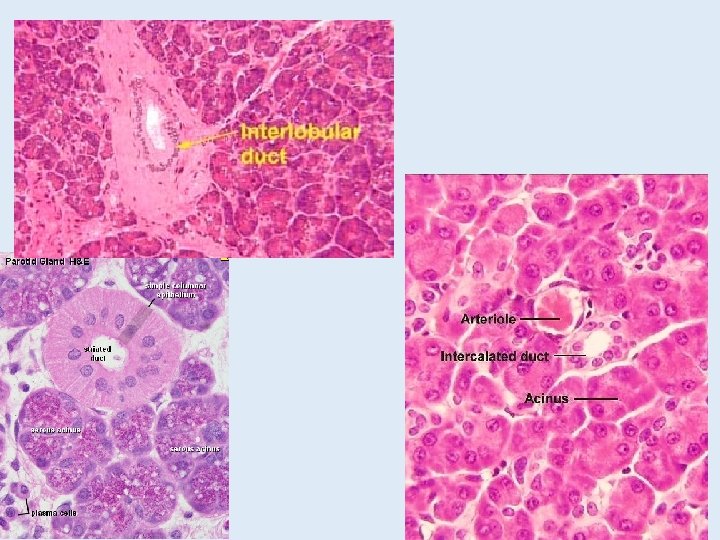
Salivary glands • Submandibular gland • Mixed gland Intercalated duct Striated duct Interlobular duct
Structure of the wall in the GI tract • Mucosa • Epithelium (1) • Lamina propria (2) • Muscularis mucosae (3) • Submucosa (4)(submucosal plex. ) • Muscularis externa (myenteric plex. ) • Inner circular (5) • Outer longitudinal (6) • Adventitia / Serosa (7)
Esophagus • Non-keratinized stratified squamous epith. • Cardia-glands in the lamina propria • Muscularis mucosae is longitudinal • Glandulae esophageae in submucosa • Muscularis externa • Adventitia in the thoracic cavity, serora in the abdominal cavity
Esophagus
Stomach • Simple columnar epith. Gastric pits • Lamina propria: long, tubular glands • Mucous secreting neck cells • Chief cells on the base – pepsinogen • Parietal cells – gastic acid • Muscularis mucosae • Inner circular&outer longitudinal layers • Submucosa • Muscularis externa • 3 layers: inner obilque, middle circular, outer longitudinal • Serosa
Stomach
Histology of the small intestine Structures helping absorbtion (with surface enlargment): 1. Circular folds(plicae circulares): More prominent in the duodenum and in the proximal part of the jejunum. They disappear at the terminal part of the ileum. 2. Villus (villi intestinales): projecting parts of the mucosa. They are more numerous in the duodenum and in the jejunum. Size: 0, 5 -1, 5 mm 3. Microvilli: Projections on the apical surface of the epithelial cells. 3 1 2
Histology of the small intestine villi Lieberkühn cripts (intestinal galands) epithelium Mucosa lamina propria muscularis mucosae Submucosa Muscularis externa Serosa circular longitudinali
Paneth cells Ileum
Peyer’s patches