Respiratory system Barbora Konen basa konecnagmail com www

Respiratory system Barbora Konečná basa. konecna@gmail. com www. imbm. sk

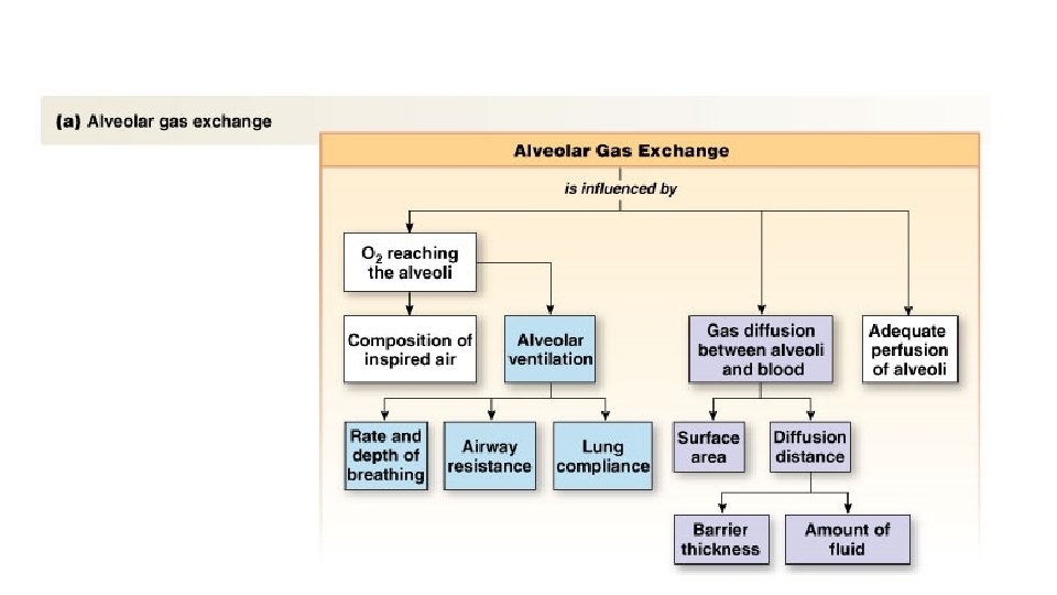
Primary functions RS • • Brings O 2 and eliminates CO 2 Keeps p. H of an organism Protection from inhaled pathogens and irritating substances Vocalization • 4 steps of respiration: 1. Ventilation – exchange of gases between lungs and background 2. Outside breathing – exchange of gases between alveoli and blood 3. Transport – towards or away from tissues (by cardiovascular system) 4. Inner breathing – change of gases between blood and tissue

The Lungs and Thoracic Cavity The respiratory system is divided into upper and lower regions. Pharynx Nasal cavity Vocal cords Upper respiratory system Tongue Esophagus Larynx Trachea Lower respiratory system Left lung Right bronchus Diaphragm © 2016 Pearson Education, Ltd. Left bronchus

CONDUCTING ZONE No gas changes • • Upper airways- nose, pharynx, larynx Trachea Primary bronchi Secundary bronchi Tertialy bronchi Bronchioli Terminal bronchioli Transient zone • • Respiratory bronchioli Ductuli alveolares Sacculi alveolares Alveoli pulmonis RESPIRATORY ZONE Exchange of respiratory gases Dead space the space or volume of those parts of respiratory system where is no exchange of gases between air and blood (Vd=150 ml) • TOTAL (PHYSIOLOGICAL) DEAD SPACE 1. ANATOMIC DEAD SPACE - The respiratory passageways conducting to and from alveoli 2. ALVEOLAL DEAD SPACE - The volume of non-functional alveoli - Together anatomic and alveolal dead space = 150 m. L • Functions od conducting zone 1. Warming and humidification of the inspired air 2. Filtration (particles > 1 µm) 3. Cleaning (mucus)

The Lungs and Thoracic Cavity The Bronchi and Alveoli Branching of airways creates about 80 million bronchioles. On external view, the right lung is divided into three lobes, and the left lung is divided into two lobes. Larynx Apex The trachea branches into two primary bronchi. Superior lobe Left primary bronchus Cartilage ring Middle lobe The primary bronchus divides 22 more times, terminating in a cluster of alveoli. Inferior lobe Trachea Secondary bronchus Inferior lobe Base Cardiac notch Bronchiole Alveoli © 2016 Pearson Education, Ltd.

Epithelial cells lining the airways and submucosal glands secrete saline and mucus. Cilia move the mucus layer toward the pharynx, removing trapped pathogens and particulate matter. Dust particle Mucus layer traps inhaled particles. Ciliated epithelium Watery saline layer allows cilia to push mucus toward pharynx. Cilia Goblet cell secretes mucus. Movement of mucus Nucleus of columnar epithelial cell Mucus layer Lumen of airway Submucosal gland Basement membrane © 2016 Pearson Education, Ltd.

The Bronchi and Alveoli Structure of lung lobule. Each cluster of alveoli is surrounded by elastic fibers and a network of capillaries. Branching of airways creates about 80 million bronchioles. Larynx The trachea branches into two primary bronchi. Bronchiole Bronchial artery, nerve, and vein Branch of pulmonary vein Elastic fibers Capillary beds The primary bronchus divides 22 more times, terminating in a cluster of alveoli. Smooth muscle Trachea Left primary bronchus Cartilage ring Branch of pulmonary artery Lymphatic vessel Secondary bronchus Alveoli Bronchiole Alveoli © 2016 Pearson Education, Ltd.

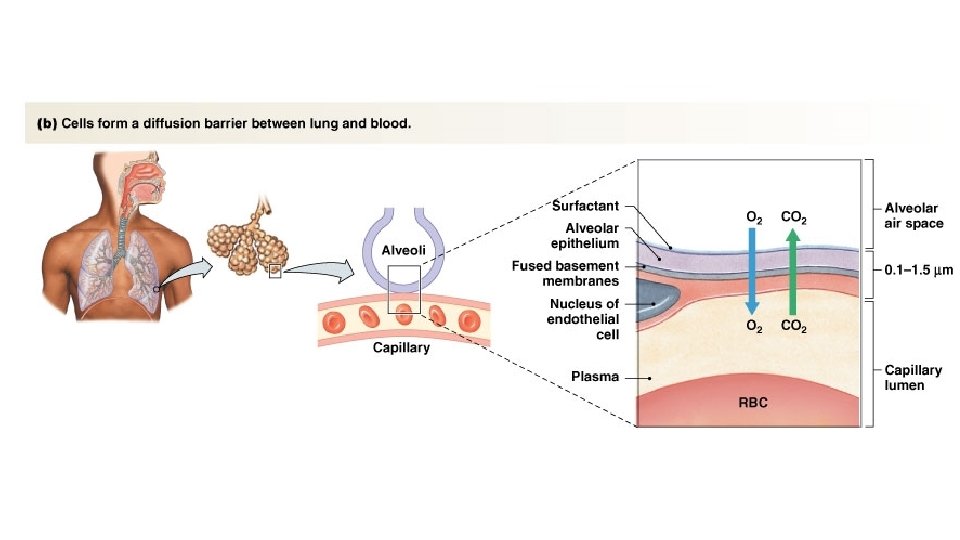
The Bronchi and Alveoli Alveolar structure Capillary Exchange surface of alveoli Elastic fibers Nucleus of endothelial cell Alveolar epithelium RBC Type I alveolar cell for gas exchange Capillary Endothelium Endothelial cell of capillary Plasma 0. 11. 5 μm Type II alveolar cell (surfactant cell) synthesizes surfactant. Alveolar air space Surfactant Limited interstitial fluid Alveolar macrophage ingests foreign material. Fused basement membranes Blue arrow represents gas exchange between alveolar air space and the plasma. © 2016 Pearson Education, Ltd. Alveolus
 formed by type II alveolar")
Pulmonary surfactant - is a surface-active lipoprotein complex (phospholipoprotein) formed by type II alveolar cells - reduces surface tension - increases the lung compliance (is the ability of lungs and thorax to expand) - stabilizes the system of lung alveoli - prevent the collapse - prevents lung to dry out - enables the conduction of air

Alveolar structure Pneumocyte II. type Pneumocyte I. type Alveoli Surfactant Phobic part Water layer on the Surface of alveolar cells Surfactant Phyllic part

• Respiratory gases pass through alveolar and capillary endothelium (in between there are fused basal membranes ) Note the following different layers of the respiratory membrane: 1. a layer of fluid lining the alveolus and containing surfactant that reduces the surface tension of the alveolar fluid 2. the alveolar epithelium composed of thin epithelial cells 3. an epithelial basement membrane 4. a thin interstitial space between the alveolar epithelium and the capillary membrane 5. a capillary basement membrane that in many places fuses with the alveolar epithelial basement membrane 6. the capillary endothelial membrane

The Lungs and Thoracic Cavity Muscles of the thorax, neck, and abdomen create the force to move air during breathing. Sternocleidomastoids Scalenes Internal intercostals External intercostals Diaphragm Abdominal muscles Muscles of inspiration of expiration © 2016 Pearson Education, Ltd.

At rest: Diaphragm is relaxed. During inspiration, the dimensions of the thoracic cavity increase. Pleural space Sternum Vertebrae Rib Diaphragm Inspiration: Thoracic volume increases. Side view: “Pump handle” motion increases anterior-posterior dimension of rib cage. Movement of the handle on a hand pump is analogous to the lifting of the sternum and ribs. Vertebrae Rib Diaphragm contracts and flattens. Expiration: Diaphragm relaxes, thoracic volume decreases. © 2016 Pearson Education, Ltd. Sternum Front view: “Bucket handle” motion increases lateral dimension of rib cage. The bucket handle moving up and out is a good model for lateral rib movement during inspiration.

Pressure changes during ventilation • Pleural pressure • between lung and chest pleura • normally, the pressure is always negative in comparison to atmospheric and alveolar pressures • about - 5 cm of H 2 O • during inspiration, the expansion of chest cage pulls the surface of the lung and creates even more negative pressure (-7, 5 cm of H 2 O)

Pressure changes during ventilation • Alveolar pressure inside the alveoli when airway opened, the pressure is the same as atmospheric pressure 0 cm of H 2 O during inspiration the pressure in alveoli falls to negative, about -1 cm of H 2 O, what is enough to inhale about 0, 5 L of air • during expiration, the pressure change is opossite, the alv. pressure rises to +1 cm of H 2 O • inspiration lasts about 2 s, expiration about 3 s • •

Pressure changes during ventilation • Transpulmonary pressure • pressure difference between the alveolar pressure and the pleural pressure (outer surfaces of the lungs) • measure the elastic forces in the lungs, that tends to collapse the lungs at each point of expansion – recoil pressure • rises during inspiration • Transpulmonary pressure = alveolar pressure – pleural pressure

Pressure changes during ventilation Transpulmonary pressure

Compliance of the lungs • the extent to which lungs expand for each unit increase in transpulmonary pressure • the normal value of both lungs is about 200 ml/cm of H 2 O • 2 components • inspiratory compliance curve • expiratory compliance curve

In the normal lung at rest, pleural fluid keeps the lung adhered to the chest wall. P 3 mm Hg Intrapleural pressure is subatmospheric. Ribs Pleural fluid Visceral pleura Parietal pleura Diaphragm Elastic recoil of the chest wall tries to pull the chest wall outward. Elastic recoil of lung creates an inward pull. © 2016 Pearson Education, Ltd.

Pneumothorax. If the sealed pleural cavity is opened to the atmosphere, air flows in. The bond holding the lung to the chest wall is broken, and the lung collapses, creating a pneumothorax (air in the thorax). P Patm Knife Air Lung collapses to unstretched size. Pleural membranes © 2016 Pearson Education, Ltd. The rib cage If the sealed pleural cavity is opened expands slightly. to the atmosphere, air flows in.

CO 2 Airways Alveoli of lungs CO 2 enters alveoli at alveolar-capillary interface. Oxygen enters the blood at alveolarcapillary interface. O 2 CO 2 Pulmonary circulation CO 2 is transported dissolved, bound to hemoglobin, or as HCO 3–. Oxygen is transported in blood dissolved in plasma or bound to hemoglobin inside RBCs. Systemic circulation CO 2 diffuses out of cells. Oxygen diffuses into cells. Cells ATP © 2016 Pearson Education, Ltd. CO 2 Cellular respiration determines metabolic CO 2 production. O 2 Nutrients
 Pulmonary vein (oxygenated blood)")
Pulmonary artery (deoxygenated blood) Pulmonary vein (oxygenated blood)
 • Apnoe")
Some clinical terms • Eupnoe • Normal breathing (normal frequency and depth) • Apnoe • Cessation of breathing • Tachypnoe • Higher frequency of breathing (does not describe depth) • Hyperpnoe • Deep breathing (does not describe frequency) • Dyspnoe • Worsened breathing • Anoxia • Absence of O 2 • Hypoxia • Inadequate oxygen supply to the tissues • Ischemia • Inadequate blood perfussion through the tissues

Some clinical terms • Hyperventilation • Increased amount of ventilated air • Hypoventilation • Decreased amount of ventilated air
. •")
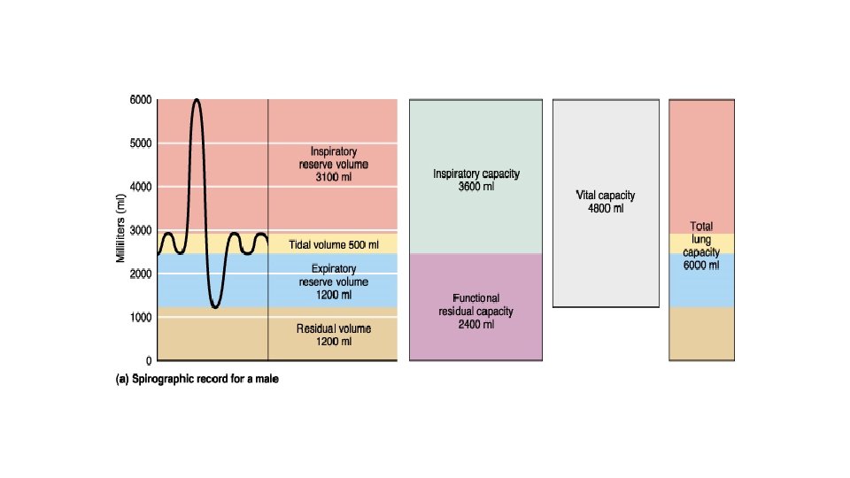
Respiratory volumes • Amount of air which is quietly exchanged (about 500 ml). • This is tidal volume – TV • The amount of air, which can be inspired after normal inspiration - inspiratory reserve volume (IRV), about 3000 ml. • Expiratory reserve volume (ERV) –amount of air that can be expired after normal expiration, about 1200 ml • After maximal expiration, there still is some air in the lungs residual volume (RV), about 1200 ml

Respiratory capacities • Inspiratory capacity – amount of air, that can be inspired IC = TV + IRV (about 3500 ml) • Functional residual capacity – amount of air that stays in lung after normal expiration FRC = ERV + RV (about 1700 ml) • Vital capacity – maximal amount of air that can be expire after maximal inspiration VC = TV+IRV+ERV (about 4800 ml) • Total lung capacity – sum of all lung capacities TLC = VC + RV (about 6000 ml)
 • Described as heavy/short")
Dyspnoe • Symptome • Subjective feeling (we cannot measure it!) • Described as heavy/short breath • Respirations frequency, p. O 2, levels of blood gases do not have to correlate with the feeling • Discrepancies between the need and the ability of an organism to ensure respiration

Causes of dyspnoe • Nervousness • Obstruction • Bronchospasm • Hypoxemia • Pleural effusion • Pneumonia • Edema • Pulmonary embolism • Thick mucus secretion • Anaemia • Metabolism • Family / financial / emotional / etc. issues
 • Progressive decline in the lung function characterized by")
Chronic obstructive pulmonary disease (COPD) • Progressive decline in the lung function characterized by poorly reversible airway obstruction • Smoking • 3 subtypes/parts • Chronic bronchitis • Emphysema • Asthma

COPD • Continuous progression, airflow linked with immune response in airways
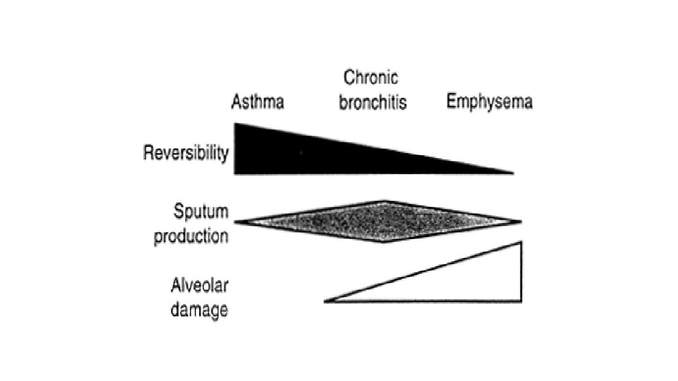
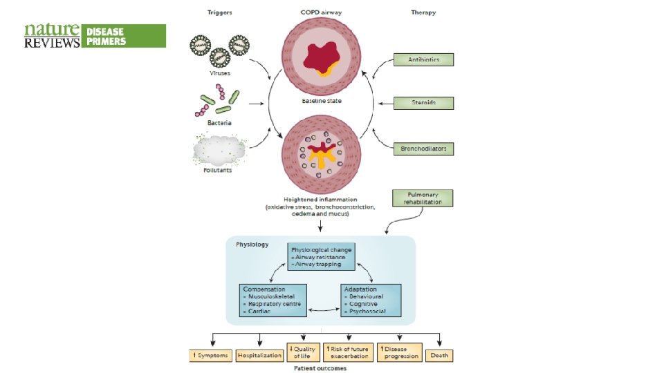

Chronic bronchitis • Chronic persistent cough lasting for at least 3 months for two consecutive years in patients where other causes of cough were excluded • Bigger bronchial submucosis glands (hyperplasia) • Terminal airways – easily susceptible to be obstructed with mucus • Increased airflow resistance • Abraded airways and decreased activity of the mucociliary apparatus increase the risk of infection

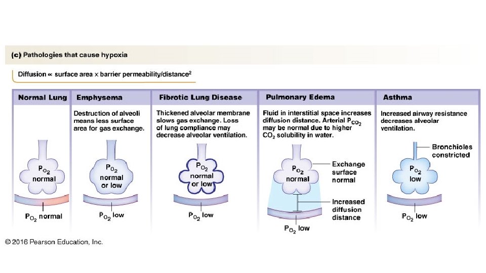
Emphysema Loss of elasticity - resistance of airways - ventilation - airway collapse during exhalation air capture - retention CO 2 less air - O 2 CO 2 Alveolar space increases - diffusion - collapse of alveoli or their rupture - surface for gas exchange - compression of lung capillaries - perfusion - hypoxemia

It can be caused by smoking, polluted air and environmental and working environments The main feature is loss of lung elasticity and reduction of elastic tissue due to alveolar destruction Destruction of elastic tissue leads to loss of elasticity of the lungs during expiration, and effort is needed to breathe Subsequently, it may destroy the airways / capillaries. Tissue destruction due to protease formation or lack of anti-proteases

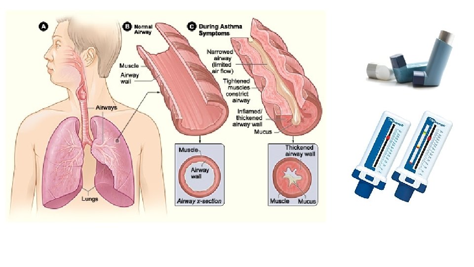
Asthma Chronic inflammatory obstruction of bronchi characterized by episodic, reversible bronchospasms with dyspnoea of the expiratory type as a result of the exacerbated response by bronchoconstriction to various stimuli (allergies) Multicellular answer

Asthma • Today it is believed that asthma is the result of a combination of genetic predisposing factors and the external environment • Main triggers: – Cigarette smoke – Polluted air – Animals – Virus respiratory infections – Alergens of cockroaches – Weather changes
, guinea pig,")
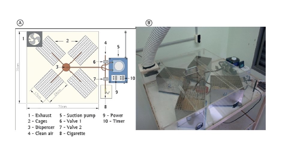
COPD animal models • Exposure to cigarette smoke mice, rats (relat. resistant), guinea pig, dog • Exposure to inflammatory stimuli (LPS) IN application • Exposure to proteolytic enzymes (elastase) IT application • Combination • Genetic modification

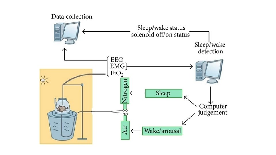
Sleep apnea syndrome - SAS Abnormal breathing pauses during sleep Types of SAS Obstructive (OSAS) Respiratory effort is maintained but the air supply is missing in the nose and mouth (larynx, . . . ) Linked with obesity Central (CSAS) Flow and respiratory effort is stopped The problem is at the respiratory center level Associated e. g. with heart failure Mixed SAS Combination of OSAS and CSAS

Obstructive SAS • OSAS is the most common disorder that can be observed in sleep centers and is responsible for mortality and morbidity more than any other sleep disorder • OSAS is defined as repeated episodes of complete or partial airway obstruction during sleep. As a result, the patient has restless sleep and excessive daytime fatigue / sleep.

New treatment options

SAS animal models • Natural models of sleep apnea • Intermittent hypoxia • Combined (intermittent hypoxia + hypercapnia) • Airway obstruction (tracheal band)

Pulmonary embolism • Embolization of the blood vessels of the lung through blood clots, air, fat, amniotic fluid • Occurs due to disorders that accelerate blood clotting: • Immobility (stasis in veins) • DIC (disseminated intravascular coagulation) • Systemic lupus erythematosus • A thrombus from a pregnant woman's pelvis is the most common cause of embolism in pregnant women

Pulmonary embolism • Virchow’s triad • Stasis of blood in veins • Vascular wall disorder • Hypercoagulation • 4 categories • massive – for example major pulmonary artery involvement • pulmonary infarction – death of a part of a tissue • without infarction – not so serious embolus • multiple pulmonary embolus

Animal models of pulmonary embolism • Farmacological – Thrombine, collagen+adrenaline • Photochemical methods – Green laser damages cell wall oxidative stress damage on blood vessel wall thrombus on the damaged endothelium

Acute Respiratory Distress Syndrome ARDS • Pulmonary edema resulting not from cardiac failure • Progressive refractory hypoxemia (not responding to compensatory mechanisms or treatment) • Severe dyspnoea • Diffusion of bilateral infiltrates • Physiological changes • Damage to the lung endothelium causes increased lung permeability • The fluid escapes into interstitium, causing pulmonary edema • Complications of hospitalized patients • Serious medical - surgical problem • It does not have to be related to lung damage • Mortality is about 50 -60%

Etiology of ARDS • • Not one exogenous or endogenous factor, several causes The exact mechanism is unknown Direct and indirect causes Conditions associated with possible developments • The most often • Out of the lungs • Gram (-) sepsis • Trauma • Lungs • • Aspiration AIDS Drowning Pulmonary embolism

ARDS - pathophysiology • Physiological changes • Pneumocytes type II damage causes an increase in surface tension and atelectasis • Damage to the alveolocapillary membrane, inflammation, substances accumulate at the site of damage, thus reducing gas exchange • Consequences • Ventilation-perfusion anomalies • Reduced lung compliance • Increased work / effort when breathing
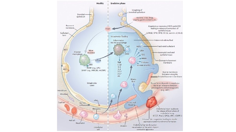
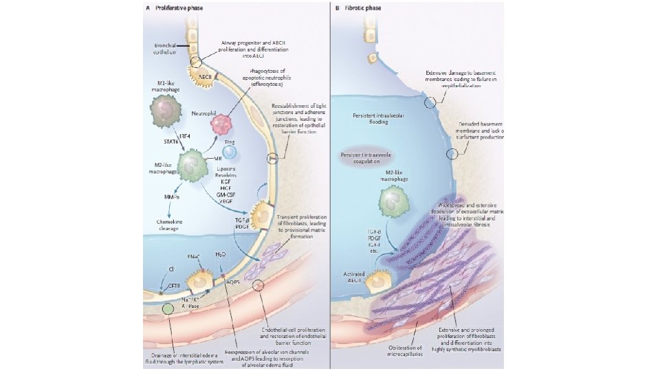

Damage/trauma Reduction of blood flow to the lungs Blood clotting HISTAMINE, BRADYKININE, SEROTONINE release, release of complemet, inflammatory mediators Inflammation of alveoli Increase in permeability of alveoli Decrease of sufactant production Pulmonary edema Collapse of alveoli Decrease of gas changes HYPOXEMIA METABOLIC ACIDOSIS

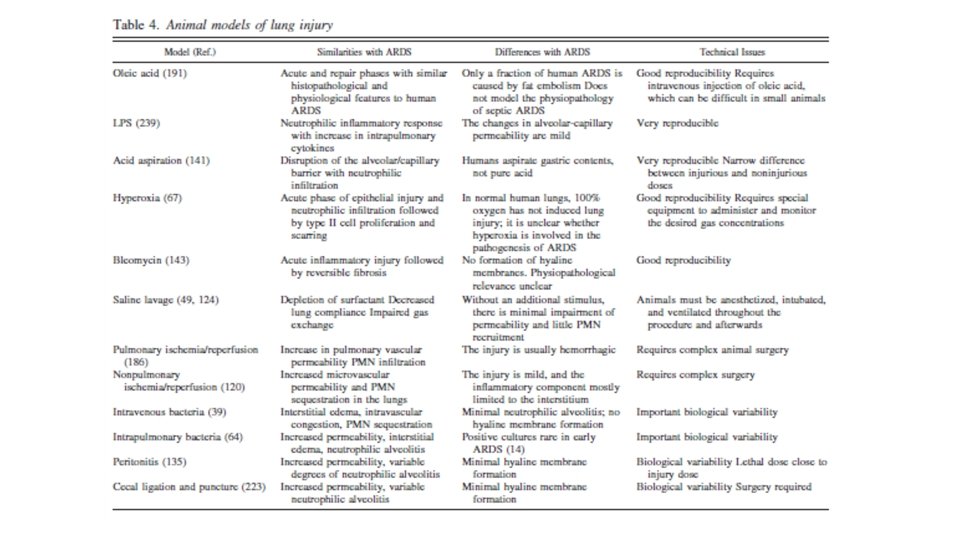
Animal models of ARDS Acute lung injury Oleic acid Pulmonary ischemia/reperfusion LPS Nonpulmonary ischemia/reperfusion Acid aspiration Intravenous bacteria Hyperoxia Intrapulmonary bacteria Bleomycin Peritonitis Saline lavage Cecal ligation and puncture

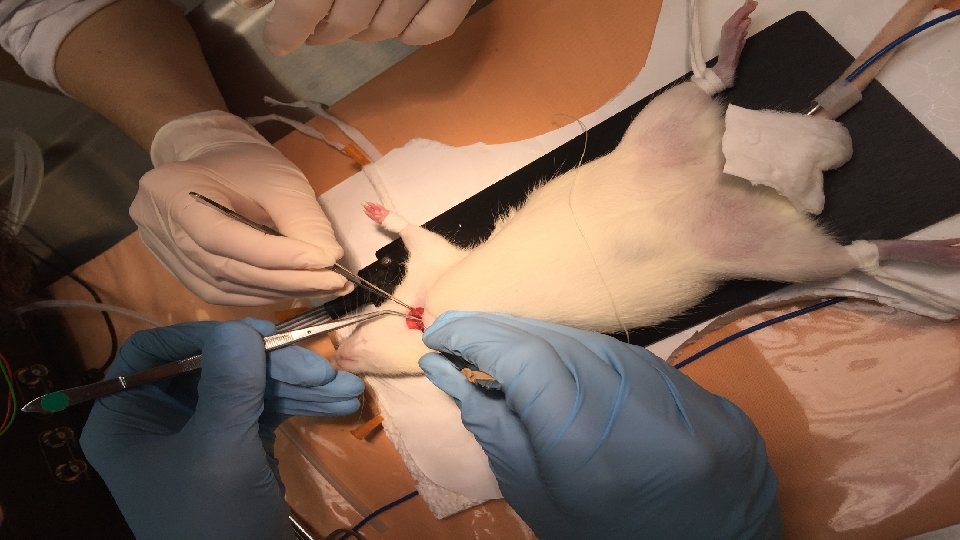
Animal model of sepsis and measurement of surface tension of surfactant • LPS to trachea • Regulation of airflow • They take blood • They monitor inflammation markers • They do lavage of lungs and measure surfactants with surfactometer

basa. konecna@gmail. com
- Slides: 63