Respiratory System Anatomy Physiology II Tony Serino Ph


, nasal")


")
 • Secondary")
 bronchi lead to each")


 • Red is inhibitory • Black is excitatory")


- Slides: 41
Respiratory System Anatomy & Physiology II Tony Serino, Ph. D. Biology Department Misericordia University
Respiration • External Respiration – The exchange of gas between the blood and external environment (usually includes ventilation) • Internal Respiration – The exchange of gas between the blood and the tissues • Cellular Respiration – Burning of fuel to produce energy within cells • Ventilation (Breathing) – Movement of air in and out of the lungs
Respiratory Organs – Divided into: • Upper Respiratory Tract – Includes: nostrils (nares), nasal cavity, and nasopharynx • Lower Respiratory Tract – Includes: larynx, trachea, bronchi, and lungs – Conducting Air passages include: nares to terminal bronchioles • Move air to respiratory membrane • Condition the air – Moisten, Warm, Clean
Upper Respiratory Tract
Beginning of Lower Respiratory Tract
Larynx
Trachea
Mucous Membrane (pseudostratified columnar epithelium)
Bronchi • Primary bronchi lead to to each lung (left and right) • Secondary (lobar) bronchi lead to each lung lobe (3 on right and 2 on left)
Bronchi Branches Tertiary Bronchi Primary Bronchi Secondary Bronchi Tertiary (segmental) bronchi lead to each lung bronchopulmonary segment Bronchi continue to divide at least 20 more times.
Broncho-pulmonary Segments
Right Lung Left Lung
Bronchioles • Air passages less than 1 mm in diameter are bronchioles. • The terminal bronchioles are the last of the purely conducting air passages.
Alveoli highly specialized for Gas Exchange • Lots of Surface Area • Highly vascular • Thin walls
Alveolus
P = pressure to collapse T = surface tension r = radius Role of surfactant is to decrease surface tension in alveoli.
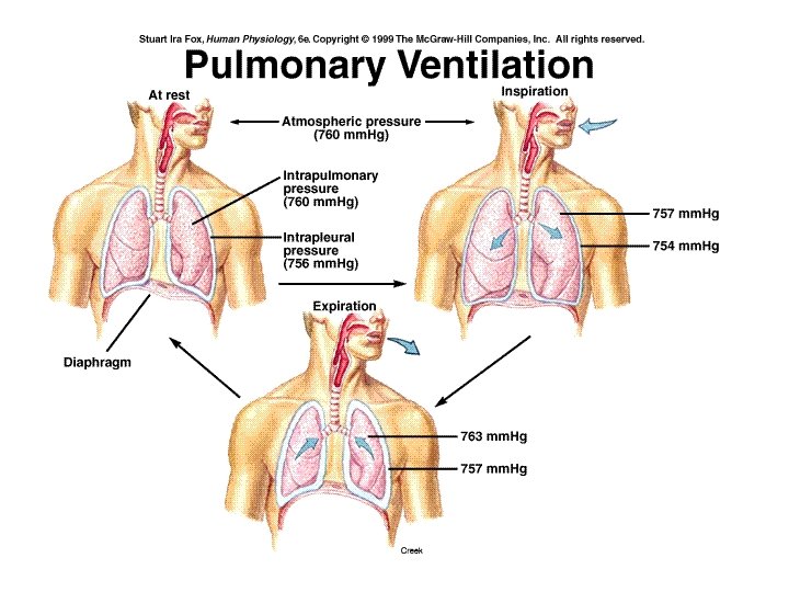
Pressures affecting Breathing
Inspiration
Expiration
Pressure changes around lung
Only used during rapid breathing.
Lung Volumes
Normal Lung Volumes
Partial Pressure Favors Resp. Gas Movement
Time to Complete O 2 Saturation in Pulmonary Capillaries
Oxygen Content of Blood Plasma Whole Blood PO = 100 mm. Hg Oxyhemoglobin O 2 PO = 100 mm. Hg 2 Oxygen 2 Total Volume of Oxygen = 0. 3 ml Plasma + 20 ml whole blood
Hemoglobin
Oxyhemoglobin Dissociation Curve
Hemoglobin Affinity for Oxygen: Effect of Temperature Affinity decreases with increasing Temperature
Hemoglobin Affinity for Oxygen: Effect of p. H Affinity decreases with increasing acidity (i p. H)
Gas Exchange in Lungs
Gas Exchange in Tissues
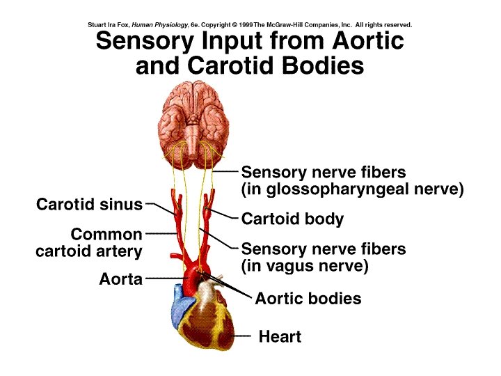
Neural Control Voluntary control located in cerebral cortex and acts through the corticospinal tract. of Breathing Involuntary located in pons and medulla acting through the spinal cord in the roots of the phrenic nerve (C 3 -C 5) and thoracic cord roots of the external (inspriation(I)) and internal (expiration(E)) intercostal nerves PRG –pontine resp. group (formerly the apneustic and pneumotaxic centers) –play role in smoothing between insp. and exp. , especially during sleep, vocalization and exercise. VRG and DRG – ventral and dorsal resp. group of the medulla. DRG primarily responsible for inspiration; VRG mixture of I and E neurons contains Pre-Botzinger complex which may be pacemaker cells for respiration
Neural control of Breathing PRG (smoothing) • Red is inhibitory • Black is excitatory (Insp. center) DRG Hering-Breuer Reflex I neurons Ext. Intercostals & diaphragm Lung Stretch Chemoreceptors VRG (Resp. pacemaker? ) E neurons Intercostals
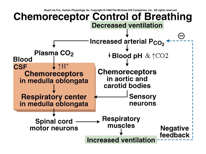
Medulla sensitive to H+ directly from CSF; indirectly to CO 2
Factors Effecting Respiratory Centers
CO 2 Drive
COPD