Respiratory system Anatomy 1 2 3 4 5


")


 Laryngeal cancer")
 Obstructive airway diseases")
")

 Using inhalers , Using discs , Nebulizers Technique")


 despite high O")


- Slides: 30
Respiratory system
Anatomy
1. 2. 3. 4. 5. 6. 7. Nose Pharynx Larynx Trachea Bronchiole Lungs
Function Gas exchange (O 2/ CO 2)
Clinical features of resp. disease • Symptoms • signs
symptoms • • • Cough Dyspnea Chest pain Wheeze Hemoptysis
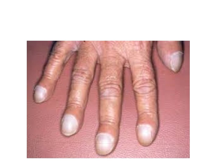
Signs • Cyanosis • Tachypnea • Clubbing
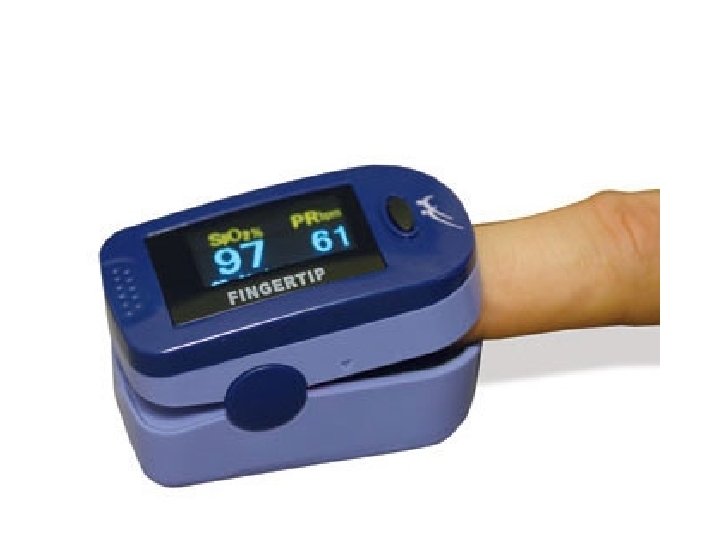
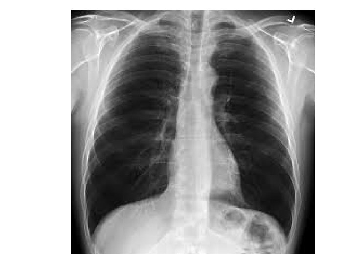
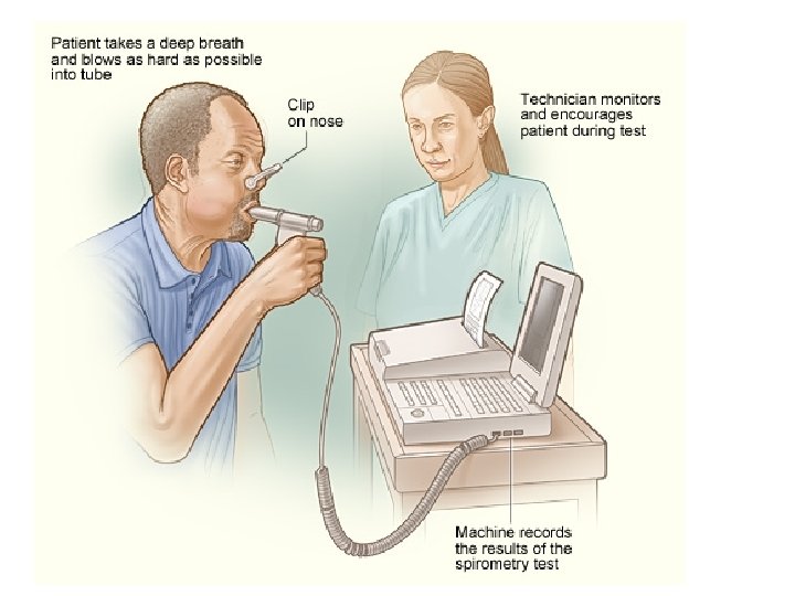
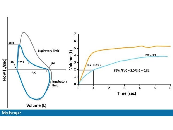
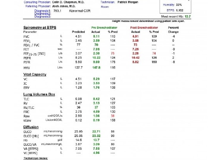
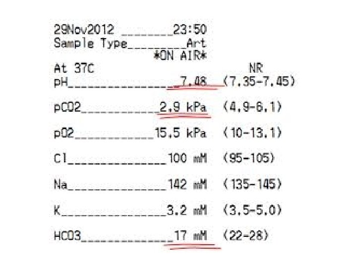
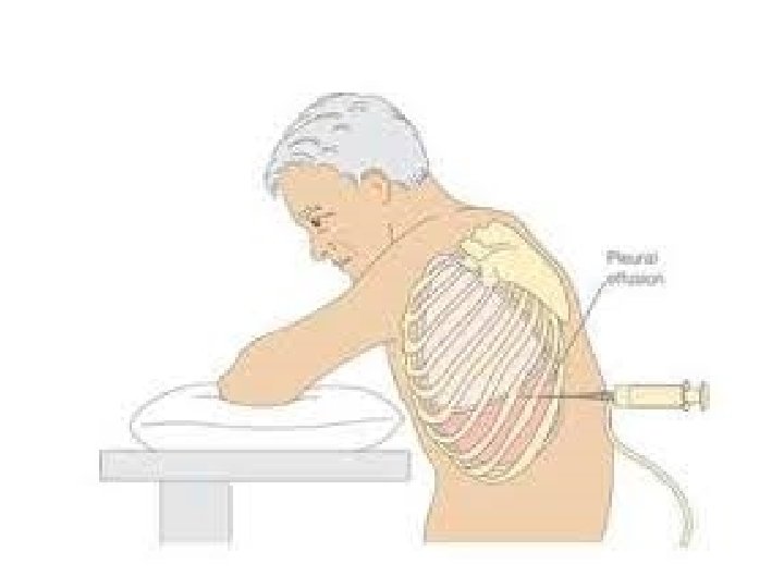
Investigations • • Sputum exam Oximetry Chest x ray spirometry Pulmonary function test Blood gas analysis Pleural aspirate and biopsy
Blood gas analysis
Nursing management • • Rhinitis Epistaxis Suffocation (foreign body in the larynx) Laryngeal cancer and tracheostomy care
• Bronchial asthma • Chornic obstructive pulmonary disease(copd) Obstructive airway diseases
Bronchial asthma • Oxygen therapy – high flow brochodilators (inhalors , nebulizer , disc)
COPD • Oxygen – low concentration • Brochodilators
Oxygen (mask, cannula, others) Using inhalers , Using discs , Nebulizers Technique
Cardiopulmonary resuscitation • CPR = 15: 2
Endotracheal intubation • Indications 1. 2. 3. 4. 5. Hypoxia Hypercapnea Laryngeal edema Avoiding aspiration Mechnical ventilation
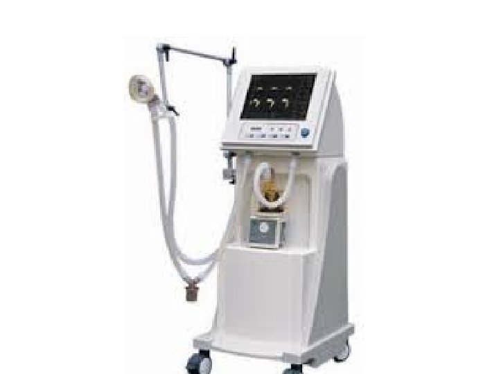
Mechanical ventilation Indications 1. Hypoxia (pa. O 2< 50 mm. Hg) despite high O 2 treatment. 2. PH < 7. 25 (acidosis), not corrected by high oxygen therapy. 3. Vital capacity < 2 times of Tidal volume. 4. Negative inspiratory force < 25 cm water. 5. Respiratory rate > 35 cycle per minute
Modes of mech. ventilations 1. Controlled ventilation – for comatose patient. 2. Intermittant mandatory ventilation – tired 3. Synchronized intermittant mechanical ventilation (SIMV), nearly universal , weaning 4. PEEP (positive end expiratory pressure)-COPD 5. CPAP (continuous positive airway pressure)for Sleep apnea patient. 6. others
Setting • Tidal volume = 10 – 15 ml / kg • Use lowest effective O 2 (Fi. O 2) to mentain Pa. O 2 80 -100 mm. Hg • Use SIMV mode • Adjust sensitivity of the ventilator to 2 mm negative inspiratory force , • Monitor every 20 minutes • Weaning from the ventilator.