RESPIRATORY SYSTEM 3 Main Divisions 1 Conducting Zone


 • Bronchioles-smooth muscle,")

. Increase in mucus production • Irritants")
- Slides: 12
RESPIRATORY SYSTEM
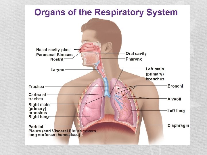
3 Main Divisions 1. Conducting Zone Structures: nose, pharynx, larynx, trachea, bronchioles -Passage for air to move in and out of lungs -no gas exchange 2. Respiratory Zone Structures: bronchioles, alveolar ducts, alveolar sacs, alveoli -Allows for O 2 to be exchanged for CO 2 3. Supporting Structures: diaphragm (inhalation), external intercostals (inhalation), internal intercostals (exhalation), ribs, coastal cartilage, sternum
Nasal Cavity • Anterior Nares • Posterior Nares • Nasal Conchae Bones • Cribform Plate of Ethmoid Bone
Pharynx • Nasopharynx: opening of eustachian tube, soft palate, uvula • Oropharynx • Laryngopharynx
Larynx • Hyoid Bone • Epiglottis Cartilage • Thyroid Cartilage-Adams Apple • Vocal Folds-true vocal cords
Trachea • Trachea-windpipe • C-rings of hyaline cartilage keep the air passage open
Lungs • Right and left separated by heart and rest of mediastinum (contains all the chest organs except the lungs) • Right lung- 3 lobes, superior, middle, and inferior divided by oblique and horizontal fissures • Left lung- 2 lobes (room for heart), superior and inferior divided by oblique fissure • Apex at top, base at bottom
Lungs • Located in pleural cavities of thorax • Parietal and visceral pleura - Visceral pleura: outer covering of lungs -Parietal pleura: line walls of thoracic cavity -Pleural Cavity: fluid filled
Bronchi • Left and right sides of lung (primary, secondary, tertiary) • Bronchioles-smooth muscle, constrict or dilate airways -Pulmonary Arteries: O 2 poor blood -Pulmonary Veins: O 2 rich blood
Bronchi Cont. • Terminal Bronchiole- end of conducting zone • Respiratory Bronchiole- where gas exchange starts • Alveoli- O 2, CO 2 exchange, supplied with blood capillaries
Asthma • Chronically inflamed, hypersensitive bronchial passages (airways). Increase in mucus production • Irritants such as dust mites, dander, fungi, • Causes coughing, wheezing, chest tightness (tightening of muscles), shortness of breath • Treatment: inhalers , -albuterol: rescue inhaler, relaxes constricted muscles -corticosteroid: decrease swelling, easier to breath