Respiratory practical block Lung Type I pneumocyte Alveolar

Respiratory practical block

Lung Type I pneumocyte Alveolar space Capillary lumen Type II pneumocyte Endothelium

The respiratory acinus • Cartilage is present to level of proximal bronchioles • Beyond terminal bronchiole gas exchange occurs • The distal airspaces are kept open by elastic tension in alveolar walls

T R Ad R As A Microscopic section of normal lung showing terminal bronchiole, respiratory bronchiole, alveolar duct, alveolar sac, and alveoli.

1 -Lobar pneumonia

A closer view of the lobar pneumonia demonstrates the distinct difference between the upper lobe and the consolidated lower lobe.
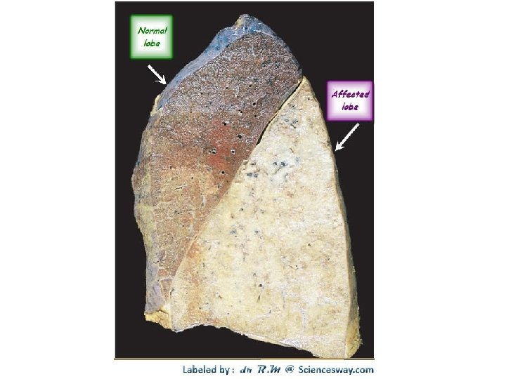
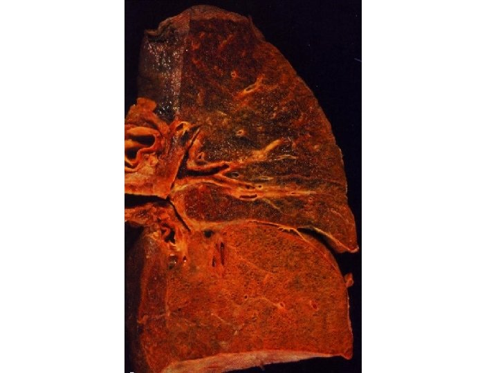
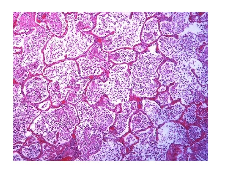
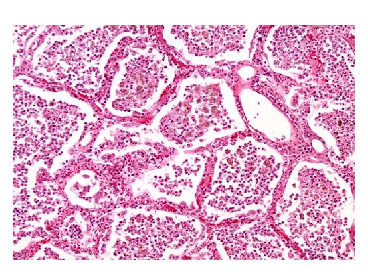

Lobar pneumonia: Section of the lung shows diffuse consolidation: All the alveoli are filled with fibrinous exudate containing fibrin threads, polymorphs, macrophages and red cells. Alveolar walls are congested. Pleura is covered by fibrinous exudate.

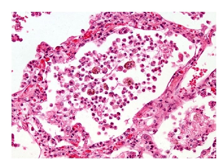
2 -Bronchopneumonia
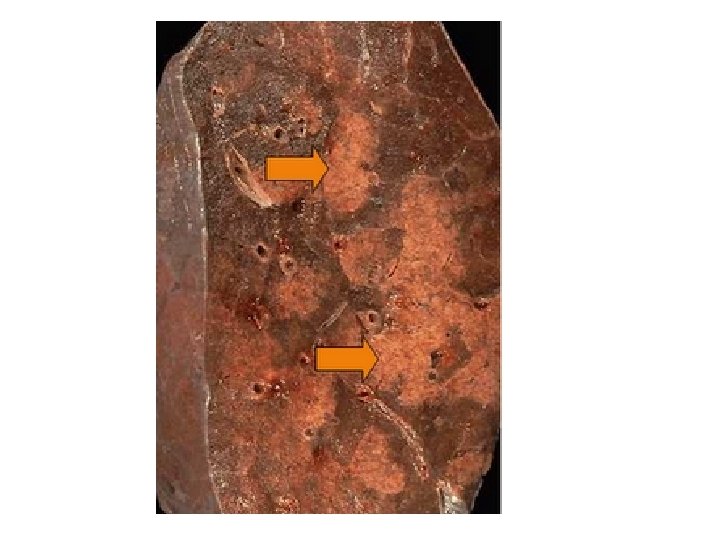
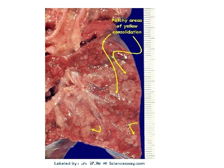
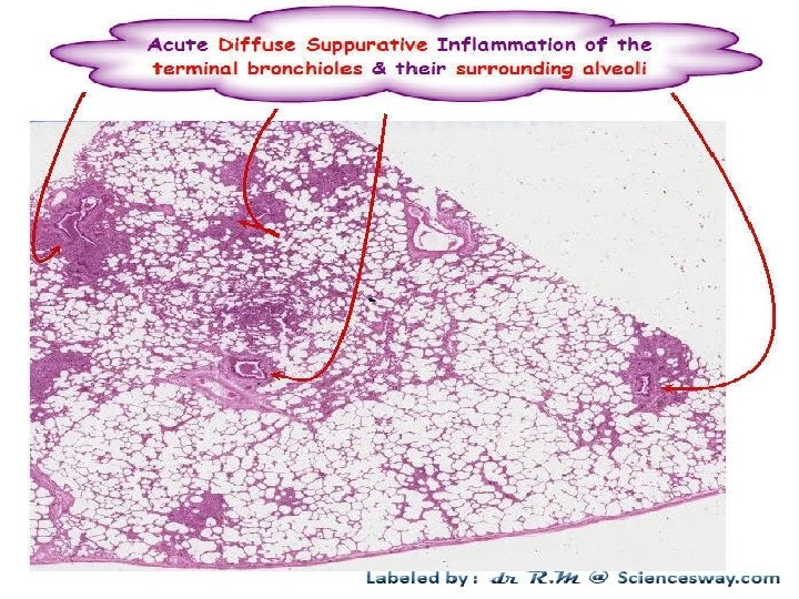

Bronchopneumonia: Section of the lung shows foci of inflammatory consolidation surrounding bronchioles: Bronchioles are filled with an inflammatory purulent exudate and show ulceration of mucosa, focal inflammation and necrosis of walls. Alveoli surrounding the bronchiole are filled with fibrin threads , polymorphs and few macrophages. Surrounding lung parenchyma shows congestion and edema.

3 - Tuberculosis of the lung

Ghon’s Complex
")
Organ: lung Dx : Caseous necrosis (tuberculosis)

Tuberculous Granulomas

Epitheloid cells in Granuloma
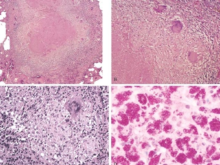

MORE ACID-FAST BACILLI, AFB/Ziehl-Neelsen stain

Miliary tuberculosis of the lung : • Section of the lung shows : The alveolar septae contain many tubercles/granulomas which consist of epithelioid cells , few langhan’s giant cells and peripheral rim of lymphocytes with or without caseation

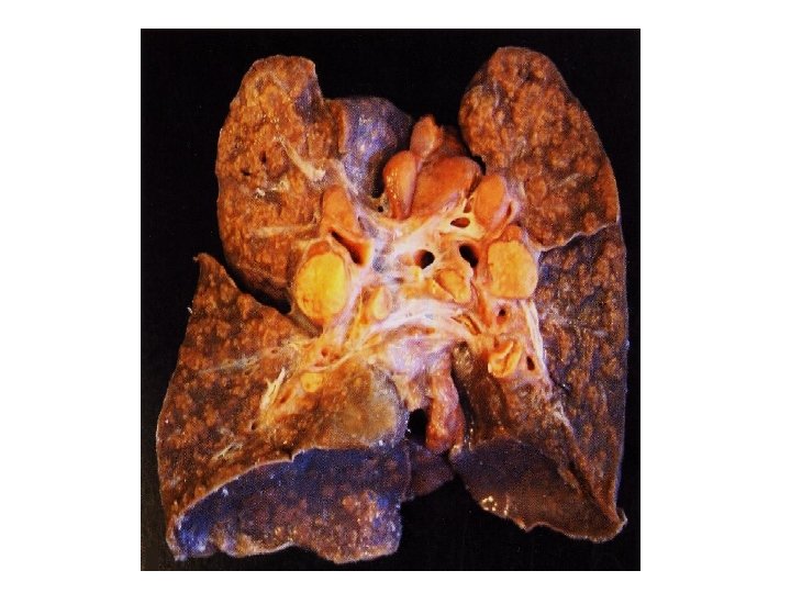
4 -Emphysema

Normal lung

Panacinar emphysema

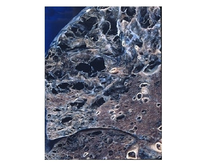
Pathology of lung showing centrilobular emphysema characteristic of smoking. Closeup of fixed, cut surface shows multiple cavities lined by heavy black carbon deposits.
")
EMPHYSEM A (LUNG)
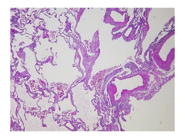
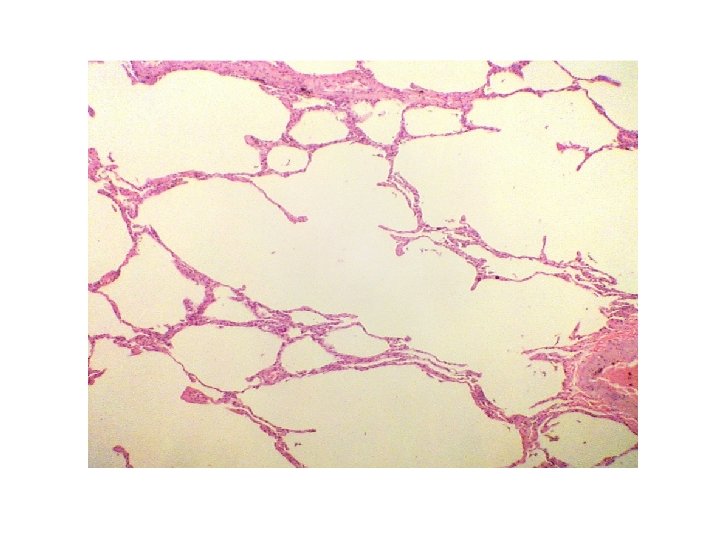
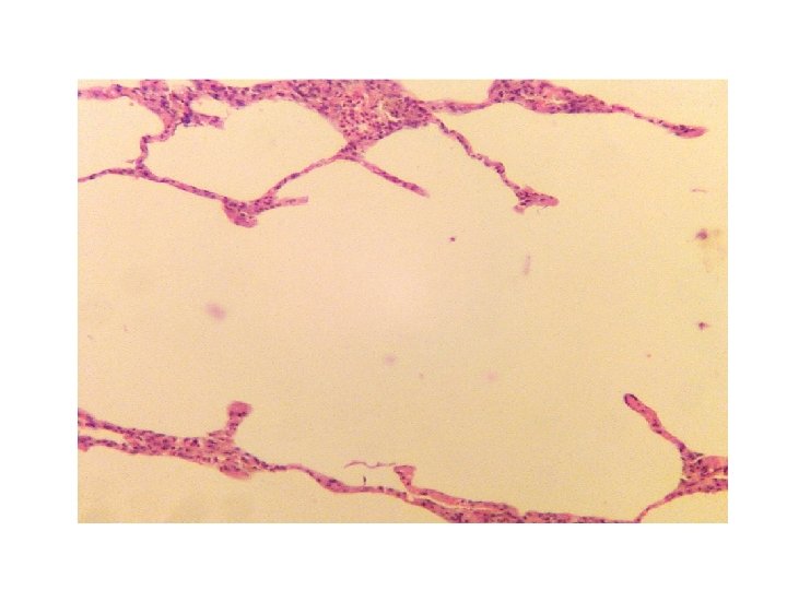

Emphysema: Section of lung shows: Increase in the size of air spaces. Decrease in number of air spaces and their walls are thinned. Some of the alveolar septae are ruptured and the ruptured septa project with in air spaces on the form of spurs. Alveolar blood vessels show reactive thickening of their walls. Ruptured emphysematous peripheral bullae with accumulation of air in the pleural cavity can cause

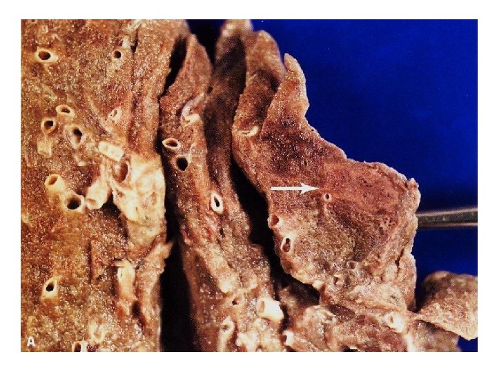
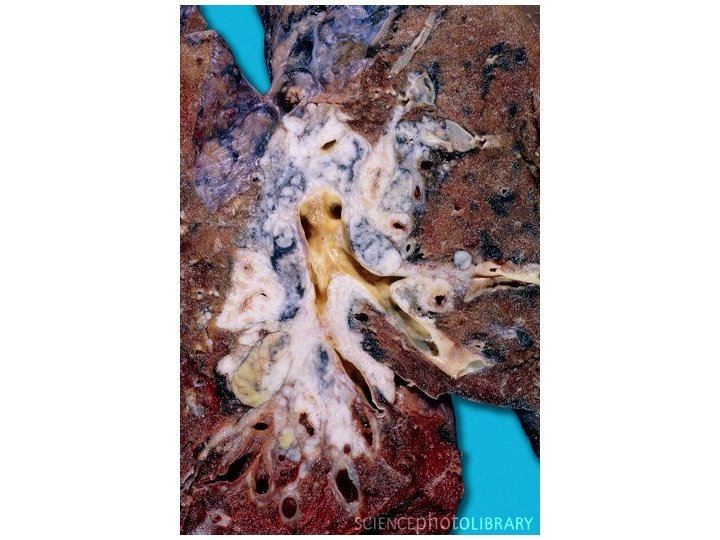
5 -Bronchiectasis

-Permanent dilatation of bronchi and bronchioles caused by destruction of muscle and elastic tissue resulting from or associated with chronic necrotizing infection -Markedly distended peripheral bronchi.
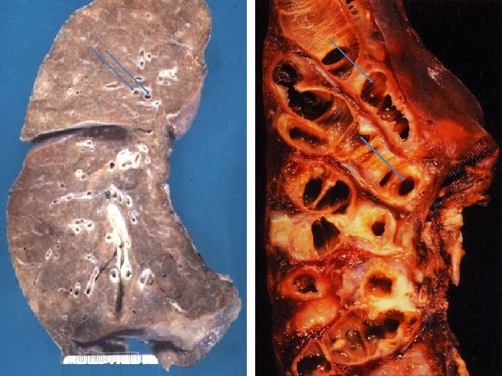

In brochiectasis, mucus production increases, the cilia are destroyed or damaged, and areas of the bronchial wall become chronically inflamed and are destroyed.

Section of a dilated bronchi with florid acute on chronic inflammation of the bronchial wall and surrounding interstitial fibrosis.

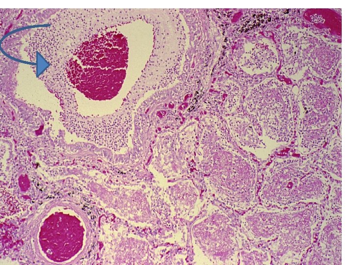
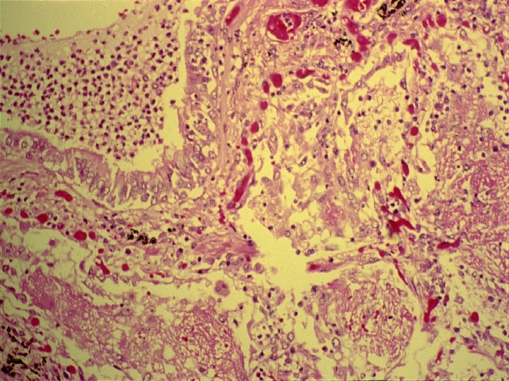
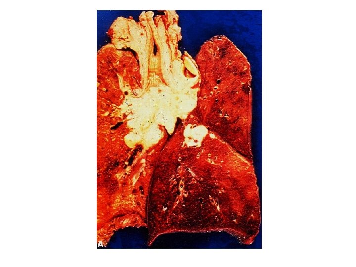
6 -Pulmonary embolus and infarction

Pulmonary embolus and infarction Longitudinal transection of lung showing a wedge shaped peripheral hemorrhagic infarction. A thrombus is seen in a major branch of pulmonary artery ( arrow head ).

TWO TYPES of lung carcinoma • NON-SMALL CELL – SQUAMOUS CELL CARCINOMA – ADENOCARCINOMA – LARGE CELL CARCINOMA • SMALL CELL CARCINOMA
~ 5% ADH (hyponatremia) ACTH (Cushing) PTH (Hyper-CA)")
SYSTEMIC effects of LUNG CANCER (PARA-NEOPLASTIC SYNDROMES)~ 5% ADH (hyponatremia) ACTH (Cushing) PTH (Hyper-CA) CALCITONIN (Hypo-CA) GONADOTROPINS SEROTONIN/BRADYKININ

7 -Squamous cell carcinoma of the lung
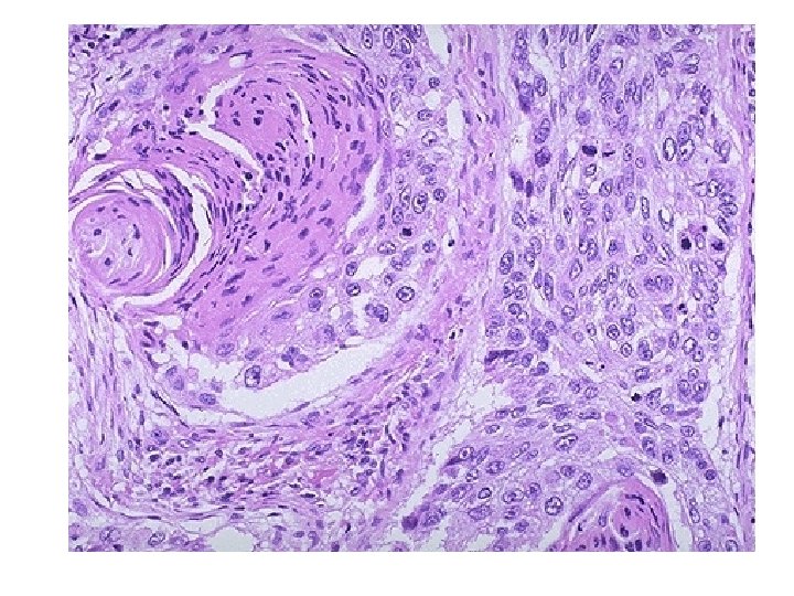
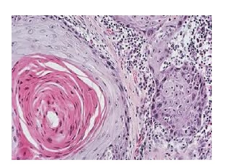
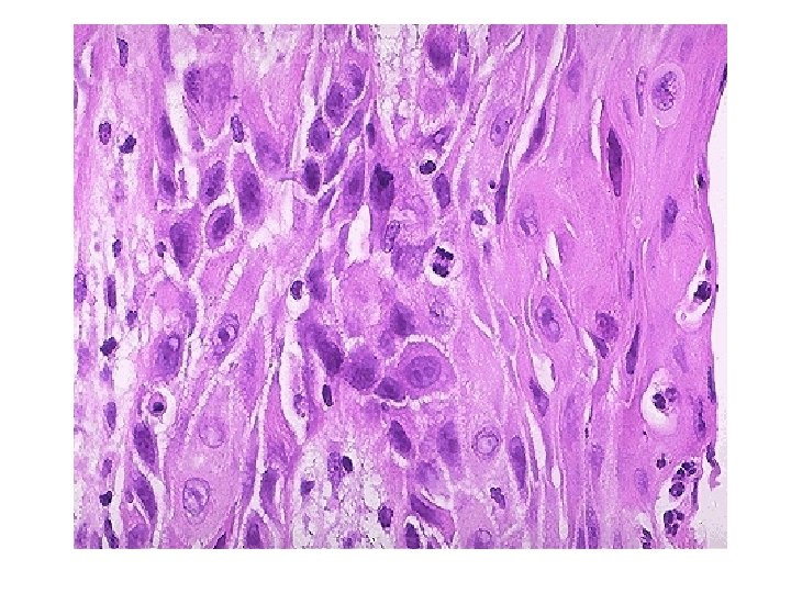

Squamous cell carcinoma of the lung: Section of the lung shows one small bronchus and tumour masses: Tumour consists of trabeculate and sheets of moderately differentiated squamous cells with little connective tissue stroma. Neoplastic squamous cells show pleomorphism, hyperchromatism, individual cell keratinization, mitoses and areas of necrosis. Peribronchial and perivascular lymphatics are occluded by tumour cells. Parathyroid-like hormone secreted inappropriately by this neoplasm.

8 -Adenocarcinoma of the lung

Adenocarcinoma, CT image

Adenocarcinoma

Adenocarcinoma and emphysema

Adenocarcinoma, microscopic

Adenocarcinoma Section of the tumour shows moderately differentiated malignant glands lined by pleomorphic and hyperchromatic malignant cells showing conspicuous nucleoli. Note the presence of tissue desmoplasia around the neoplastic glands.

9 -Small cell carcinoma of the lung

Small-cell carcinoma, microscopic

Small cell carcinoma Section of the tumour shows clusters of malignant cells which are small , round , ovale , or spindle shaped with prominent nuclear molding , finely granular nuclear chromatin (salt and pepper pattern ) , high mitotic count and focal necrosis.

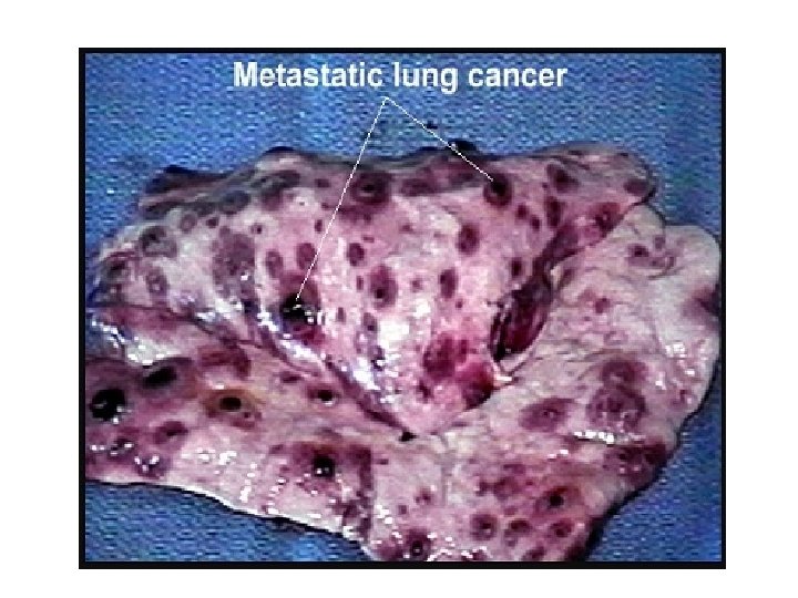
10 -Metastatic tumours of the lung

METASTATIC TUMORS • LUNG is the MOST COMMON site for all metastatic tumors, regardless of site of origin • It is the site of FIRST CHOICE for metastatic sarcomas for purely anatomic reasons!

Metastases
- Slides: 68