Respiratory Physiology heart heart circuitry heart circuitry tissues


 The walls conducting")

 -V of the")


 x breasths/min VA - alveolar ventilation (m.")
 x breasths/min VA = (500 – 125)")


")
")
")

")


")
")
")


![Shift to the right % hemoglobin saturation 100 temperature p. H [H+] 2, 3](https://slidetodoc.com/presentation_image/6648d80166fa4b61ce0a4f2b87f9aeb2/image-80.jpg "Shift to the right % hemoglobin saturation 100 temperature p. H [H+] 2, 3")
![Shift to the left % hemoglobin saturation 100 temperature p. H [H+] 2, 3](https://slidetodoc.com/presentation_image/6648d80166fa4b61ce0a4f2b87f9aeb2/image-81.jpg "Shift to the left % hemoglobin saturation 100 temperature p. H [H+] 2, 3")


- Slides: 88
Respiratory Physiology
heart
heart circuitry
heart circuitry tissues
lung heart circuitry tissues
lung heart circuitry tissues cell
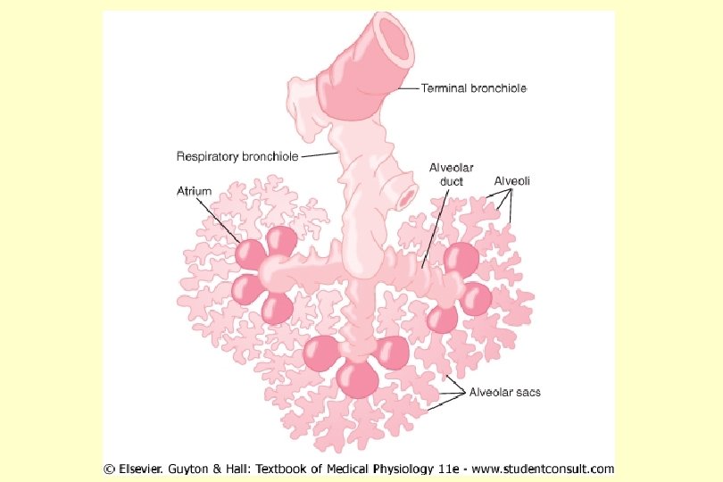
STRUCTURE of respiratory system - Conducting zone - Respiratory zone
STRUCTURE of respiratory system - Conducting zone (nose, larynx, trachea, bronchioles) The walls conducting airways contain smooth muscle -Sympathetic -Parasympathetic - 2 receptors relaxation, dilatation of the airway
STRUCTURE of respiratory system - Conducting zone Anatomic dead space -V of the conducting airways Physiologic dead space -V of the lungs that does not participapate in gas exchance
STRUCTURE of respiratory system - Conducting zone Physiologic dead space (VD) -V of the lungs that does not participapate in gas exchance Pa. CO 2 – PECO 2 VD = VT x --------Pa. CO 2 VT – tidal volume Pa. CO 2 – PCO 2 of arterial blood PECO 2 – PCO 2 of mixed expired air
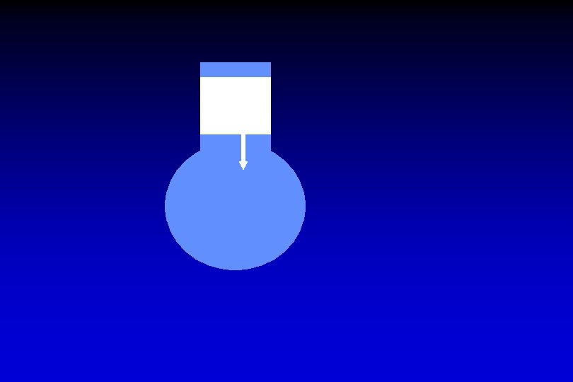
STRUCTURE of respiratory system Respiratory zone Alveoli Lung ~ 300 x 106 alveoli
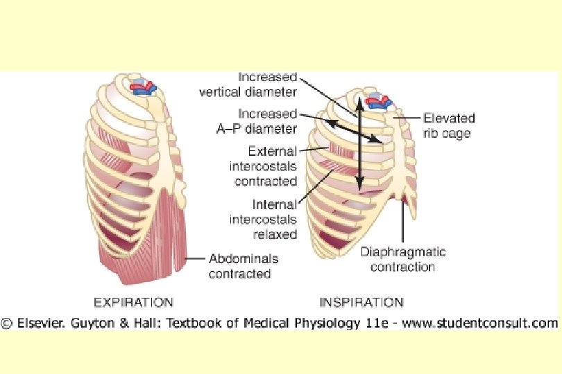
diaphragm inspiration expiration
inspiration – mm. intercosales externi
expiration – mm. intercosteles interni
inspiration – mm. intercosales externi expiration – mm. intercosteles interni
inspiration – mm. intercosales externi expiration – mm. intercosteles interni
inspiration – mm. intercosales externi expiration – mm. intercosteles interni
inspiration – mm. intercosales externi expiration – mm. intercosteles interni
inspiration – mm. intercosales externi expiration – mm. intercosteles interni
inspiration – mm. intercosales externi expiration – mm. intercosteles interni
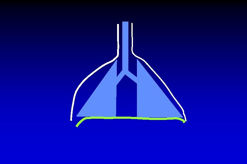
pleura parietalis diaphragm inspiration expiration
pleura visceralis diaphragm inspiration expiration
pleura parietalis transmural pressure pleura visceralis diaphragm inspiration expiration
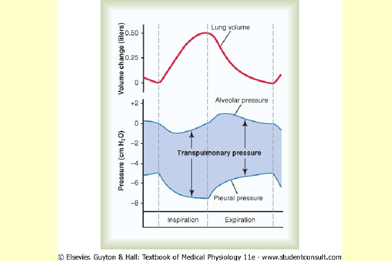
pleura parietalis pleura visceralis transpulmonal pressure diaphragm inspiration expiration
pleura parietalis pleura visceralis intrapulmonal pressure diaphragm inspiration expiration
pleura parietalis transmural pressure pleura visceralis intrapulmonal pressure transpulmonal p. diaphragm inspiration expiration
pleura parietalis transpulmonal p. pleura visceralis diaphragm inspiration expiration
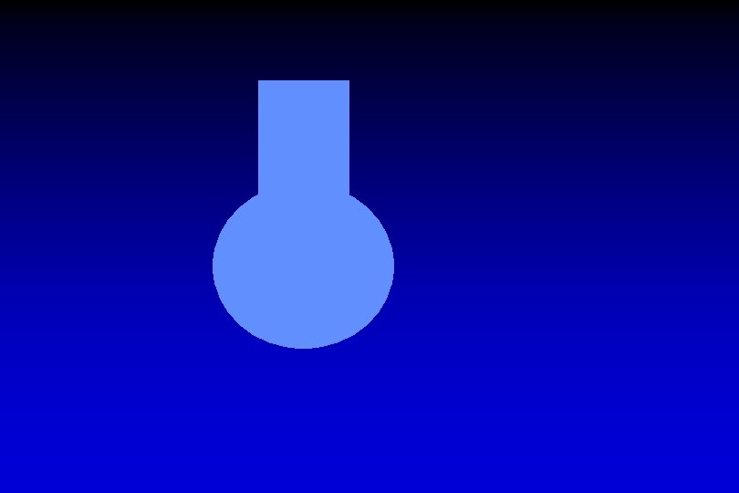
PNEUMOTHORAX pleura parietalis transmural p. pleura visceralis bránice - diaphragma inspirace expirace
alveoli pleura parietalis transmural p. pleura visceralis intrapulmonal pressure transpulmonal p. diaphragm inspiration expiration
V ventilation
Physiologic dead space Is the volume of air in the lungs that does not participate in gas exchance (i. e. e it is dead) Anatomic dead space – is the volume of conducting airways Functional dead space volume – which is made up of alveoli that do not participate in gas exchance (alveoli that are ventilated, but are not perfused by pulmonary capilary blood)
Physiologic dead space Pa. CO 2 – PECO 2 V D = VT x -----------Pa. CO 2 VD VT Pa. CO 2 PECO 2 – physiologic dead space (m. L) – tidal volume (m. L) – PCO 2 of arterial blood (mm. Hg) - PCO 2 of expered air (mm. Hg)
Physiologic dead space Pa. CO 2 – PECO 2 V D = VT x -----------Pa. CO 2 40 - 30 VD = 500 x -------40 = = 500 x 0. 25 125
Alveolar ventilation VA = (VT – VD) x breasths/min VA - alveolar ventilation (m. L/min) VT - tidal volume (m. L) VD - physiologic dead space (m. L/min)
Alveolar ventilation VA = (VT – VD) x breasths/min VA = (500 – 125) x 16 = 375 x 16 = 6000 m. L/min
lung heart circuitry tissues cell
diffusion
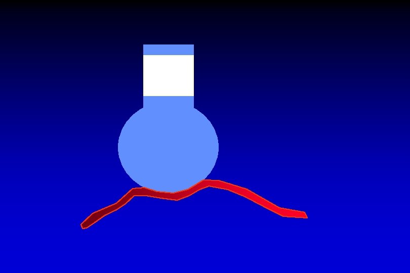
perfusion
ventilation diffusion perfusion
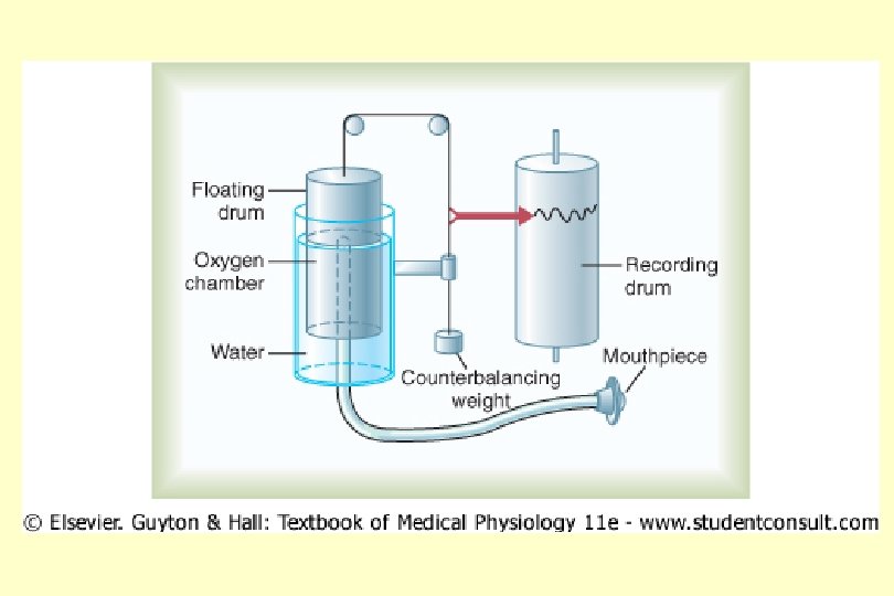
ventilation - static volums - dynamic volums
STATIC VOLUMS
DYNAMIC VOLUMS Physiological value: V FEV 1 ---- x 100 VC FEV 1 1 s VC t > 80 %
DYNAMIC VOLUMS V OBSTRUCTION FEV 1 = VC VC Restriction VC = FEV 1 1 s t
Triggers Infection inhaled allergens inhaled irritants food allergens inducing stimuli mental stress medicines
respiratory irritant factors physical activity preventing infection sick people, vaccination, intensive treatment sufficient fluid intake eat small portions breathing exercises
Differential diagnosis
potentiation of beta-adrenergic receptor bronchospasmolytic effect cholinergic antagonism reduction in mucus production steroid - anti-inflammatory effect - block the formation of antibodies - stabilization of lysosomes block - formation and release of histamine
DYNAMIC VOLUMS V RESTRICTION = FEV 1 VC FEV 1 VC 1 s t
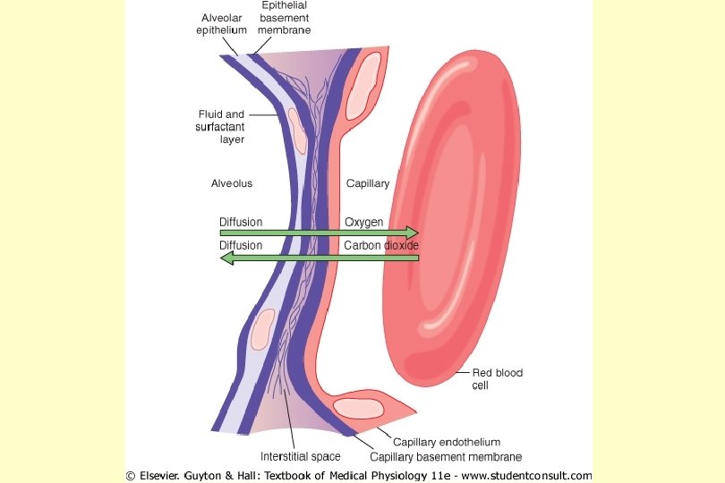
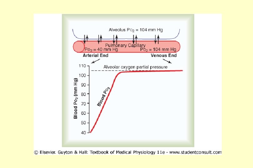
difusion alveolo-capillary membrane (surfactant)
difusion alveolo-capillar membrane (surfactant)
perfusion (heart)
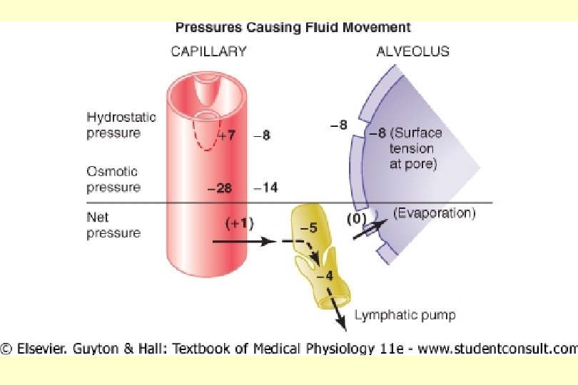
Pulmonary edema - cardiogenic - noncardiogenic
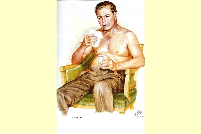
Cyanosis (right ventricle)
Causes of cyanosis Central cyanosis Decreased arterial saturation Hemoglobin abnormalities Peripheral cyanosis Reduced cardiac output Cold exposure Redistribution of blood flow from extremities Arterial obstruction Venous obstruction
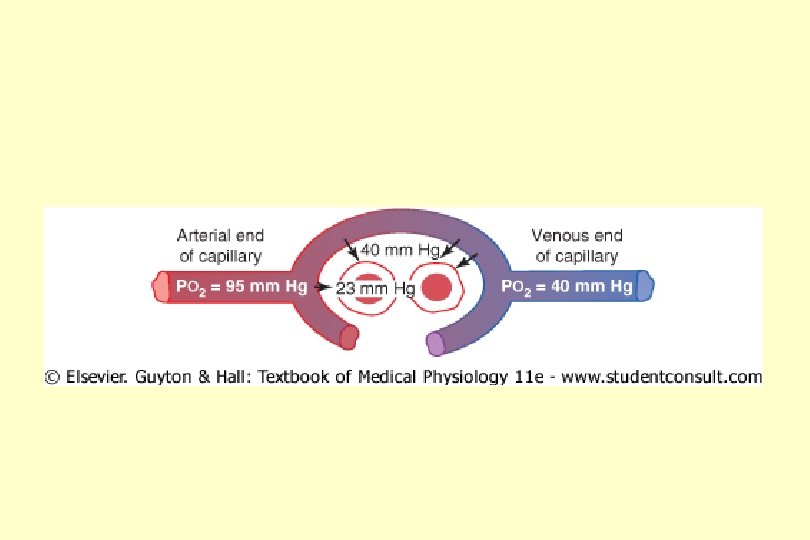
lung heart circuitry tissues cell
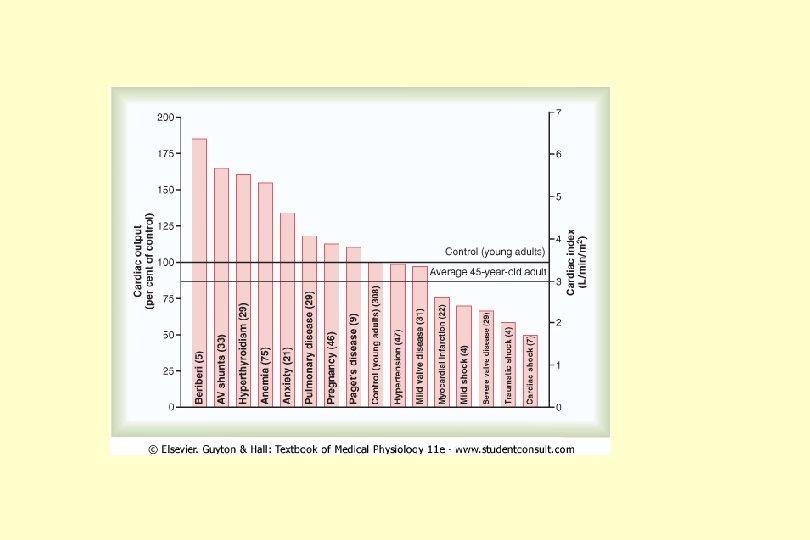
perfusion (anemia)
perfusion (emboli)
perfusion (emboli)
dg. pulmonal emboli
Dg. pulmonal emboli
Bohrs effects % hemoglobin saturation 100 p. O 2
Bohrs effect – O 2 hemoglobin dissociation curve % hemoglobin saturation 100 0 25 50 75 100 p. O 2
Shift to the right % hemoglobin saturation 100 0 25 50 75 100 p. O 2
Shift to the right % hemoglobin saturation 100 temperature p. H [H+] 2, 3 - DPG 0 25 50 75 100 p. O 2 čas
Shift to the left % hemoglobin saturation 100 temperature p. H [H+] 2, 3 - DPG 0 25 50 75 100 p. O 2
Bohr effect % hemoglobin saturation 100 0 25 50 75 100 p. O 2
Values of p. O 2 and corresponding values of percent saturation of hemoglobin p. O 2 mm. Hg 10 20 25 saturation (%) 25 35 50 30 40 50 60 80 100 60 75 85 90 96 98
lung heart circuitry tissues cell