Respiratory infections Atypical pathogens in community acquired pneumonia


 and Atypical pathogens • CAP-<50% of cases have identifiable cause")


 • Jerry, a 7 month old child,")


")

- Slides: 34
Respiratory infections Atypical pathogens in community acquired pneumonia and Whooping cough Natalie Neu, MD
Respiratory tract anatomy
Community acquired pneumonia (CAP) and Atypical pathogens • CAP-<50% of cases have identifiable cause • Atypical- differ from classic symptoms of pneumococcal pneumonia and clinical picture different, more indolent, longer duration and may involve upper and lower respiratory tract • Atypicals usually don’t respond to penicillin
Clinical scenario 1 • Myra is a 21 year old medical student living in the dorm room studying for exams • She goes to student health complaining of low grade fever, headache, non-productive cough, sore throat and general malaise • Her exam reveals mild fine inspiratory ralesnothing impressive • The Dr sends her for an xray that reveals bilateral infiltrates
Mycoplasma • Does not have a cell wall • Cell membrane contains sterols not present in other bacteria • Special enriched media needed for growth • Laboratory cultures rarely done- diagnosis usually by serology (Ig. G) • Bedside test- cold agglutinins
Mycoplasma- pathogenesis and immunity • P 1 - protein attachment factor- facilitates attachment to sialic acid receptors of respiratory epithelium and RBC surface • Remains extracellular • Causes local destruction of cilia, interferes with normal airway clearance which leads to mechanical irritation and persistent cough • Acts as a super antigen stimulating PNM’s and macrophages to release cytokines (TNF , IL 1, and IL 6)
Walking pneumonia • Lacks seasonal pattern, spread by droplet secretions • Common in children and young adults • Mild respiratory symptoms • Complications: otitis media, erythema mulitforms, hemolytic anemia, myocarditis, pericarditis, neurologic abnormalities • Treatment: erythromycin
Erythema multiforme
Clinical scenario 2 • JM 10 week old infant born to a 16 year old mom • Pregnancy history limited due to lack of prenatal care but baby born full term, no complications, left hospital 2 days • Seen by pediatrician at 2 weeks old with eye discharge was given eye drops • Returned to ER: RR 60, cough but no fever • Xray done and bloods drawn
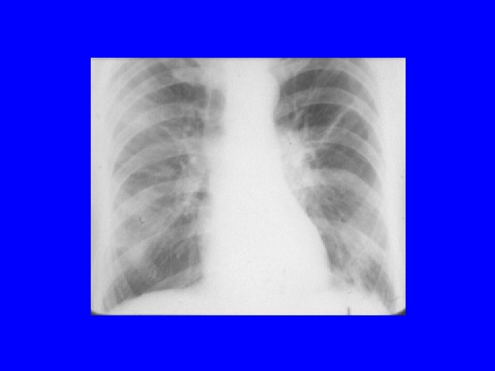
Chlamydia trachomatis xray
Chlamydial pneumonias: trachomatis, pneumoniae, psittaci • Intracellular parasites- use host high energy phosphate compounds • Trilaminar outer membrane which contains LPS • Two phase life cycle- Elementary body (infectious) and reticulate body (divides by binary fission in the host)
Chlamydial pneumonias • • Infect non-ciliated columnar cells Multiply in alveolar macrophages Perivascular and peribronchiolar infiltrates Clinical symptoms due to host immune response • Immunity not long-lasting • Diagnosis by serology- four fold rise in titer
C. trachomatis pneumonia • • Neonatal infection presents at 1 -3 months of age Staccato-like cough, rapid respiratory rate NO FEVER Evaluation: minimal chest findings, xray hyperinflation and diffuse infiltrates, peripheral eosinophilia • Associations: atherosclerotic heart disease • Treatment: erythromycin • Prevention: maternal screening
C. pneumoniae • • Single strain- TWAR Prolong incubation period Common in school age children Indolent course-sore throat, chronic cough, no fever • Chest xray variable (lobar, diffuse, bilateral) • Diagnosis: PCR and serology • Treatment: macrolide, doxycycline, levofloxacin
C. psittaci • History: Parrot exposure • Mild clinical respiratory symptoms, fever, rash • Concomitant symptoms: cns- headache, confusion, cranial nerve palsy, seizures; hepatitis; pericarditis • Xray-consolidation, reticular nodular pattern, adenopathy • Titers: > 1: 64 diagnostic • Treatment: doxy, tetracycline, erythromycin
Clinical scenario 3 • Charlie is a 68 year old retired plumber who recently underwent a renal transplantation • Felt great and was tinkering around his house updating his bathroom fixtures • Came for follow up visit complaining of high fever, cough, chills and his wife said that he was acting confused at times • Laboratory studies reveal WBC 35, 000 with left shift, LDH >1000 • Chest xray reveals multilobar process
Legionella species
The 1976 Legionnaire’s Convention, Philadelphia, PA • 29/180 patients died due to pneumonia • Identification of a gram negative bacilli • Epidemiologic link to being in the lobby of Hotel A; historical link to 1966 outbreak in a psychiatric hospital • National panic- worries about biologic and chemical warfare- media frenzy • 6 months to identify the organism
Legionella pneumophila and micdadei • 2 -6% community acquired pneumonias • Risk: immunocompromised, hospitalized, and outbreak situations • Gram negative bacilli- don’t stain with common reagents • Fastidious and grow on supplemented media • Organisms contaminate water sources: air conditioning systems and water tanks
Legionella: pathogenesis and immunity • Intracellular pathogen- multiply in macrophages and monocytes • Proteolytic enzymes kill the infected respiratory cells leading to formation of microabscesses • Immunity- Cell mediated immunity (T cells) needed for immune response
Legionnaires disease • Incubation period up to 10 days • Clinical- influenza like illness or severe manifestation= pneumonia • Fever (105), rigors, cough, headache • Multilobular infiltrates and microabscesses • Extrapulmonary manifestations: CNS, diarrhea, abdominal pain, nausea • High white counts, abnormal liver, renal panel • High mortality-15 -20% depending on host
Legionella: Diagnosis, prevention and treatment • Urine antigen detection assays- EIA for L. pneumophila only • Serology >1: 128 positive however late development of antibodies • Culture on special media • Treatment: macrolide or levofloxacin • Prevention: hyperclorination, super heating, continuous copper-silver ionization
Clinical scenario 4 (Loyola Univ Medical Center) • Jerry, a 7 month old child, comes to clinic with a running nose, sneezing and slightly irritable • Diagnosed with URI • Returns 2 weeks later because he is turning blue with coughing spells. Spells are worse at night, seems to have spasms and then he “whoops” for air. • Examination reveals mildly dehydrated, not distressed, clear lung exam • WBC reveals leucocytosis with lymphocytosis
Bordetella pertussis
Bordetella pertussis “Whooping cough” • • • Fastidious, gram negative coccobacilii Pertussis, parapertussis, and bronchiseptica Spread by respiratory droplets Rapid multiplication in mucus membrane No bacteremia Toxins cause local tissue damage
Binding and uptake by phagocytic cells
Pertussis toxin
Toxin production and disease manifestation
G protein and ADP riboyslation
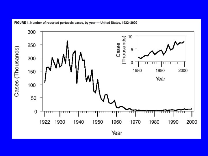
Pertussis • Incidence declined due to vaccine • Affects children under 1 and adults with waning immunity • Incubation period 7 -10 days • Three stages of disease: catarrhal, paroxysmal, convalescent • Diagnosis: special media- Bordet-Gengou- blood, charcoal, and starch. Nasopharyngeal culture • Serologic testing: acute and convalescent titers
Toxins and pathogenesis Pertussis toxin- increased CAMP Increased resp secretions and mucus (paroxysmal stage) Adenylate cyclase and hemolysin toxin Inhibit leukocyte chemotaxis, phagocytosis, and killing Local tissue destruction Heat-labile toxin Tracheal cytotoxin Lipid A and Lipid X Destroys ciliated epith cells, IL-1 (fever), NO (kills epithelial cells) Activate alternative complement, cytokine release
Pertussis clinical symptoms