Respiratory infections Arranged by Lec Hadeel Dalman Respiratory

Respiratory infections Arranged by: Lec. Hadeel Dalman

Respiratory tract infections Upper respiratory tract infections Lower respiratory tract infections
")
Upper respiratory tract infections • Colds and flu • Influenza • Sore throat (pharyngitis) • Acute epiglottitis • Otitis media • Acute sinusitis

Lower respiratory infections • Acute bronchitis and acute exacerbations of COPD • Bronchiolitis • Pneumonia • Severe acute respiratory syndrome • Cystic fibrosis

For each case you need to know the followings: • Definition • Causative organisms • Clinical features • Diagnosis • Treatment

Upper respiratory tract infections

Influenza • True influenza is caused by one of the influenza viruses (influenza A, B or rarely C). • Characterised by severe malaise and myalgia • Potentially complicated by life-threatening secondary bacterial infections such as staphylococcal pneumonia. • No Coryzal symptoms
 • include")
Treatment Prophylaxis • influenza vaccine… Prevention and Treatment • neuraminidase inhibitors (NAIs) • include agents such as zanamivir and oseltamivir. • The anti-Parkinsonian drug amantadine, which has activity against influenza A virus, is not recommended.
")
Coryzal symptoms • Sore throat or throat irritation. • Runny nose (increased mucus production) or postnasal drip. • Sneezing. • Nasal and sinus blockage (thick mucus and debris) or congestion with or without sinus pressure. • Headache. • Cough. • Mild fever. • Watery eyes or redness and/or itchiness of eyes.
 • Most cases are viral • Epstein–Barr virus (EBV), which causes")
Sore throat (pharyngitis) • Most cases are viral • Epstein–Barr virus (EBV), which causes glandular fever • The only common bacterial cause of sore throat is Streptococcus pyogenes (β-haemolytic Streptococcus),

Clinical features • sore throat often associated with fever and the usual symptoms of the common cold. • In more severe cases----marked inflammation of the pharynx with a whitish exudate on the tonsils, plus enlarged and tender cervical lymph nodes. • May accompaniments with otitis media, peritonsillar abscess and sinusitis.

Diagnosis ØThroat Swab • to distinguish the streptococcal sore throat from viral infections. • directed towards detecting β-haemolytic streptococci. ØSerological test • to detect antibodies to EBV

Treatment • Viral sore throat is directed at symptomatic relief, for example with rest, antipyretics and aspirin gargles. • Bacterial sore throat need Antibiotic treatment also reduces the incidence of non-suppurative complications

Streptococcal Sore Throat there are three treatment strategies: 1. give antibiotics to all patients with suspected streptococcal infection and do not investigate unless symptoms persist 2. give antibiotics to all patients with suspected streptococcal infection but stop them if a throat swab is negative, or 3. wait for throat swab culture results before starting antibiotics.
")
Antibiotics effective against S. pyogenes include • Penicillins, • Cephalosporins • Macrolides (less effective)

Acute epiglottitis • A medical emergency of rapidly progressive cellulitis of the epiglottis and adjacent structures. • Local swelling has the potential to cause rapid-onset airway obstruction, • Common patient is a child between 2 and 4 years old.
 mainly, • Pneumococci, streptococci and staphylococci")
ØCausative organisms: • Haemophilus influenzae type b (Hib) mainly, • Pneumococci, streptococci and staphylococci (less). ØClinical feature: • Fever • Difficulty speaking and breathing. • Drooling because of impaired swallowing. ØDiagnosis: • By visualization of the epiglottis as ‘cherry-red’. • By Microbiological confirmation by culturing the epiglottis and the blood (but not until the airway is secure).

Treatment • Treatment of choice is a cephalosporin (third-generation cephalosporin such as cefotaxime or ceftriaxone). • High-dose parenteral amoxicillin may be substituted (amoxicillin resistance among encapsulated H. influenza)
 is a common condition")
Otitis Media • Inflammation of the middle ear (otitis media) is a common condition seen most frequently in children under 3 years of age. • Caused by influenza virus and rhinoviruses (mainly) • S. pneumoniae and H. influenza • Moraxella catarrhalis and S. pyogenes account for a smaller proportion of cases,
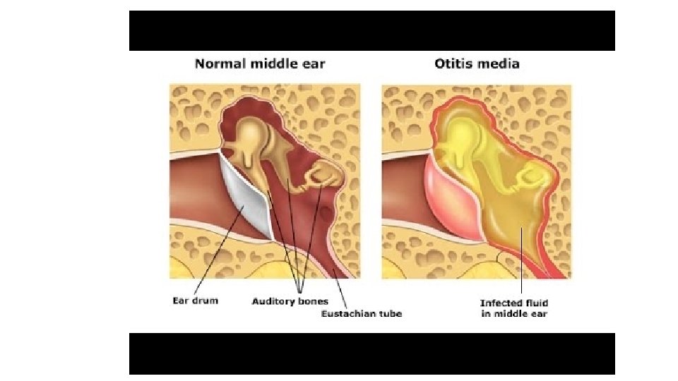

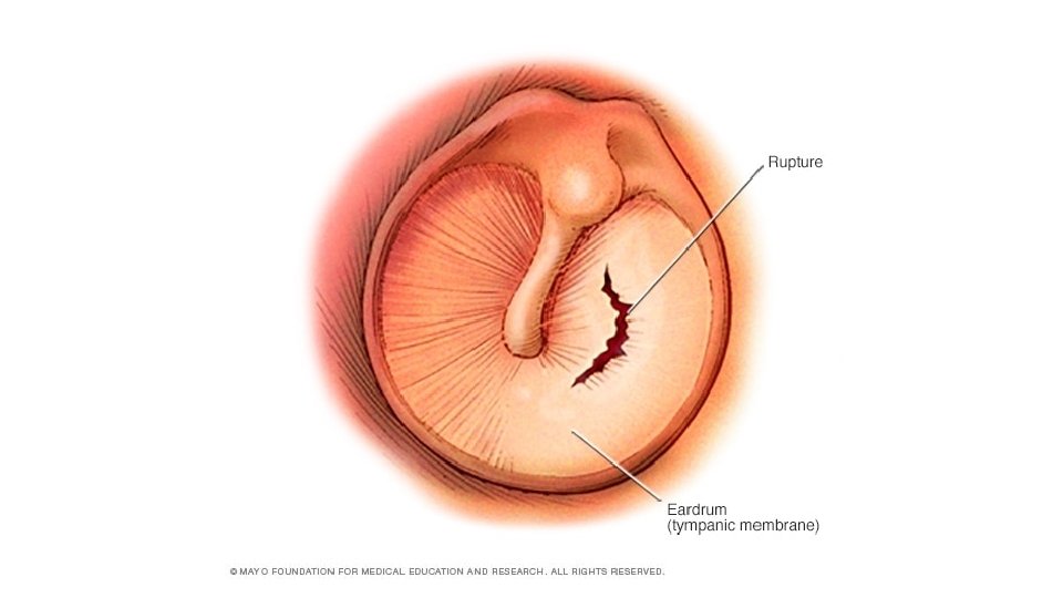
Clinical features • Ear pain, which may be severe. • If the drum perforates, the pain is relieved and a purulent discharge may follow. • There may be a degree of hearing impairment plus non-specific symptoms such as fever or vomiting. • Complications include mastoiditis, meningitis and, particularly, septicaemia and disseminated infection.

Diagnosis • The diagnosis of otitis media is essentially made clinically and laboratory investigations have little role to play. • A swab of the external auditory canal---- if the drum is perforated. For this reason, a causative • Organism is rarely isolated and treatment has to be given empirically.

Treatment • If antibiotic treatment is to be given, it should be effective against the three main bacterial pathogens: S. pneumoniae, H. influenza and S. pyogenes. • Amoxicillin or Ampicillin • Later-generation Cephalosporin

Acute Sinusitis • Normally, the paranasal sinuses are sterile but they can become infected following damage to the mucous membrane which lines them. • Usually occurs following a viral URTI but is sometimes associated with the presence of dental disease. • Same organisms which cause otitis media S. aureus,

Clinical features & Diagnosis • Facial pain and tenderness, • Often accompanied by headache and a purulent nasal discharge. • Complications include frontal bone osteomyelitis, meningitis and brain abscess. ØDiagnosis: • No specific test. • Therapeutic sinus washouts may yield specimens for microbiological culture.

Treatment • Since the causative organisms are the same as those found in otitis media, the same recommendations for treatment apply. • Amoxicillin/clavulanate or Doxycycline • If associated with dental disease, and in such cases, the addition of metronidazole

Lower respiratory infections

Acute bronchitis • Bronchitis means inflammation of the bronchi. • Acute bronchitis, which is usually infective. • Chronic bronchitis, which is a chronic inflammatory condition characterized by thickened, edematous bronchial mucosa with mucus gland hypertrophy and usually caused by smoking. • The importance of chronic bronchitis is that it renders the patient more susceptible to acute infections and more likely to suffer respiratory compromise as a result.

ØCausative organisms: • Viruses such as rhinovirus, coronavirus, adenovirus and influenza virus. • Bacteria such as Bordetella pertussis, Mycoplasma pneumoniae and Chlamydophila pneumonia, S. pneumoniae, H. influenzae and M. catarrhalis ØClinical features: • Cough (productive of purulent sputum), that is, • Phlegm (yellow or green), the color reflecting the presence of pus cells. • Wheezing and breathlessness ØDiagnosis: • A sputum sample should be sent for bacteriology, as this will allow antibiotic sensitivity tests to be performed on potential pathogens.

Treatment EMPIRIC ANTIBIOTIC TREATMENT: • First-line agents • Doxycycline • Amoxicillin • Second-line agents • Co-amoxiclav • Clarithromycin • Cefixime

Bronchiolitis • Characterised by inflammatory changes in the small bronchi and bronchioles, but not by consolidation. • It is recognised as a disease of infants in the first year of life, in whom a small degree of airway narrowing can have a dramatic effect on airflow. • Caused by: • respiratory syncytial virus (RSV), • h. MPV, parainfluenza viruses, • rhinoviruses, adenoviruses • M. pneumonia.

• Clinical Feature: • Fever and • Coryzal symptoms • Progresses to wheezing, respiratory distress and hypoxia • Diagnosis: • Immunofluorescence and/or • Viral culture of respiratory secretions, • Treatment: • Supportive consists of oxygen, adequate hydration and ventilatory assistance • Severe cases of respiratory syncytial virus disease may be treated with ribavirin, administered by nebuliser.

Pneumonia ØPneumonia is defined as inflammation of the lung parenchyma, that is, of the alveoli rather than the bronchi or bronchioles, of infective origin and characterised by consolidation. ØPneumonia is often classified clinically into • Lobar pneumonia, • Bronchopneumonia, • Atypical pneumonia.
 • Causative organisms ØS. pneumoniae, the pneumococcus, which can cause both")
Community-acquired pneumonia (CAP) • Causative organisms ØS. pneumoniae, the pneumococcus, which can cause both lobar and bronchopneumonia, and non-capsulate strains of H. influenzae which usually give rise to bronchopneumonia. ØViral, Influenza can cause a primary viral pneumonia as well as be complicated by secondary bacterial (particularly staphylococcal) pneumonia, • chickenpox can be complicated by primary varicella pneumonia particularly in adults, and • Cytomegalovirus is capable of causing a variety of infections, including pneumonia,

Atypical Pneumonias • A heterogeneous group of diseases which nevertheless have several clinical features in common and which are clinically distinct from the classic picture of pneumococcal pneumonia. • The atypical pneumonias are characterised clinically by fever, systemic symptoms and a dry cough, radiologically by widespread patchy consolidation in both lungs and biochemically by abnormalities in liver enzymes and perhaps evidence of inappropriate antidiuretic hormone secretion, evident as a low plasma sodium.

Clinical features • Pneumococcal lobar pneumonia presents with a cough, initially dry but later producing purulent or blood-stained, rust-coloured sputum, together with dyspnoea, • fever and pleuritic chest pain. • The peripheral white blood cell count is usually raised and the patient may be bacteraemic. • The chest X-ray shows consolidation confined to one or more lobes (or segments of lobes) of the lungs.

Clinical features • Bronchopneumonia presents with productive cough and breathlessness, and • patchy consolidation on the chest X-ray usually in the bases of both lungs. • This disease is very common and is typically seen in patients with severe COPD or in those who are frail and terminally ill. In fact, pneumonia has been described as the old man's friend because it is a relatively painless cause of death.
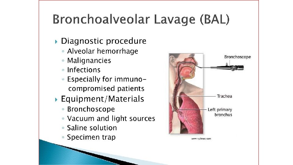
 • Bronchoscopy and")
Diagnosis • Sputum culture (dependent upon the quality of the specimen) • Bronchoscopy and bronchoalveolar lavage. (Lavage fluid, being uncontaminated by mouth flora) • Blood cultures • Plasma and urine testing for pneumococcal antigen • Viruses may be detected by immunofluorescence, by viral culture or by polymerase chain reaction (PCR),

Empiric treatment Targeted treatment
 • Nosocomial pneumonia accounts for 10– 15% of all hospital-acquired infections,")
Hospital-acquired pneumonia (HAP) • Nosocomial pneumonia accounts for 10– 15% of all hospital-acquired infections, • Usually presenting with sepsis and/or respiratory failure. • Up to 50% of cases are acquired on intensive care units. • Predisposing features include stroke, mechanical ventilation, chronic lung disease, recent surgery and previous antibiotic exposure.
 and")
ØCausative organisms: • Gram-negative bacilli (Enterobacteriaceae, Pseudomonas spp. • and Acinetobacter spp. ) and S. aureus, including MRSA. ØDiagnosis: • Sputum is commonly sent for culture (it may be contaminated by mouth flora. If the patient has received antibiotics, the normal mouth flora is often replaced by resistant organisms such as staphylococci or Gram-negative bacilli, making the interpretation of culture results difficult). • Bronchoalveolar lavage is often more helpful. • Blood cultures may be positive.

The choice of antibiotics will be influenced by: preceding antibiotic therapy, the duration of hospital admission Macrolide would be added if Legionnaire's disease was suspected and, Metronidazole would be required for suspected anaerobic infection.

Aspiration pneumonia • Initiated by inhalation of stomach contents contaminated by bacteria from the mouth. • Risk factors include alcohol, hypnotic drugs and general anaesthesia, • Make a patient vomit while unconscious. • Gastric acid is very destructive to lung tissue and leads to severe tissue necrosis. • Damaged tissue is then prone to secondary infection often with abscess formation. • Treatment with metronidazole plus amoxicillin is usually adequate,
 • Caused by a coronavirus (SARS-associated coronavirus). • Clinically")
Severe acute respiratory syndrome (SARS) • Caused by a coronavirus (SARS-associated coronavirus). • Clinically it causes pneumonitis, presenting with a flu-like prodrome • progressing to dyspnoea, dry cough. • Treatment is largely supportive (ventilatory support).
 • Is an inherited, autosomal recessive disease • Is due to")
Cystic fibrosis (CF) • Is an inherited, autosomal recessive disease • Is due to a defect in the transport of ions in and out of cells. • This leads to changes in the consistency and chemical composition of exocrine secretions, • In the lungs is manifest by the production of very sticky, tenacious mucus which is difficult to clear by mucociliary action. • The production of such mucus leads to airway obstruction with resulting infection.

ØInfecting organisms • S. aureus, H. influenzae are the most common In infants and young children, • P. aeruginosa forolder than 5. • Gram-negative bacteria are seen, such as Escherichia coli. ØClinical features • • • persistent cough and copious and purulent sputum. Breathless fever, increased cough Eventually, chronic pulmonary infection leads to respiratory insufficiency, Cardiac failure and death.

Treatment ØIn a children • Anti-staphylococci, such as flucloxacillin or erythromycin can be used. ØP. aeruginosa • oral ciprofloxacin and a nebulised antibiotic such as colistin. ØFor chronically colonised patients, • regular prophylactic intravenous β-lactam/aminoglycoside combination such as ceftazidime plus tobramycin. • Meropenem or a quinolone are usually reserved for treatment failures or when resistant organisms are encountered.

Notes… • Patients with cystic fibrosis need larger doses of aminoglycosides because have a rapid clearance more than other patients. • Parenteral therapy as inhaled (nebulised) antibiotics use for treatment of acute exacerbations and for longer-term use in an attempt to reduce the Pseudomonas load. Agents which have been administered in this way include colistin, tobramycin and other aminoglycosides, carbenicillin and ceftazidime.
- Slides: 50