
Respiratory Emergencies East Region Washington OTEP M7 Brian
Respiratory Emergencies East Region (Washington) OTEP M-7 Brian Reynolds, MD Deaconess Medical Center Spokane, WA
Respiratory Emergencies ü We are going to cover material for ALL levels of training ü YOU CAN ONLY PRACTICE AT THE LEVEL YOU HAVE BEEN CERTIFIED
Topics ü Anatomy and function of the Respiratory System ü Patient Assessment ü Airway Management
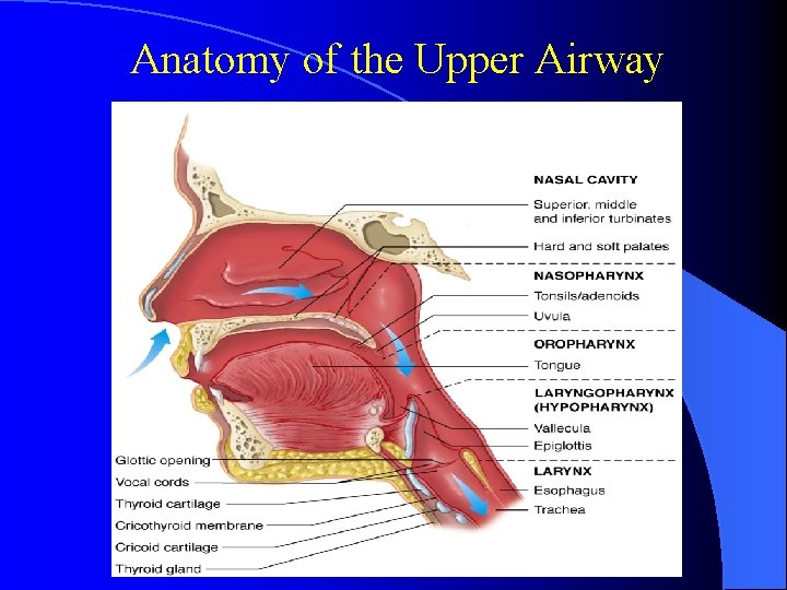
Anatomy of the Upper Airway
Upper Airway ü Nasal cavity ü Oral cavity ü Pharynx
Nasal Cavity üNares üMucous membranes ü Sinuses
Oral Cavity ü ü ü Cheeks Hard palate Soft palate Tongue Gums Teeth
Pharynx ü Nasopharynx ü Oropharynx ü Laryngopharynx
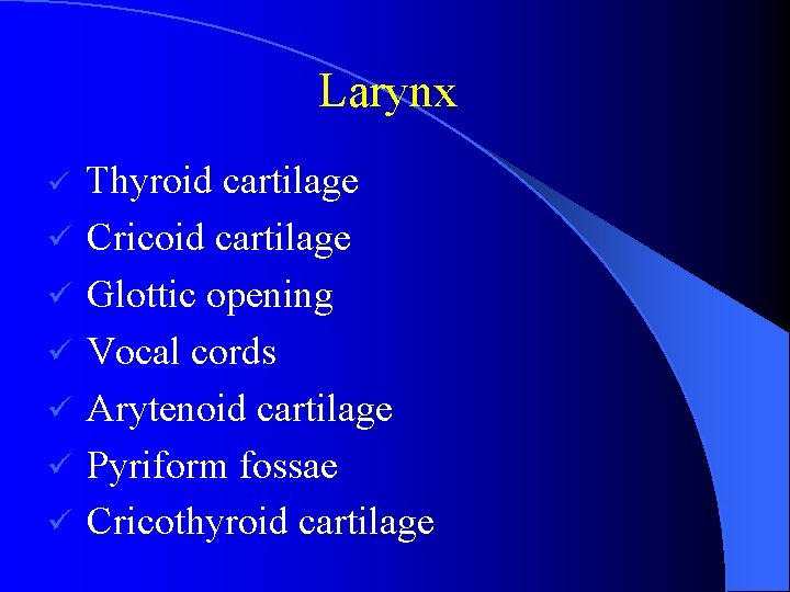
Larynx ü ü ü ü Thyroid cartilage Cricoid cartilage Glottic opening Vocal cords Arytenoid cartilage Pyriform fossae Cricothyroid cartilage
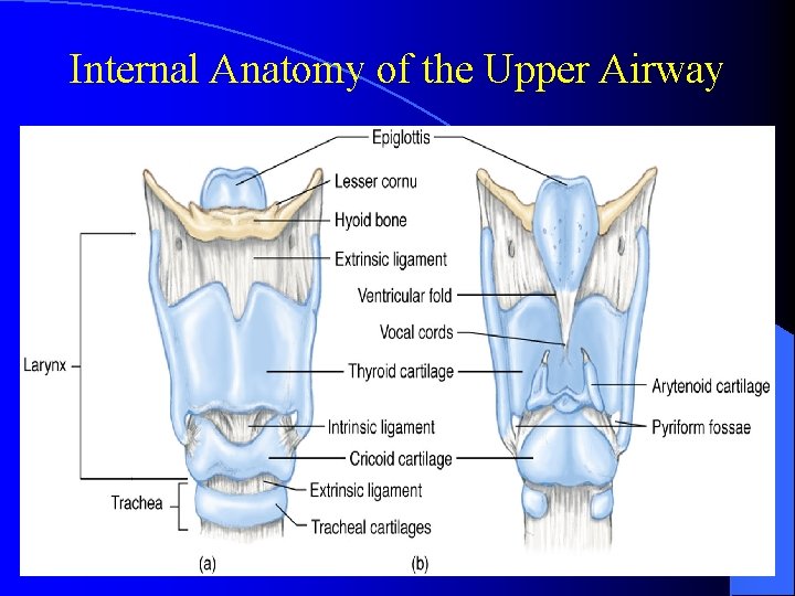
Internal Anatomy of the Upper Airway
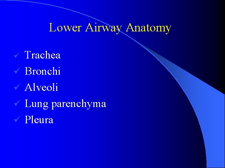
Lower Airway Anatomy ü ü ü Trachea Bronchi Alveoli Lung parenchyma Pleura
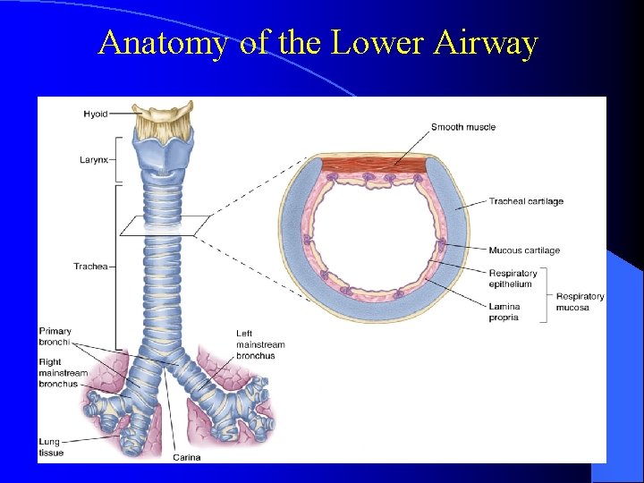
Anatomy of the Lower Airway
Definitions ü Atelectasis – collapse of small segments of lung ü Hypoxia – lack of oxygen ü Hypoxemia blood – lack of oxygen in arterial
Introduction Ventilation is the mechanical process that brings O 2 to the lungs, and clears CO 2 from the lungs ü Oxygenation is the diffusion of O 2 to the blood ü Perfusion is the flow of blood through the lungs (thus exchanging oxygen and CO 2) ü Brain stem is the involuntary regulator of respirations ü
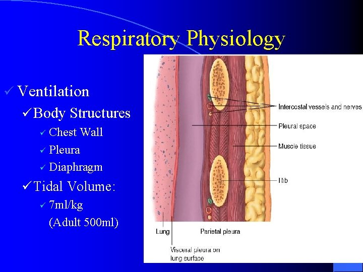
Respiratory Physiology ü Ventilation ü Body Structures Chest Wall ü Pleura ü Diaphragm ü ü Tidal Volume: ü 7 ml/kg (Adult 500 ml)
Pathophysiology ü Disruption in Ventilation ü Upper & Lower Respiratory Tracts ü Obstruction due to trauma or infectious processes ü Chest Wall & Diaphragm ü Trauma ü Pneumothorax ü Hemothorax ü Flail chest ü Neuromuscular disease
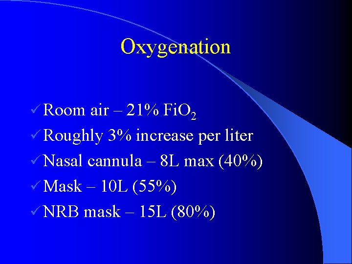
Oxygenation ü Room air – 21% Fi. O 2 ü Roughly 3% increase per liter ü Nasal cannula – 8 L max (40%) ü Mask – 10 L (55%) ü NRB mask – 15 L (80%)
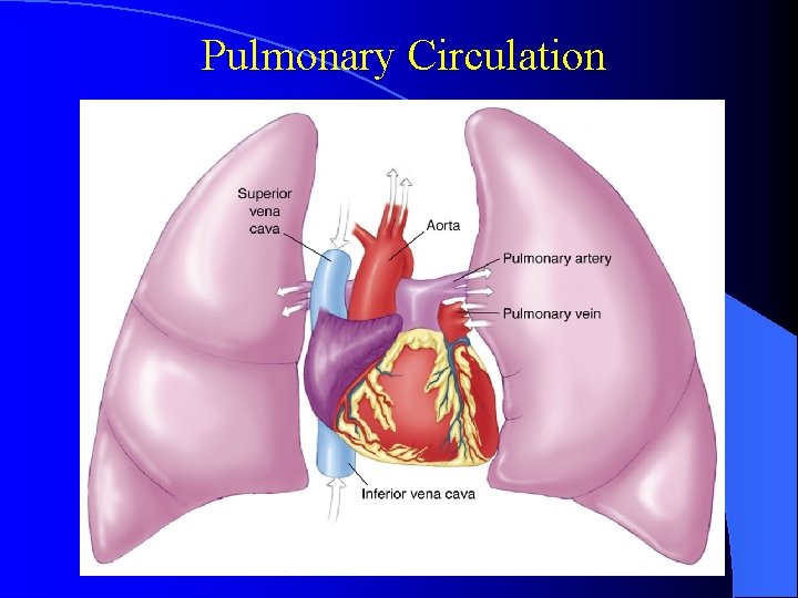
Pulmonary Circulation
Respiratory Physiology ü Pulmonary Perfusion üRequirements ü Adequate blood volume ü Intact pulmonary capillaries ü Efficient pumping by the heart üHemoglobin üCarbon Dioxide
Pathophysiology ü Disruption in Perfusion üAlteration in systemic blood flow üChanges in hemoglobin üPulmonary shunting üDamaged alveoli
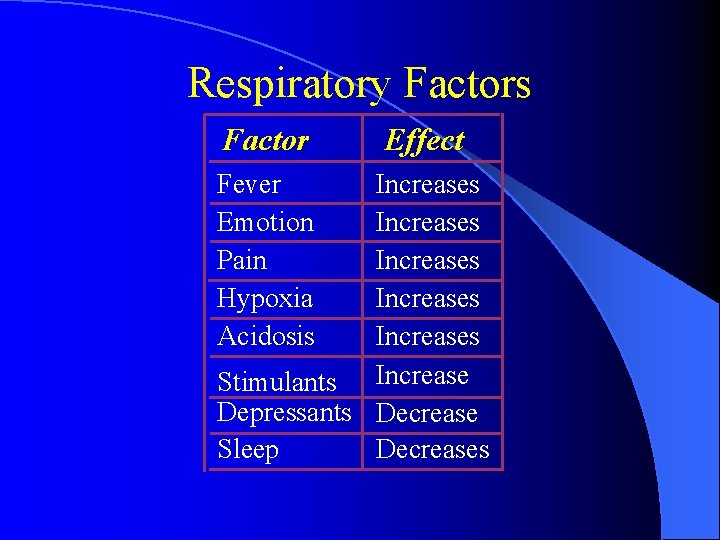
Respiratory Factors Factor Fever Emotion Pain Hypoxia Acidosis Effect Increases Increases Stimulants Increase Depressants Decrease Sleep Decreases
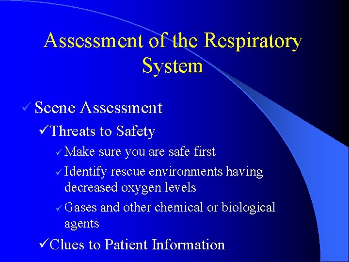
Assessment of the Respiratory System ü Scene Assessment üThreats to Safety ü Make sure you are safe first ü Identify rescue environments having decreased oxygen levels ü Gases and other chemical or biological agents üClues to Patient Information
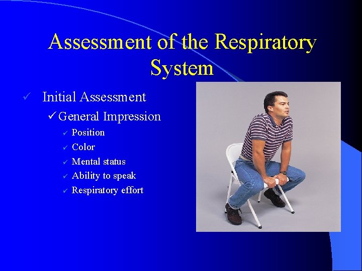
Assessment of the Respiratory System ü Initial Assessment ü General Impression ü ü ü Position Color Mental status Ability to speak Respiratory effort
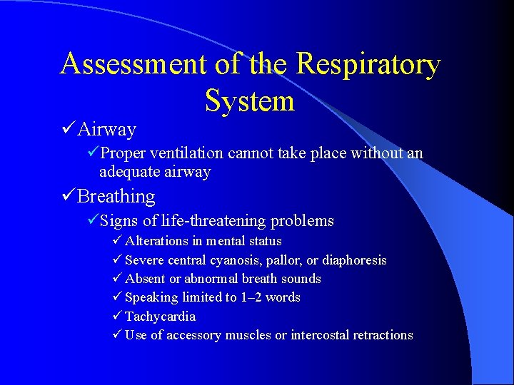
Assessment of the Respiratory System üAirway üProper ventilation cannot take place without an adequate airway üBreathing üSigns of life-threatening problems ü Alterations in mental status ü Severe central cyanosis, pallor, or diaphoresis ü Absent or abnormal breath sounds ü Speaking limited to 1– 2 words ü Tachycardia ü Use of accessory muscles or intercostal retractions
Abnormal Respiratory Patterns Kussmaul’s respirations: ü Deep, slow or rapid, gasping; common in diabetic ketoacidosis Cheyne-Stokes respirations: ü Progressively deeper, faster breathing alternating gradually with shallow, slower breathing, indication brain stem injury
Abnormal Respiratory Patterns Agonal respirations: üShallow, slow, or infrequent breathing, indicating brain anoxia
Focused History & Physical Exam ü History ü SAMPLE History ü Paroxysmal nocturnal dyspnea and orthopnea Coughing, fever, hemoptysis ü Associated chest pain ü Smoking history or environmental exposures ü ü Similar Past Episodes
Focused History & Physical Exam ü Physical Examination ü Inspection ü Look for asymmetry, increased diameter, or paradoxical motion ü Palpation ü Feel for subcutaneous emphysema or tracheal deviation ü Percussion ü Auscultation
Focused History & Physical Exam ü Auscultation ü Normal Breath Sounds ü Bronchial, Bronchovesicular, and Vesicular ü Abnormal Breath Sounds ü Snoring ü Stridor ü Wheezing ü Rhonchi ü Rales/Crackles ü Pleural friction rub
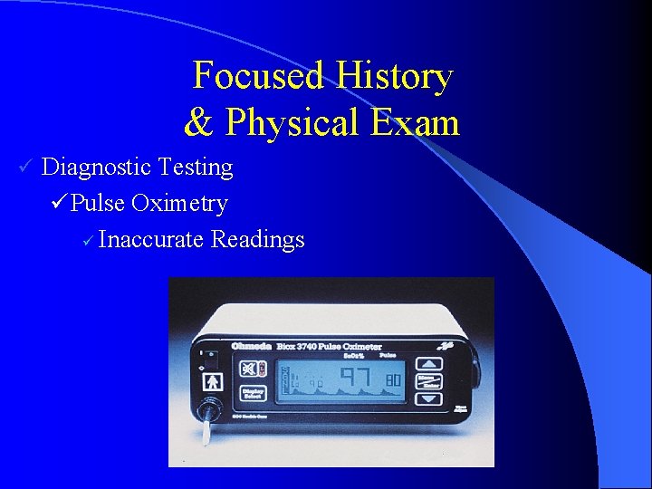
Focused History & Physical Exam ü Diagnostic Testing ü Pulse Oximetry ü Inaccurate Readings
Ausculation Listen at the mouth and nose for adequate air movement ü Listen with a stethoscope for normal or abnormal air movement ü ü Proper listening positions
Airway Obstruction ü ü ü The tongue is the most common cause of airway obstruction Foreign bodies Trauma Laryngeal spasm and edema Aspiration
Congestive Heart Failure ü Wet, crackly lung sounds ü Lower ü Must extremity edema sit and sleep upright ü Frothy, pink sputum
Obstructive Lung Disease ü Types ü Emphysema ü Chronic Bronchitis ü Asthma ü Causes ü Genetic Disposition ü Smoking & Other Risk Factors
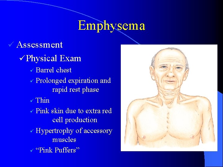
Emphysema ü Assessment ü Physical Exam Barrel chest ü Prolonged expiration and rapid rest phase ü Thin ü Pink skin due to extra red cell production ü Hypertrophy of accessory muscles ü “Pink Puffers” ü
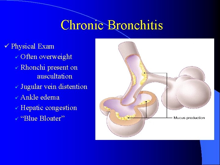
Chronic Bronchitis ü Physical Exam Often overweight ü Rhonchi present on auscultation ü Jugular vein distention ü Ankle edema ü Hepatic congestion ü “Blue Bloater” ü
Asthma ü Physical Exam ü Presenting signs may include dyspnea, wheezing, cough ü No wheezing is severe disease ü Speech may be limited to 1– 2 word sentences Look for hyperinflation of the chest and accessory muscle use/feel chest wall for crepitus ü Carefully auscultate breath sounds and measure peak expiratory flow rate ü
Pneumonia ü Infection of the Lungs ü Immune-Suppressed Patients ü Pathophysiology ü Bacterial & Viral Infections Hospital-acquired vs. community-acquired ü Alveoli may collapse, resulting in a ventilation disorder ü
Lung Cancer ü Pathophysiology ü General Majority are caused by carcinogens secondary to cigarette smoking or occupational exposure ü May start elsewhere and spread to lungs ü High mortality ü ü Types Adenocarcinoma ü Epidermoid, small-cell, and large-cell carcinomas ü
Toxic Inhalation ü Pathophysiology Includes inhalation of heated air, chemical irritants, and steam ü Airway obstruction due to edema and laryngospasm due to thermal and chemical burns ü ü Assessment ü Focused History & Physical Exam ü SAMPLE & OPQRST History ü Determine nature of substance ü Length of exposure and loss of consciousness
Carbon Monoxide Inhalation ü Pathophysiology ü Binds to Hemoglobin Prevents oxygen from binding to RBC’s ü Room air half life – 6 hrs. , HBO – 23 minutes ü ü Assessment ü Focused History and Physical Exam ü SAMPLE & OPQRST History ü Determine source and length of exposure ü Presence of headache, confusion, agitation, lack of coordination, loss of consciousness, and seizures
Pulmonary Embolism ü Pathophysiology ü Obstruction of a pulmonary artery Emboli may be of air, thrombus, fat, or amniotic fluid ü Foreign bodies may also cause an embolus ü ü Risk Factors Recent surgery, long-bone fractures ü Pregnant or postpartum ü Oral contraceptive use, tobacco use ü Immobility ü Blood disorders ü
Spontaneous Pneumothorax ü Pathophysiology ü Pneumothorax ü Can occur in the absence of blunt or penetrating trauma ü Risk factors ü Assessment ü Focused history ü ü ü SAMPLE Presence of risk factors Rapid onset of symptoms Sharp, pleuritic chest or shoulder pain Often precipitated by coughing or lifting
Hyperventilation Syndrome ü Assessment ü Focused History & Physical Exam ü SAMPLE ü Fatigue, nervousness, dizziness, dyspnea, chest pain ü Numbness and tingling in mouth, feet, and both hands Presence of tachypnea and tachycardia ü Spasms of the fingers and feet ü
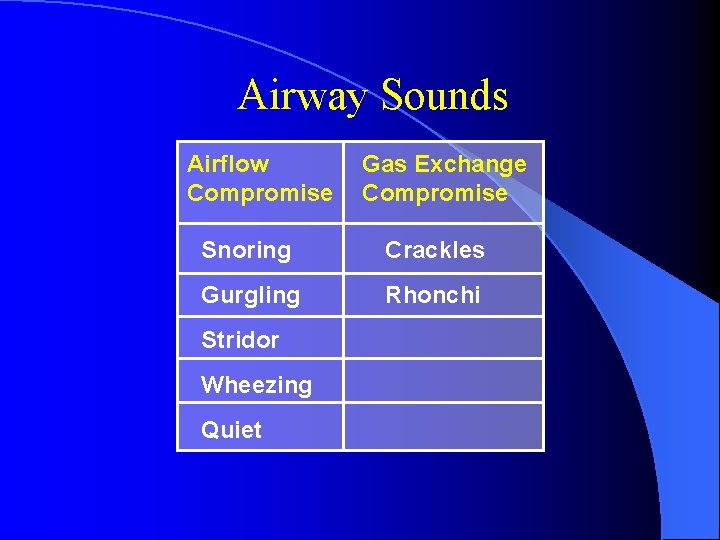
Airway Sounds Airflow Compromise Gas Exchange Compromise Snoring Crackles Gurgling Rhonchi Stridor Wheezing Quiet
Basic Mechanical Airways
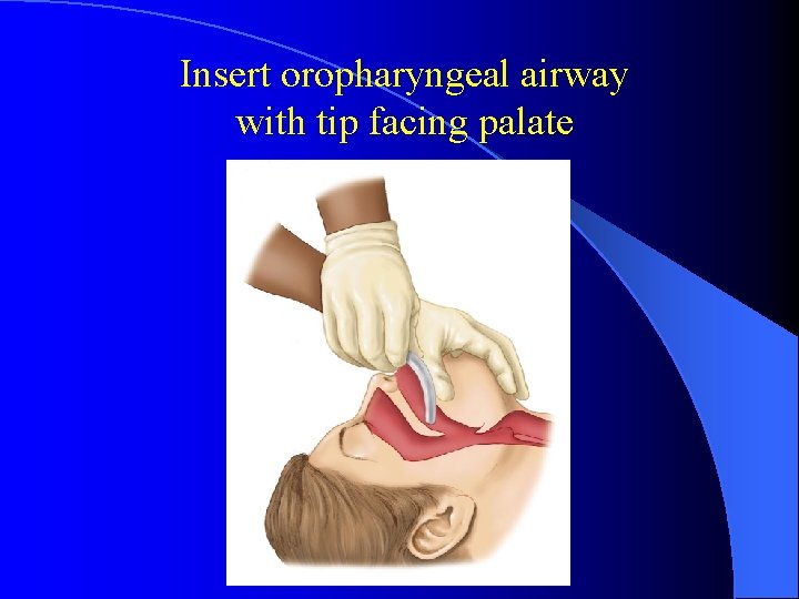
Insert oropharyngeal airway with tip facing palate
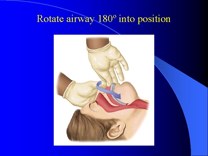
Rotate airway 180º into position
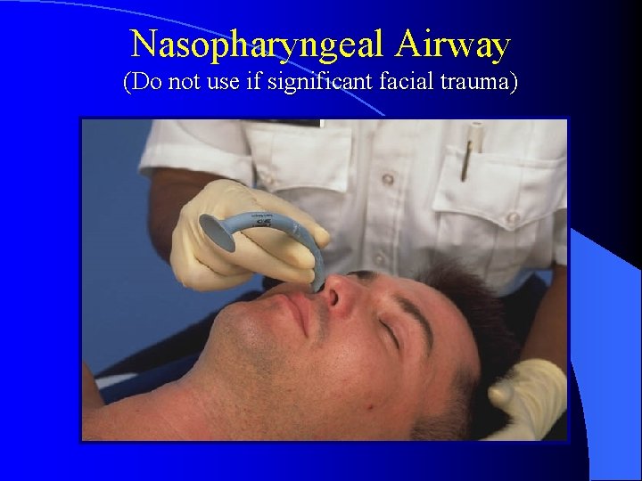
Nasopharyngeal Airway (Do not use if significant facial trauma)
Advanced Airway Management
Advanced Airway Management ü Endotracheal intubation ü Combitube ü CPAP and Bi. PAP ü CO 2 monitors – measure exhaled CO 2 ü Normal – 5 -6%
Advantages of Endotracheal Intubation ü ü ü Isolates trachea and permits complete control of airway Maximizes ventilation and oxygenation Impedes gastric distention Eliminates need to maintain a mask seal Offers direct route for suctioning
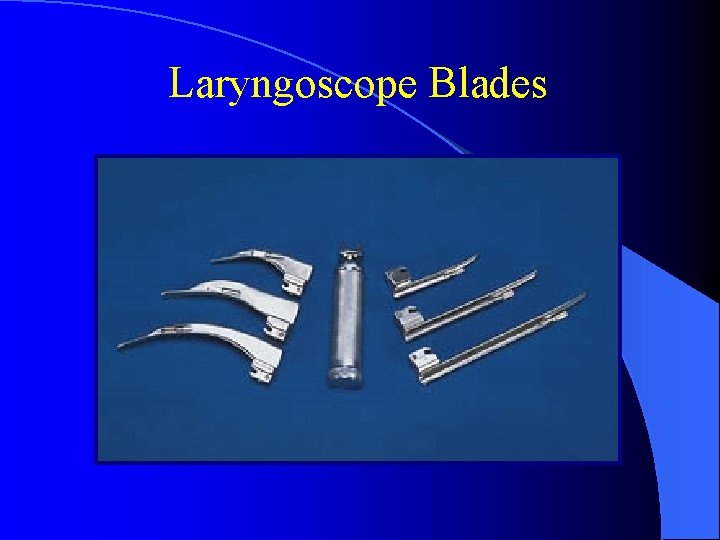
Laryngoscope Blades
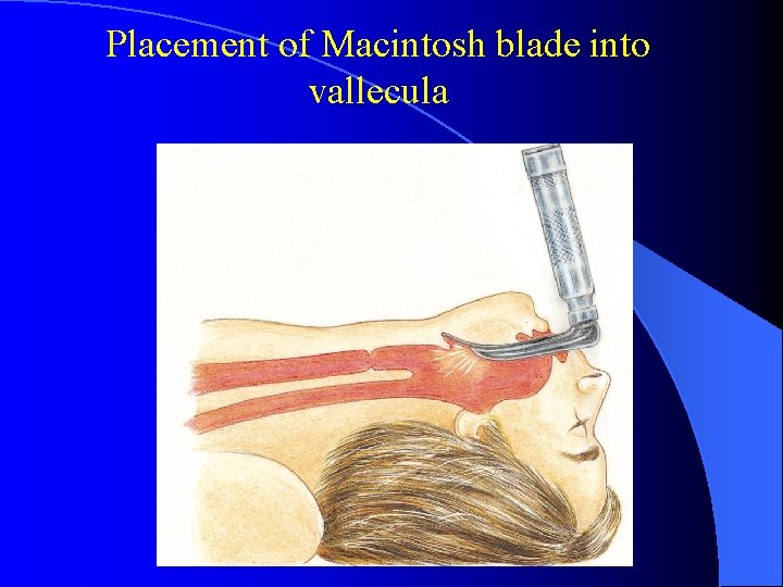
Placement of Macintosh blade into vallecula
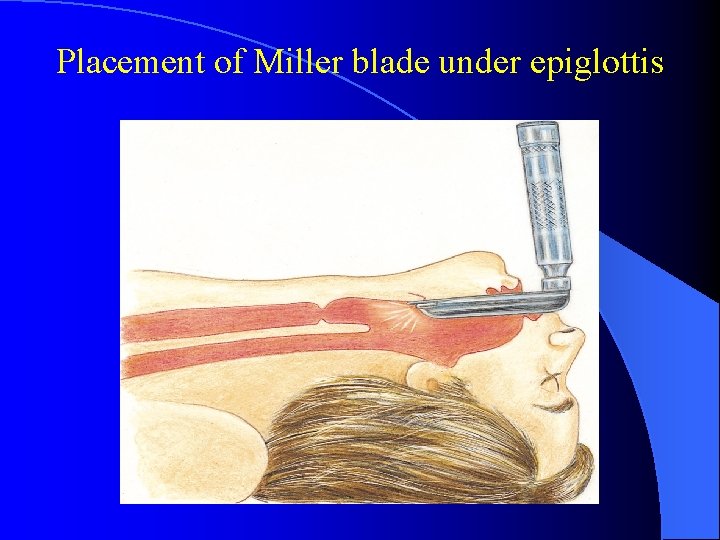
Placement of Miller blade under epiglottis
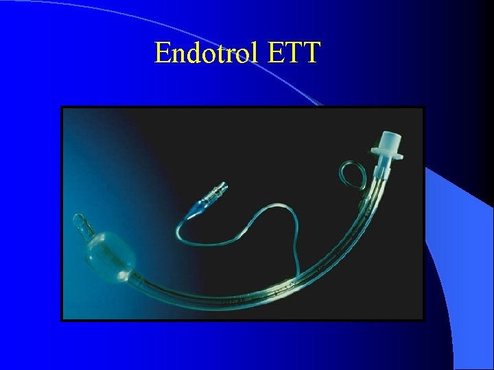
Endotrol ETT
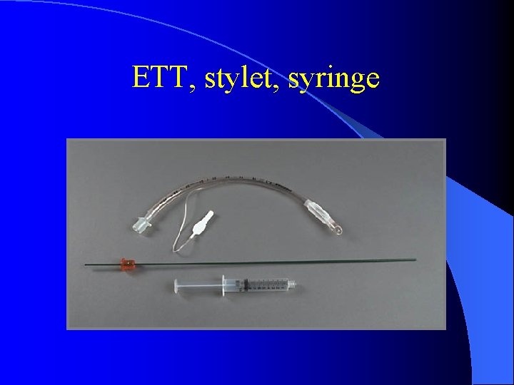
ETT, stylet, syringe
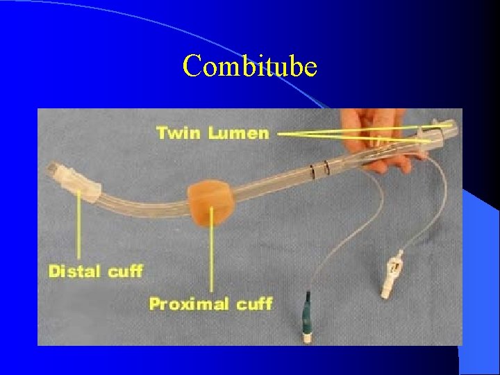
Combitube
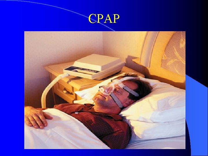
CPAP
Endotracheal Intubation Indicators ü ü ü Respiratory or cardiac arrest Unconsciousness Risk of aspiration Obstruction due to foreign bodies, trauma, burns, or anaphylaxis Respiratory extremis due to disease (Pneumothorax), hemothorax, (hemopneumothorax) with respiratory difficulty
Complications of Endotracheal Intubation ü ü ü ü Equipment malfunction Teeth breakage and soft tissue injury Hypoxia Esophageal intubation Endobronchial intubation Tension pneumothorax Extubation
Tracheostomies/Stomas ü Use patient’s supplies ü Ambu ü Treat bag attaches easily as an endotracheal tube ü Suction
Questions 1. Which one is lack of oxygen in the blood? a. Hypoxia b. Hypocarbia c. Hypoxemia d. Hypocarbemia
Questions 2. Which one is the best airway? a. Nasal cannula b. Endotracheal tube c. Oral airway d. Combitube
Questions 3. Which one is a contraindication to nasal trumpet use? a. Seizure b. Bloody nose c. DNR patient d. Significant facial trauma
Questions 4. Which one is the correct tidal volume for a 200 pound patient? a. 500 cc b. 600 cc c. 700 cc d. 800 cc
Questions 5. Which one is not an indication for endotracheal intubation? a. Respiratory failure b. Cardiac arrest c. GCS of 5 d. Hyperventilation syndrome
Now you know everything about respiratory emergencies
Questions? Renee Anderson andersr@inhs. org Garry Frey freyg@inhs. org 509 -232 -8155 FAX: 509 -232 -8344 509 -242 -4263
- Slides: 69