Respiratory distress in newborn Dr mirzarahimi Neonatal Respiratory

Respiratory distress in newborn Dr. mirzarahimi
 • Nasal flaring")
Neonatal Respiratory Distress Signs and symptoms • Tachypnea (RR > 60/min) • Nasal flaring • Retraction • Grunting • Delayed or decreased air entry • +/- Cyanosis • +/- Desaturation

r e t r a c t i o n score 0 1 2 Respiratory Rate (breaths/min) 60 60 – 80 >80 or apnea episode cyanosis None In room air In 40%oxygen Retraction None Mild Moderate to severe Grunting None Audible with a stethoscope Audible without a stethoscope clear decreased Barely audible Crying

Neonatal Respiratory Distress Etiologies Pulmonary causes - RDS Pneumonia TTN MAS Other aspiration syndrome Air leak syndrome Lung hemorrhage Lung hypoplasia Congenital malformations Systemic causes - Infections Metabolic causes Temperature Anemia Polycythemia - Congenital heart disease - Pulmonary hypertension - Neuromuscular disorder diagnosis : Hx, Phx and L. F Anatomic causes - Upper airway obstruction - Airway malformation - Space occupying lesion - Rib cage anomalies - Phrenic nerve injury
 Preterm < 6 hrs old")
Neonatal Respiratory Distress Algorithm Respiratory Distress (tachypnoea, retractions, grunt) Preterm < 6 hrs old HMD (RDS) Pneumonia Lung anomaly > 6 hrs old Term < 6 hrs old TTN Pneumonia MAS/PPHN CHD Pul. Hemorrhage Asphyxia Lung anomaly Air leak > 6 hrs old Pneumonia CHD

Respiratory Distress Syndrome

Introduction • The most frequent cause of respiratory distress in premature infants. • 60 -80% of <28 wk GA ; 15 -30% of 32 -36 wk GA ; 5% of 37 wk-term. • Classic presentation of grunting, retractions, increasing O 2 requirement, reticulogranular pattern and air bronchograms on CXR and onset < 6 hrs age

Pathogenesis Prematurity Prenatal asphyxia Reduced surfactant synthesis, storage, release Increased alveolar surface tension Progressive atelectasis Diffusion Uneven V/Q Hypoventilation gradient Hypoxemia CO 2 retention Acidosis Pulmonary vasoconstriction Hypoperfusion Capillary endothelial damage Plasma leak Fibrin

Pathology • Gross : Lung firm, red, liverlike • Microscopic : Diffuse atelectasis, pink membrane lining alveoli & alveolar ducts. Pulmonary arterioles with thick muscular coat, small lumen. Distended lymphatics • Electron microscopic : Damage / loss of alveolar epithelial cells, disappearance of lamellar inclusion bodies, swelling of capillary endothelial cells
 • Biophysical : • Deficient / absent surfactant • Abnormal pressure")
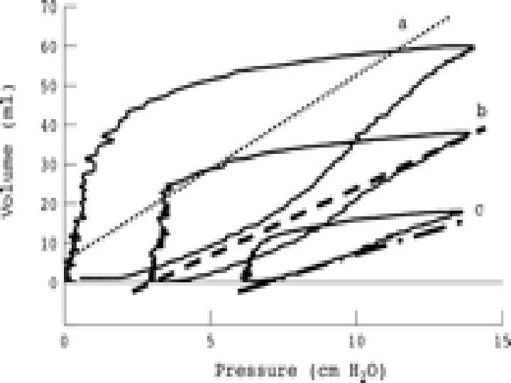
Pathology (contd. ) • Biophysical : • Deficient / absent surfactant • Abnormal pressure volume curve Normal Vol RDS Pressure • Severely reduced arterial bed with blockage near pulmonary arterioles
 • Biochemical : • Diminished surface-active phospholipid (phosphatidylcholine) • Diminished apoprotein")
Pathology (contd. ) • Biochemical : • Diminished surface-active phospholipid (phosphatidylcholine) • Diminished apoprotein content ( SP-A, B, C, D)
 • Poor lung perfusion (")
Pathophysiology • Reduced lung compliance (1/5 th -1/10 th) • Poor lung perfusion ( 50 -60% not perfused), decreased capillary blood flow • R--> L shunting ( 30 -60% ) • Alveolar ventilation decreased • Lung volume reduced • Increased work of breathing • Hypoxemia, hypercapnia, acidosis

Physiologic abnormalities • • • Lung compliance 10 -20% of norm Atelectasis…areas not ventilated Areas not perfused Decrease alveolar ventilation Reduce lung volume

Risk factor Prematurity Acidosis Hypoxia Hypercapnia Hypothermia C/S Asphyxia and stress Male Familial DM mother

signs • • tachypnea retraction grunting Nasal flaring apneic episode cyanosis extremities puffy or swollen

Chest X-ray • Ground glass appearance • Reticulogranular • With air bronchograms

Treatment • Surfactant • Prevention • rescue • Supportive • Thermal • Fluid and nutrition • oxygen • Mechanical ventilation

complications • • • Pneumothorax PDA Infection Line problems ROP Chronic lung disease

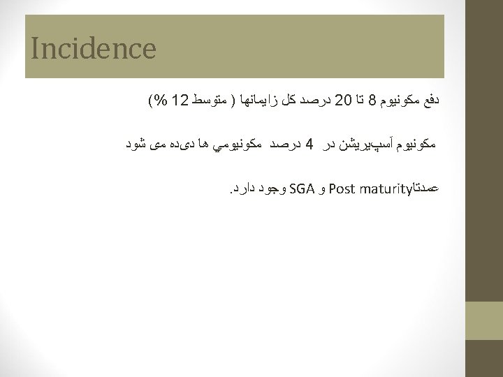
Meconium aspiration

ﺗﺮﻛﻴﺐ ﻣﻜﻮﻧﻴﻮﻡ • • Cellular particle Bile pigment Lango Mocus Vernix Pancreatic secretion One gr meconium = one mg Billirubin

Alarm of MAS 1 - Thick meconium 2 -Fetal tachycardia 3 - lack of increase heart rate during 4 -Low cord PH intra partum monitoring
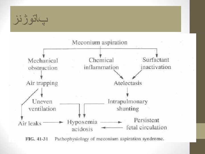

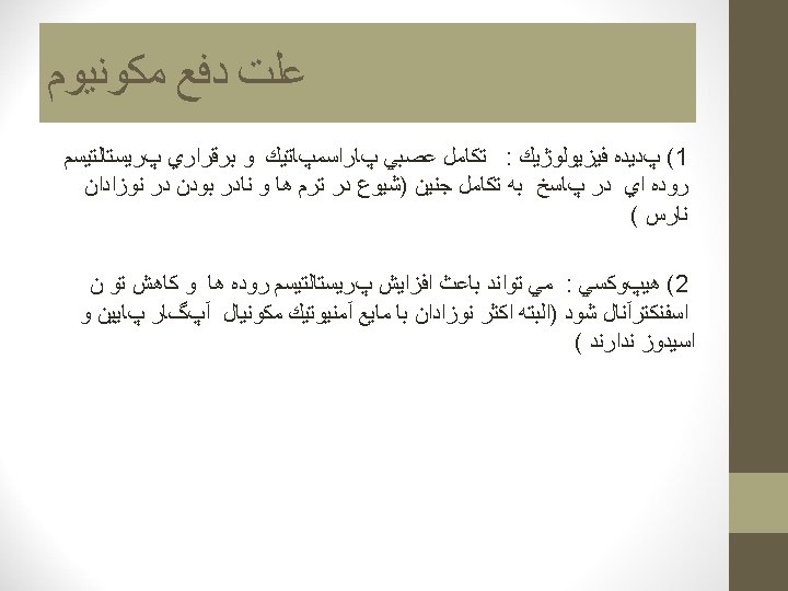
MAS complication • Partial obstruction o • complete obstruction : • Surfactant destruction • Chemical pneumonitis &Bacterial pneumonia • Asphyxia • PPHN

Clinical sign • Classic sign : Post maturity nail, skin , umblical cord are heavily stained with a yellowish pigment • Early sign (resp. Distress) : grunting & cyanosis & nasal flaring & retraction & marked tachypnea • Characteristic sign : chest overinflation and Rale

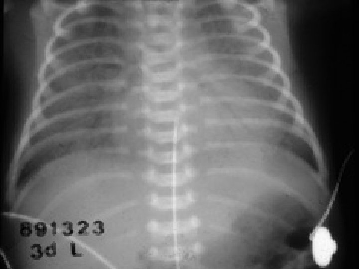
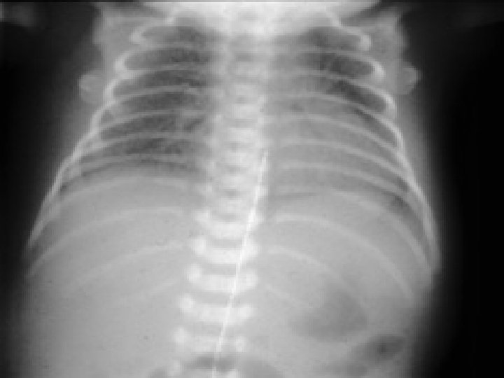
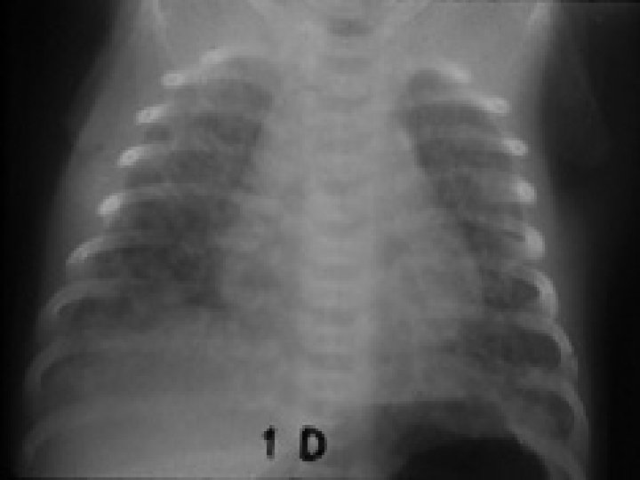
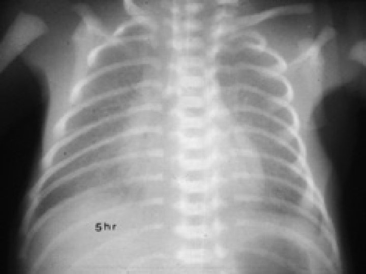
Radiography of M. A. S • Coarse , nodular , irregular pulmonary densities with areas of diminished aeration or consolidation. • Hyperinflation of the chest. • Atelectasis • Flattening of diaphragm • Cardiomegally (manifestation of the underlying prenatal hypoxia)

Chest. X. Ray

Meconium Aspiration Syndrome

Meconium Aspiration Syndrome

 Heart rate greater than 100 beat /min 2) Good muscle")
Criteria of vigorous 1) Heart rate greater than 100 beat /min 2) Good muscle tone 3) regular breathing
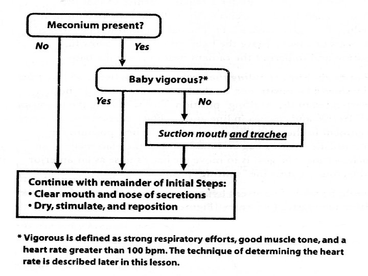
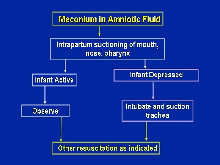

Guidelines of the baby exposed to meconium Vigorous No Immediate tracheal suction Meconium HR>100 reintubate and suction Yes Clear secretions and meconium initial resuscitation steps No meconium HR<100 PPV and suction again later The American Academy of Pediatrics Neonatal Resuscitation Program Steering Committee management guidelines of the baby exposed to meconium:
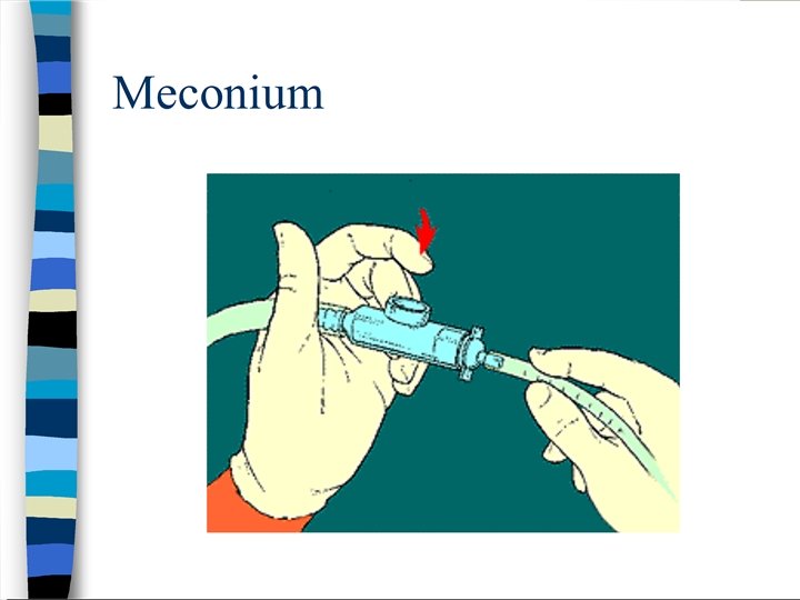

ET suction indication • Only in non vigorous baby - depressed respirations - decreased muscle tone - heart rate < 100 beats per minute • Pharyngeal suctioning of an infant before delivery of the shoulders. • Removal of meconium from hypopharynx and larynx by large-bore catheter. • Meconium aspirator attached to wall suction. • Endotracheal intubation for removal of meconium in the lower airway.

Management 1. Prevention • Monitor fetal status • Amnioinfusion • Suctioning +/- intubation and immediate suctioning • Avoid harmful techniques 2. Intervention • Optimal thermal environment & minimal handling • Respiratory care, Oxygen therapy & ECMO • Keep stable V/S • Surfactant therapy

Steroid therapy for meconium aspiration syndrome in newborn infants • The Cochrane Database of Systematic Reviews 2007 Issue 3, The Cochrane Library (ISSN 1464 -780 X • Conclusions: At present, there is insufficient evidence to assess the effects of steroid therapy in the management of meconium aspiration syndrome (no significant reduction in mortality, duration of hospital stay, Duration of mechanical ventilation, incidence of air leak, increase in duration of oxygen therapy was seen with the use of steroids)
:")
Role of antibiotics in meconium aspiration syndrome • Ann Trop Paediatr. 2007 Jun; 27(2): 107 -13. • Basu S, Kumar A, Bhatia BD. • Division of Neonatology, Department of Paediatrics, Institute of Medical Sciences, Banaras Hindu University, Varanasi, India. drsriparnabasu@rediffmail. com • CONCLUSION: Routine antibiotic therapy is not necessary for managing MAS. No significant difference – period of oxygen dependency (5. 8 vs 5. 9 days) – day of starting feeds (4. 0 vs 4. 2) – day of achievement of full feeds (9. 4 vs 9. 3) – clearance of chest radiograph (11. 7 vs 12. 9 days) – duration of hospital stay (13. 7 vs 13. 5 days)

Surfactant for meconium aspiration syndrome in full term/near term infants • Cochrane Database Syst Rev. 2007 Jul 18; (3): CD 002054 • El Shahed A, Dargaville P, Ohlsson A, Soll R. • CONCLUSIONS: In infants with MAS, surfactant administration may reduce the severity of respiratory illness and decrease the number of infants with progressive respiratory failure requiring support with ECMO. The relative efficacy of surfactant therapy compared to, or in conjunction with, other approaches to treatment including inhaled nitric oxide, liquid ventilation, surfactant lavage and high frequency ventilation remains to be tested.

PPHN prevention 1. Avoid vasoconstriction • Acidosis • Hypoxia • Metabolic disturbance 2. Prevent right to left shunt - Hypocalcemia - Hyperglycemia - Hypoglycemia

Infections

Infections • • Pneumonia & Sepsis have various manifestations including typical signs of distress as well as temperature instability. Common pathogen- Group B Streptococcus, Staph aureus, Streptococcus Pneumonia, Gm neg. rods

Infections con. . • • Risk factors- prolonged rupture of membranes, prematurity, & maternal fever. CXR- bilateral infiltrates suggesting in utero infection.

Congenital pneumonia • Sepsis risk factors • • PROM Prematurity Maternal fever, discharge, abdominal pain, leukocytosis Colonization with GBS • Same signs of RDS • X-ray

Transient Tachypnea of Newborn • • • Most common cause of respiratory distress. Residual fluid in fetal lung tissues. Risk factors- maternal asthma, c- section, male sex, macrosomia, maternal diabetes

TTN • Tachypnea immediately after birth or within two hours, with other predictable signs of respiratory distress. • Symptoms can last few hours to two days. • Chest radiography shows diffuse parenchymal infiltrates, a “ wet silhouette” around heart, or intralobar fluid accumulation

X-ray Fluid in the fissure

Transient tachypnea of newborn • • • Term Cesarian delivery Usually tachypnea without O 2 requirment Resolve in 48 -72 houres Lung fluid X-ray

Other causes • • • Congenital malformations-Pulmonary hypoplasia, congenital emphysema, esophageal atresia & diaphragmatic hernia. Neurological causes- hydrocephalus & intracranial hemorrhage. Metabolic derangements-hypoglycemia, hypocalcemia, polycythemia.

Congenital Heart disease Cyanotic Heart Disease • Tetralogy of fallot- ( VSD, Pulmonary stenosis, overriding aorta, RVH) • Tricuspid atresia • Transposition of great vessel • Total anomalous pul. venous return • Truncus arteriosus.

Hyperoxia Test • Obtain ABG–> Then place the patient on 100% O 2 for 10 minutes then repeat ABG , If the cyanosis is pulmonary , the Pa. O 2 should be increased by 30 mm of Hg. If the cause is cardiac , there will be minimal improvement in Pa. O 2.
- Slides: 62