
Resonance assignment strategies 1 The assignment problem Amino
Resonance assignment strategies 1
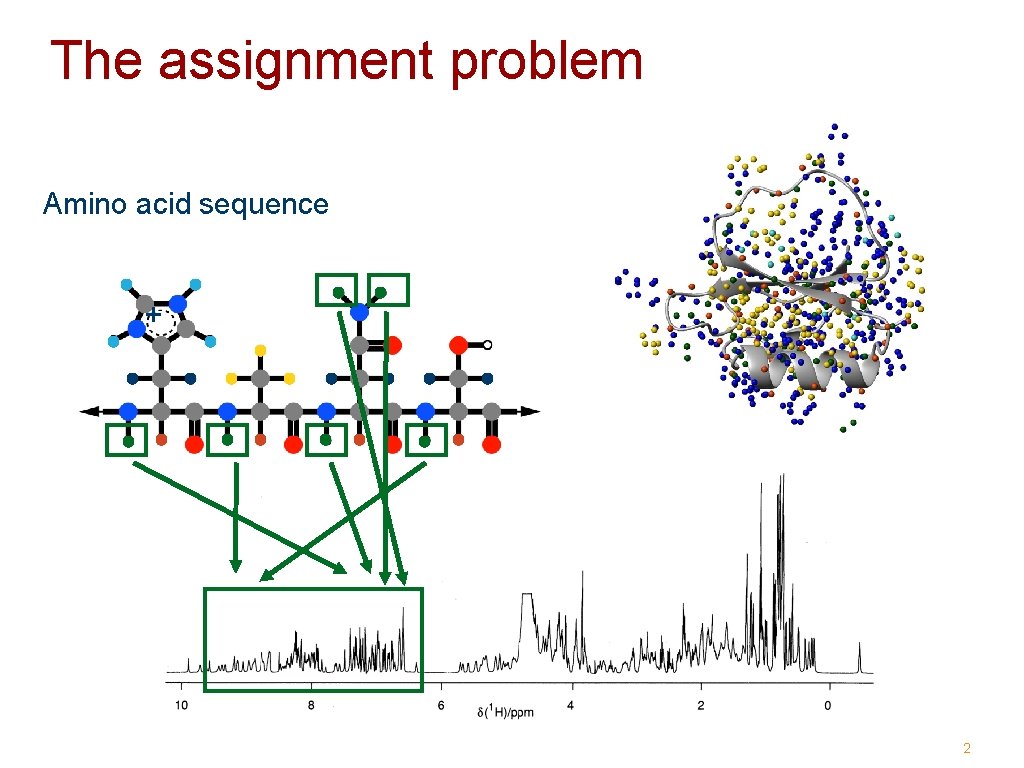
The assignment problem Amino acid sequence + 2
Assignment via 1 H NMR 3
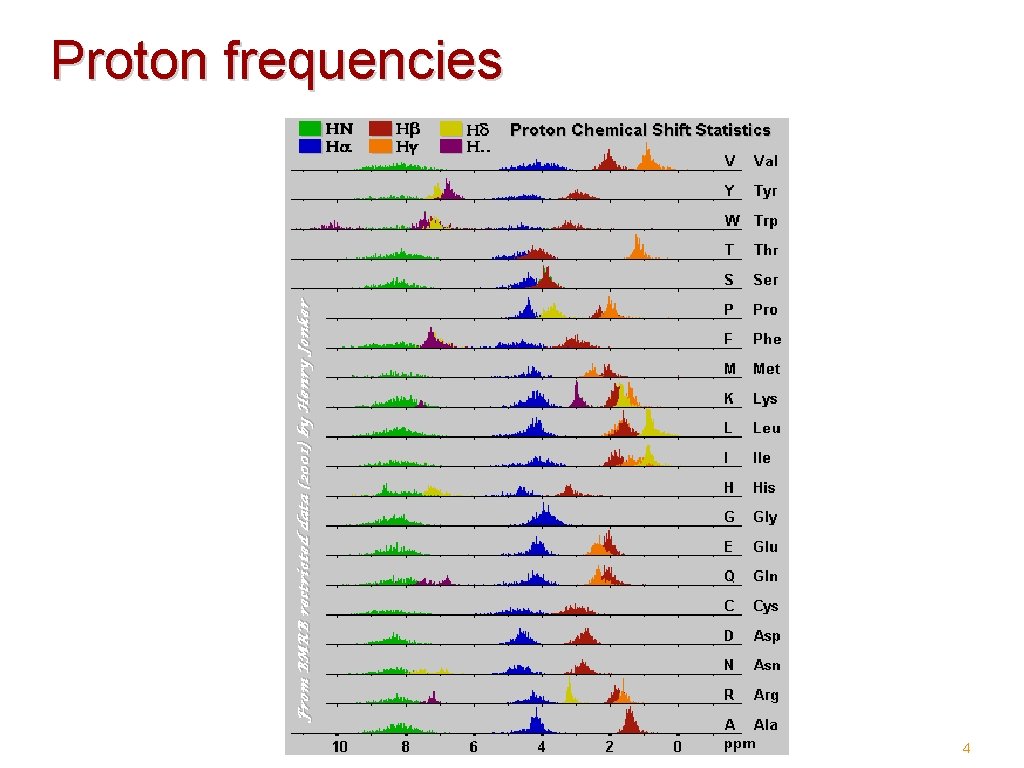
Proton frequencies 4
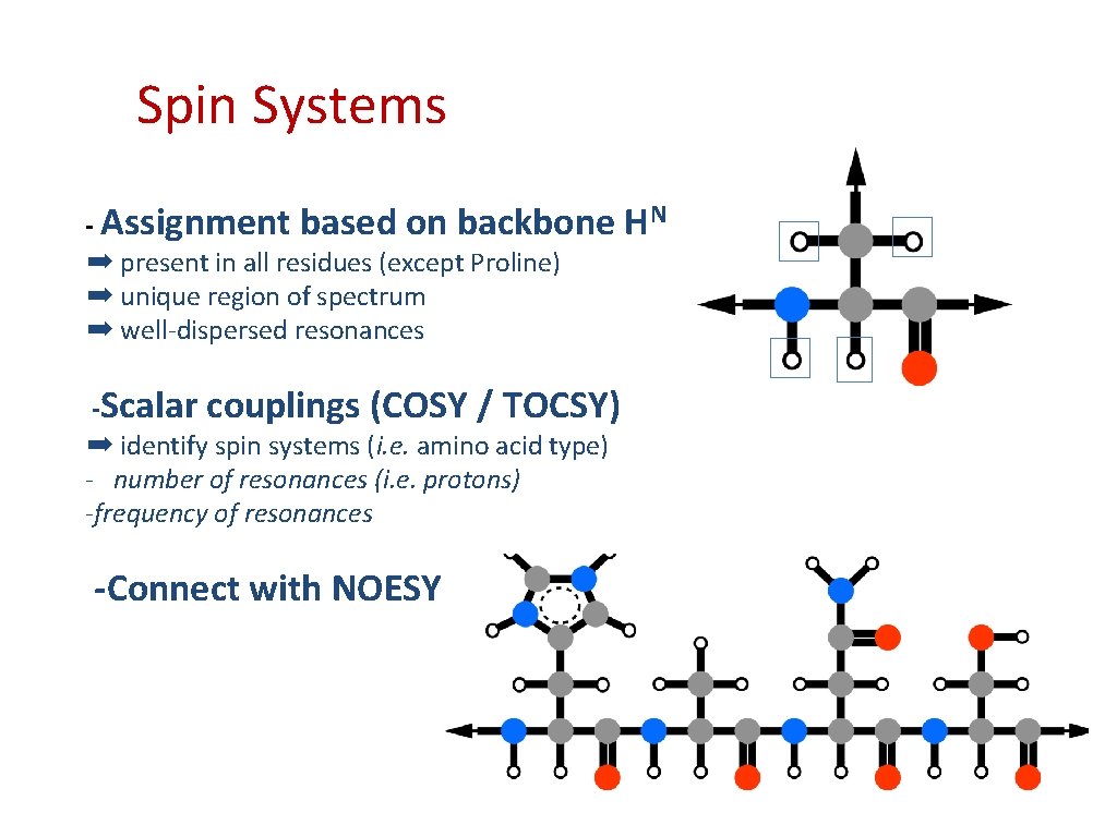
Spin Systems - Assignment based on backbone ➡ present in all residues (except Proline) ➡ unique region of spectrum ➡ well-dispersed resonances -Scalar couplings (COSY / TOCSY) ➡ identify spin systems (i. e. amino acid type) - number of resonances (i. e. protons) -frequency of resonances -Connect with NOESY HN
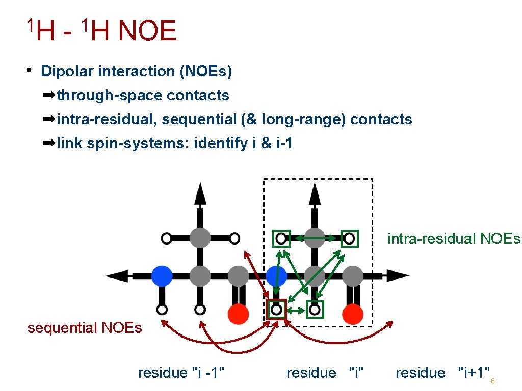
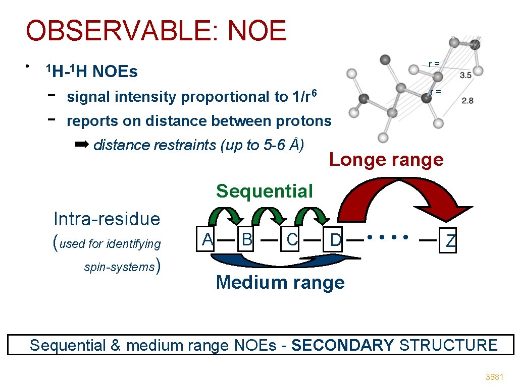
1 H - 1 H NOE • Dipolar interaction (NOEs) ➡through-space contacts ➡intra-residual, sequential (& long-range) contacts ➡link spin-systems: identify i & i-1 intra-residual NOEs sequential NOEs residue "i -1" residue "i+1" 6
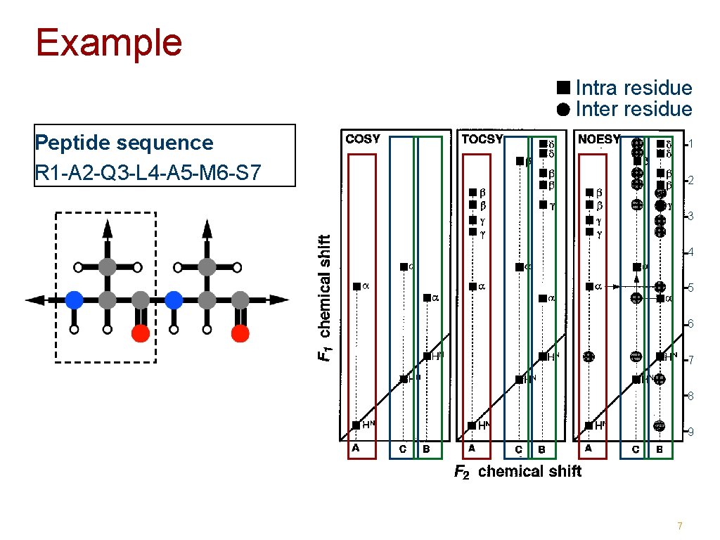
Example Intra residue Inter residue Peptide sequence R 1 -A 2 -Q 3 -L 4 -A 5 -M 6 -S 7 1 2 3 4 5 6 7 8 9 7
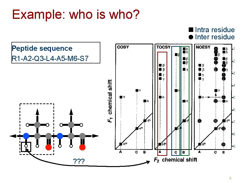
Example: who is who? Intra residue Inter residue Peptide sequence R 1 -A 2 -Q 3 -L 4 -A 5 -M 6 -S 7 1 2 3 4 5 6 7 8 9 ? ? ? 8
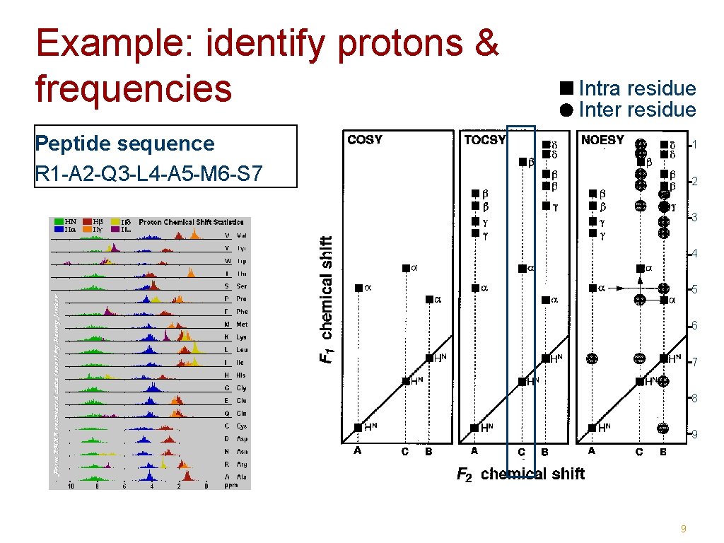
Example: identify protons & frequencies Intra residue Inter residue Peptide sequence R 1 -A 2 -Q 3 -L 4 -A 5 -M 6 -S 7 1 2 3 4 5 6 7 8 9 9
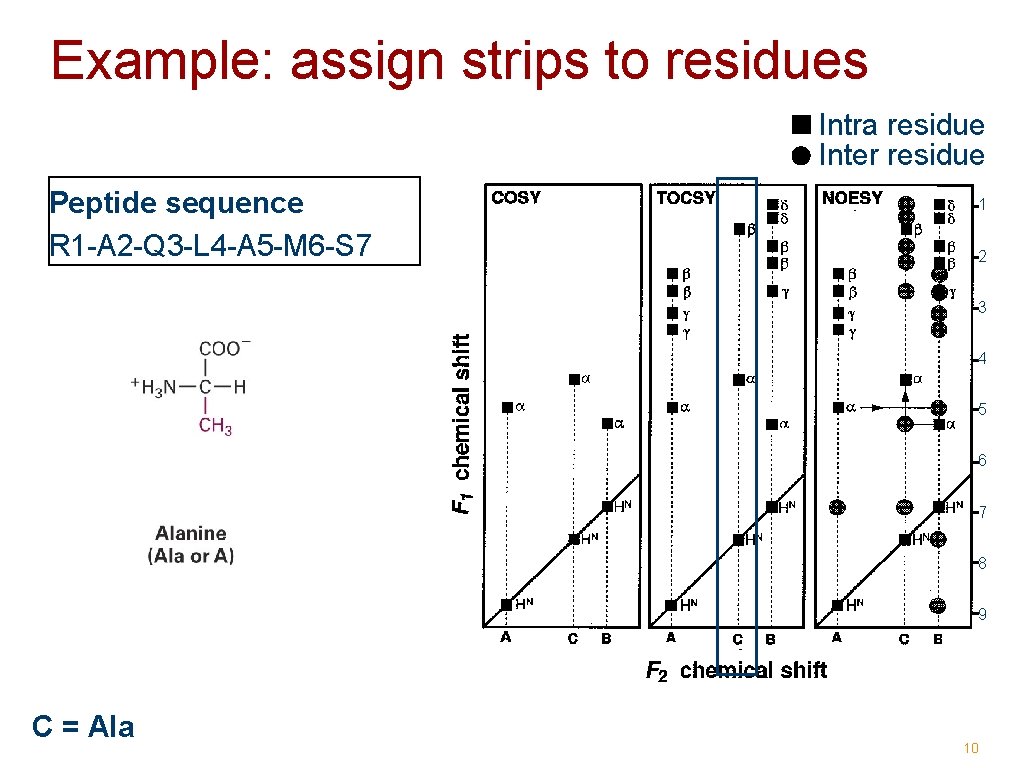
Example: assign strips to residues Intra residue Inter residue Peptide sequence R 1 -A 2 -Q 3 -L 4 -A 5 -M 6 -S 7 1 2 3 4 5 6 7 8 9 C = Ala 10
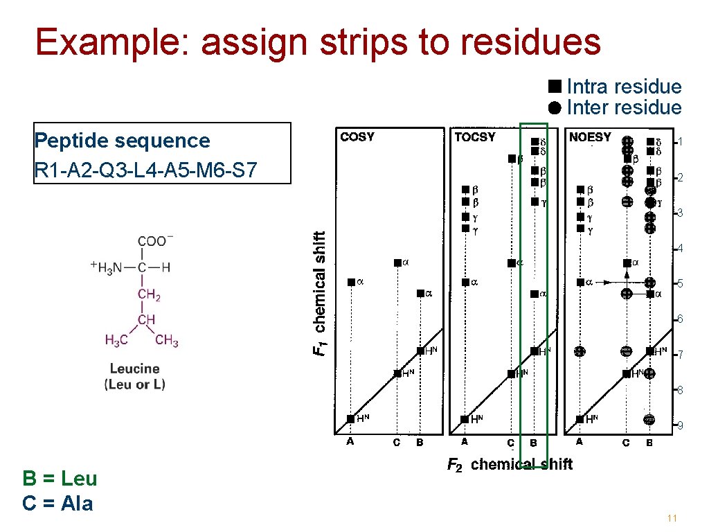
Example: assign strips to residues Intra residue Inter residue Peptide sequence R 1 -A 2 -Q 3 -L 4 -A 5 -M 6 -S 7 1 2 3 4 5 6 7 8 9 B = Leu C = Ala 11
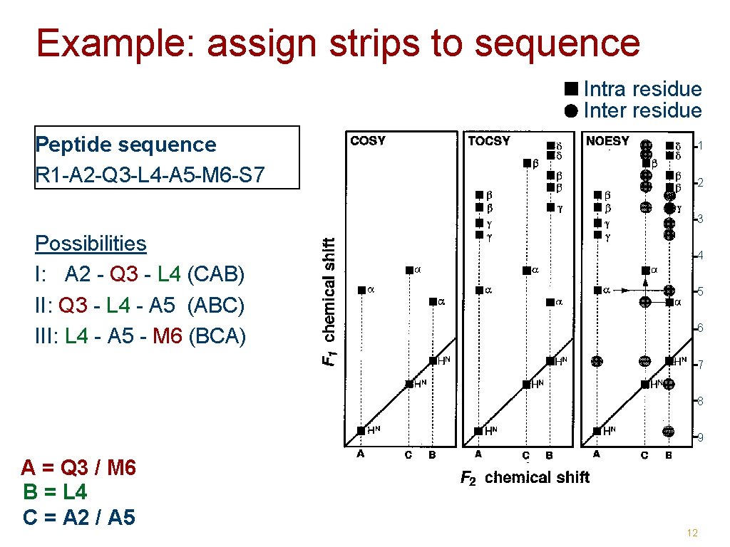
Example: assign strips to sequence Intra residue Inter residue Peptide sequence R 1 -A 2 -Q 3 -L 4 -A 5 -M 6 -S 7 1 2 3 Possibilities I: A 2 - Q 3 - L 4 (CAB) II: Q 3 - L 4 - A 5 (ABC) III: L 4 - A 5 - M 6 (BCA) 4 5 6 7 8 9 A = Q 3 / M 6 B = L 4 C = A 2 / A 5 12
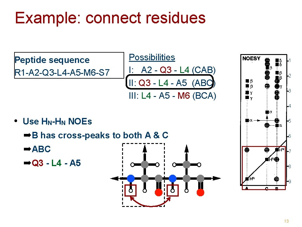
Example: connect residues Peptide sequence R 1 -A 2 -Q 3 -L 4 -A 5 -M 6 -S 7 Possibilities I: A 2 - Q 3 - L 4 (CAB) II: Q 3 - L 4 - A 5 (ABC) III: L 4 - A 5 - M 6 (BCA) 1 2 3 4 • Use HN-HN NOEs 5 ➡B has cross-peaks to both A & C 6 ➡ABC 7 ➡Q 3 - L 4 - A 5 8 9 13
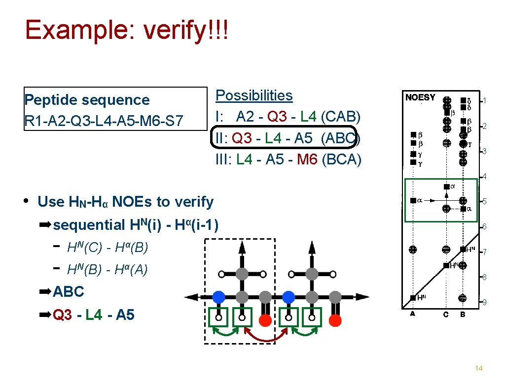
Example: verify!!! Peptide sequence R 1 -A 2 -Q 3 -L 4 -A 5 -M 6 -S 7 Possibilities I: A 2 - Q 3 - L 4 (CAB) II: Q 3 - L 4 - A 5 (ABC) III: L 4 - A 5 - M 6 (BCA) 1 2 3 4 • Use HN-Hα NOEs to verify ➡sequential HN(i) - Hα(i-1) - H (C) - H (B) - H (A) N α ➡ABC 5 6 7 8 9 ➡Q 3 - L 4 - A 5 14
Assignment via 13 and C 1 H, 15 N, 15
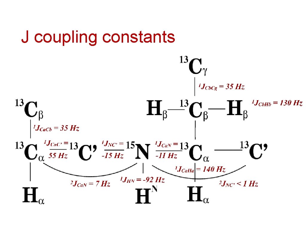
J coupling constants
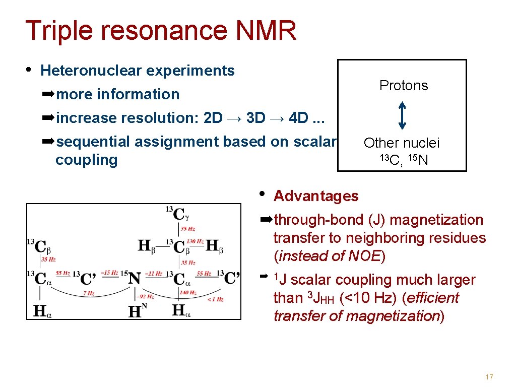
Triple resonance NMR • Heteronuclear experiments Protons ➡more information ➡increase resolution: 2 D → 3 D → 4 D. . . ➡sequential assignment based on scalar coupling • Other nuclei 13 C, 15 N Advantages ➡through-bond (J) magnetization transfer to neighboring residues (instead of NOE) ➡ 1 J scalar coupling much larger than 3 JHH (<10 Hz) (efficient transfer of magnetization) 17
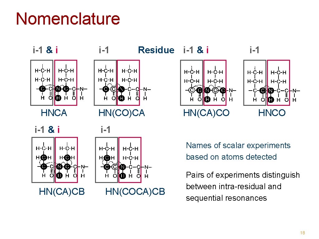
Nomenclature i-1 & i i-1 Residue i-1 & i HNCA HN(CO)CA i-1 & i i-1 HN(CA)CO HNCO i-1 Names of scalar experiments based on atoms detected HN(CA)CB HN(COCA)CB Pairs of experiments distinguish between intra-residual and sequential resonances 18
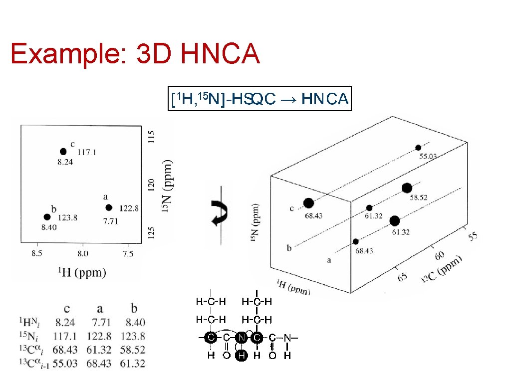
Example: 3 D HNCA
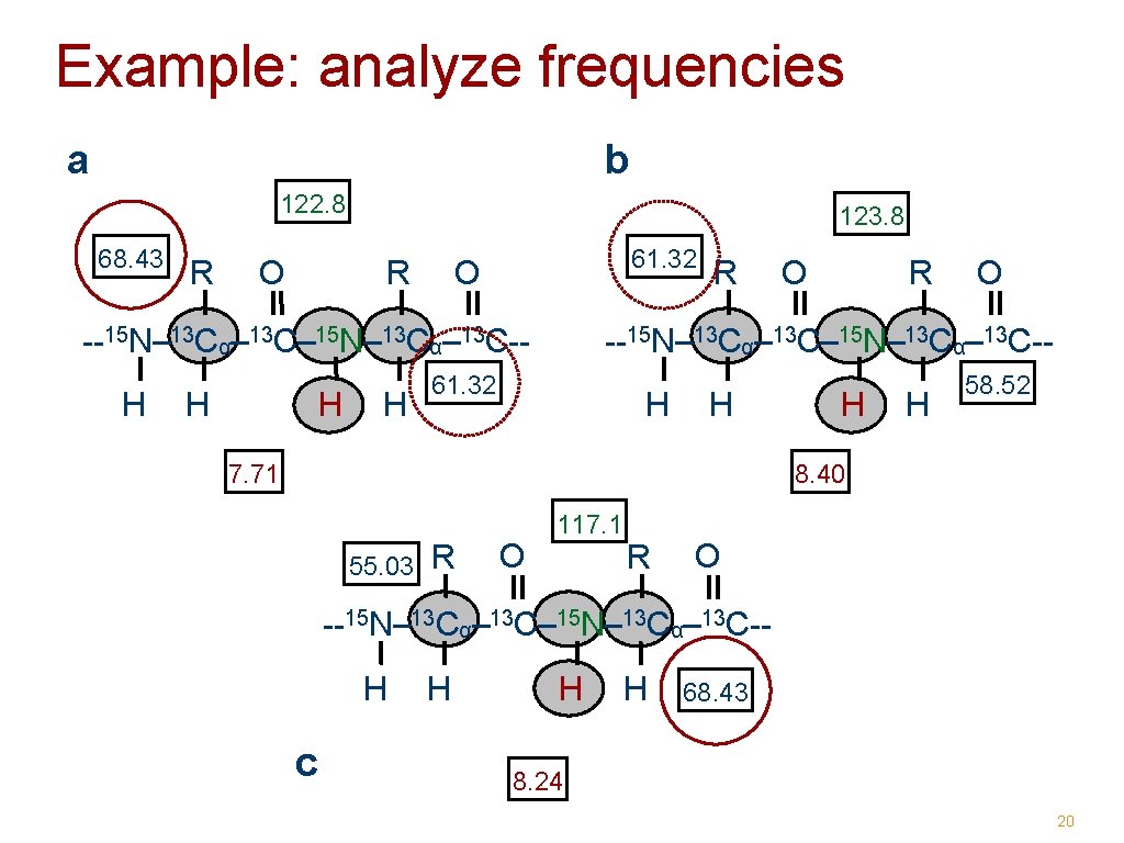
Example: analyze frequencies a b 122. 8 68. 43 R O 123. 8 61. 32 O R --15 N– 13 Cα– 13 C– 15 N– 13 Cα– 13 C-H H R O --15 N– 13 Cα– 13 C– 15 N– 13 Cα– 13 C-- 61. 32 H H 7. 71 H H 58. 52 8. 40 55. 03 R O 117. 1 R O --15 N– 13 Cα– 13 C– 15 N– 13 Cα– 13 C-H c H H H 68. 43 8. 24 20
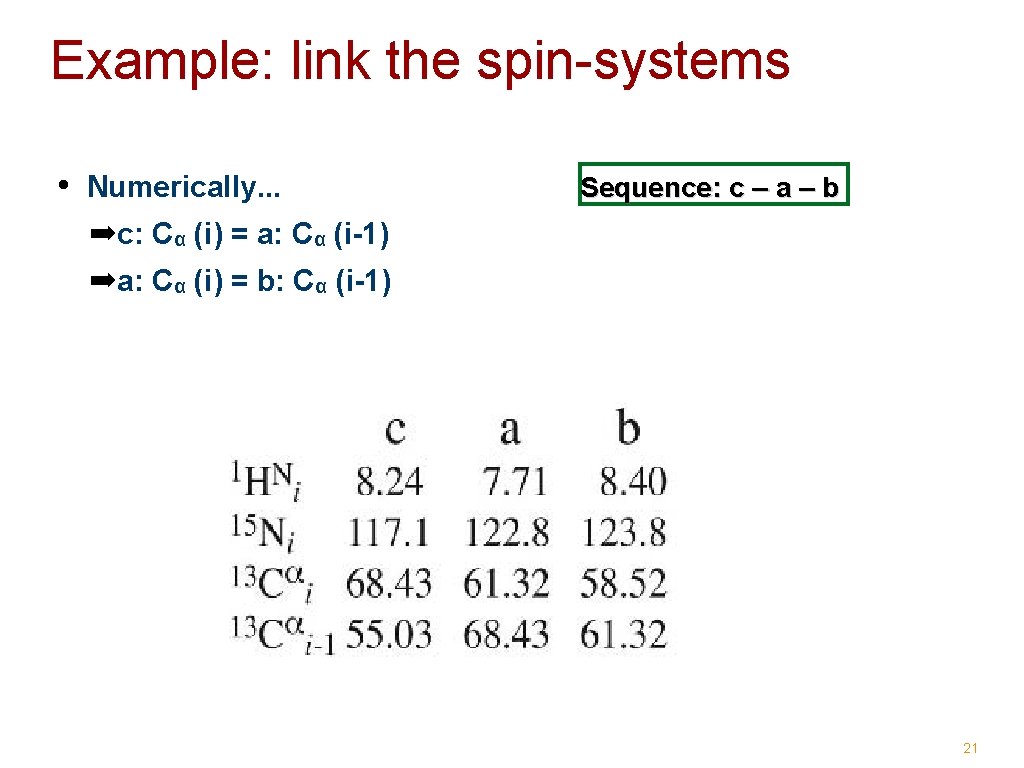
Example: link the spin-systems • Numerically. . . Sequence: c – a – b ➡c: Cα (i) = a: Cα (i-1) ➡a: Cα (i) = b: Cα (i-1) 21
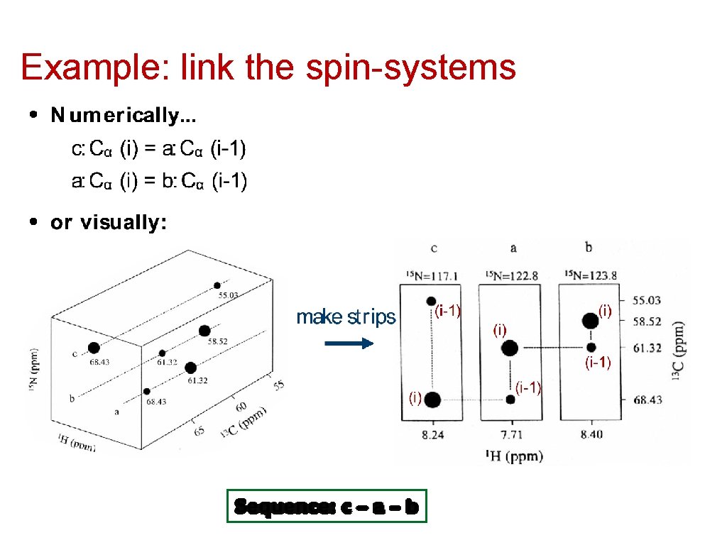
Example: link the spin-systems
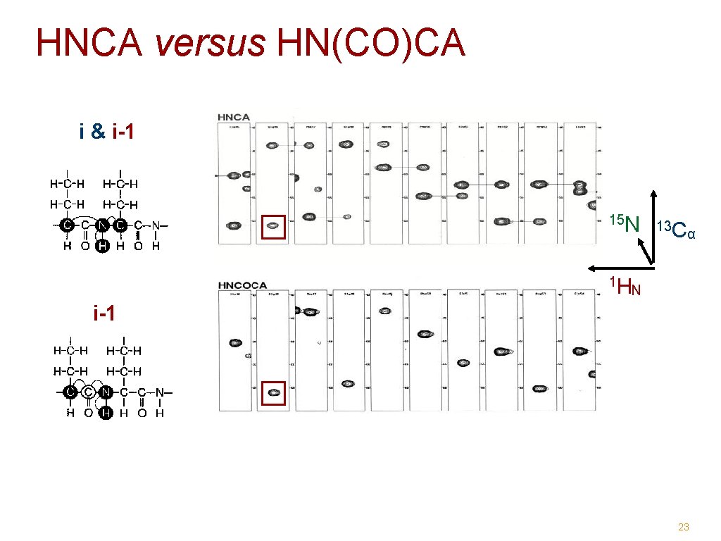
HNCA versus HN(CO)CA i & i-1 15 N 1 H 13 C α N i-1 23
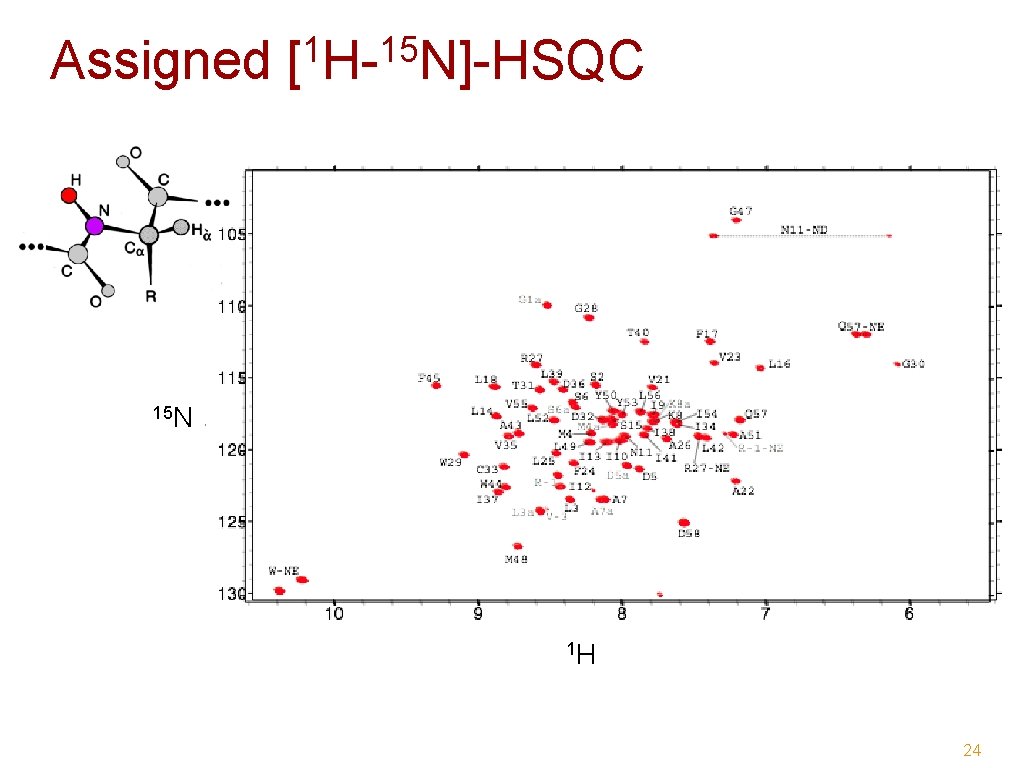
1 15 Assigned [ H- N]-HSQC 15 N 1 H 24
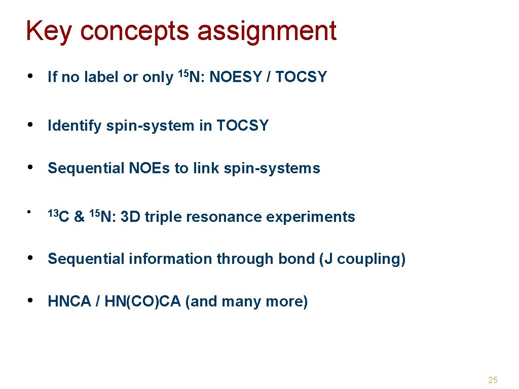
Key concepts assignment • If no label or only 15 N: NOESY / TOCSY • Identify spin-system in TOCSY • Sequential NOEs to link spin-systems • 13 C & 15 N: 3 D triple resonance experiments • Sequential information through bond (J coupling) • HNCA / HN(CO)CA (and many more) 25
NMR observables & structural restraints 26/81
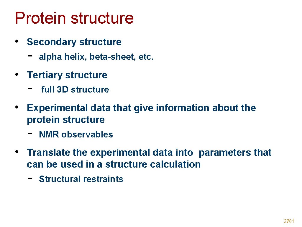
Protein structure • Secondary structure - alpha helix, beta-sheet, etc. • Tertiary structure - full 3 D structure • Experimental data that give information about the protein structure - NMR observables • Translate the experimental data into parameters that can be used in a structure calculation - Structural restraints 27/81
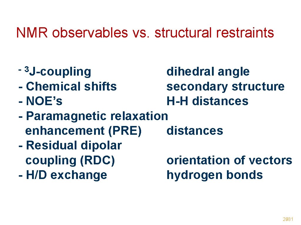
NMR observables vs. structural restraints - 3 J-coupling dihedral angle - Chemical shifts secondary structure - NOE’s H-H distances - Paramagnetic relaxation enhancement (PRE) distances - Residual dipolar coupling (RDC) orientation of vectors - H/D exchange hydrogen bonds 28/81
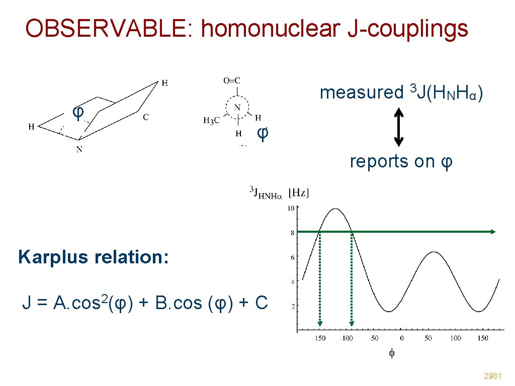
OBSERVABLE: homonuclear J-couplings φ measured 3 J(HNHα) φ reports on φ Karplus relation: J = A. cos 2(φ) + B. cos (φ) + C 29/81
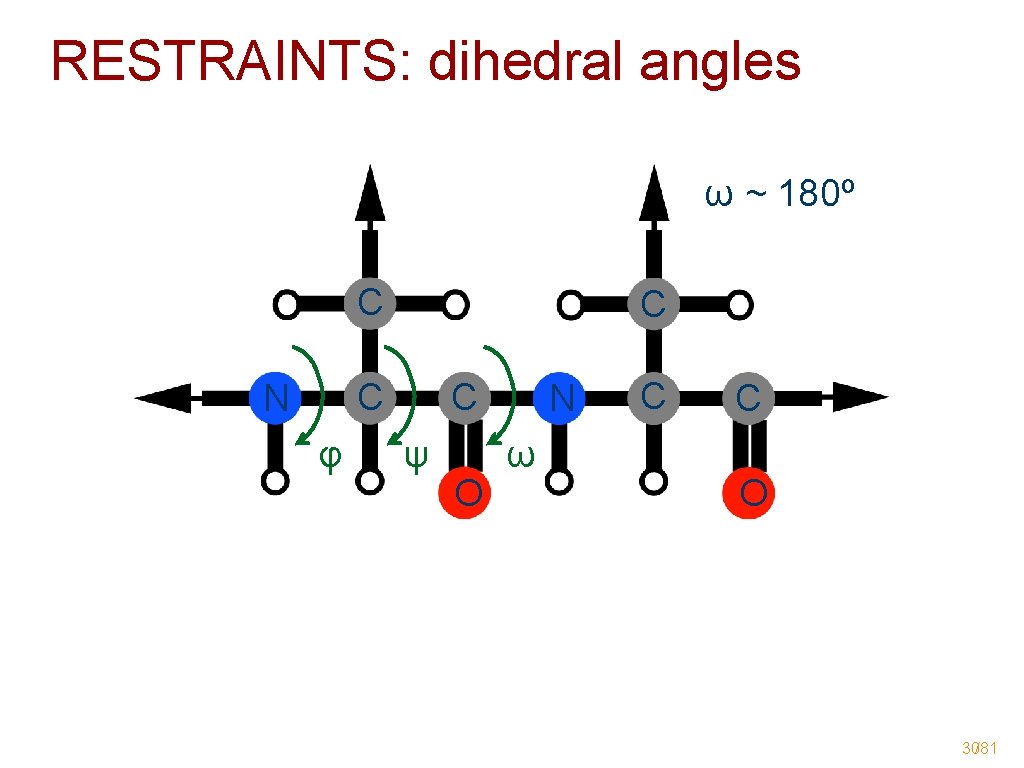
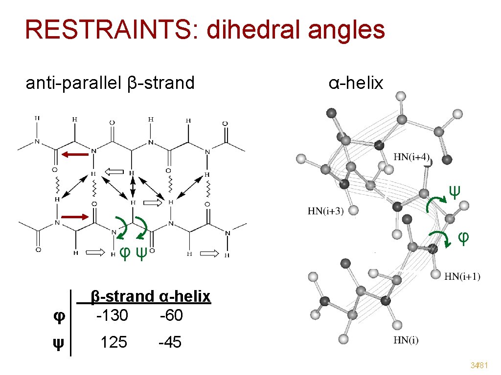
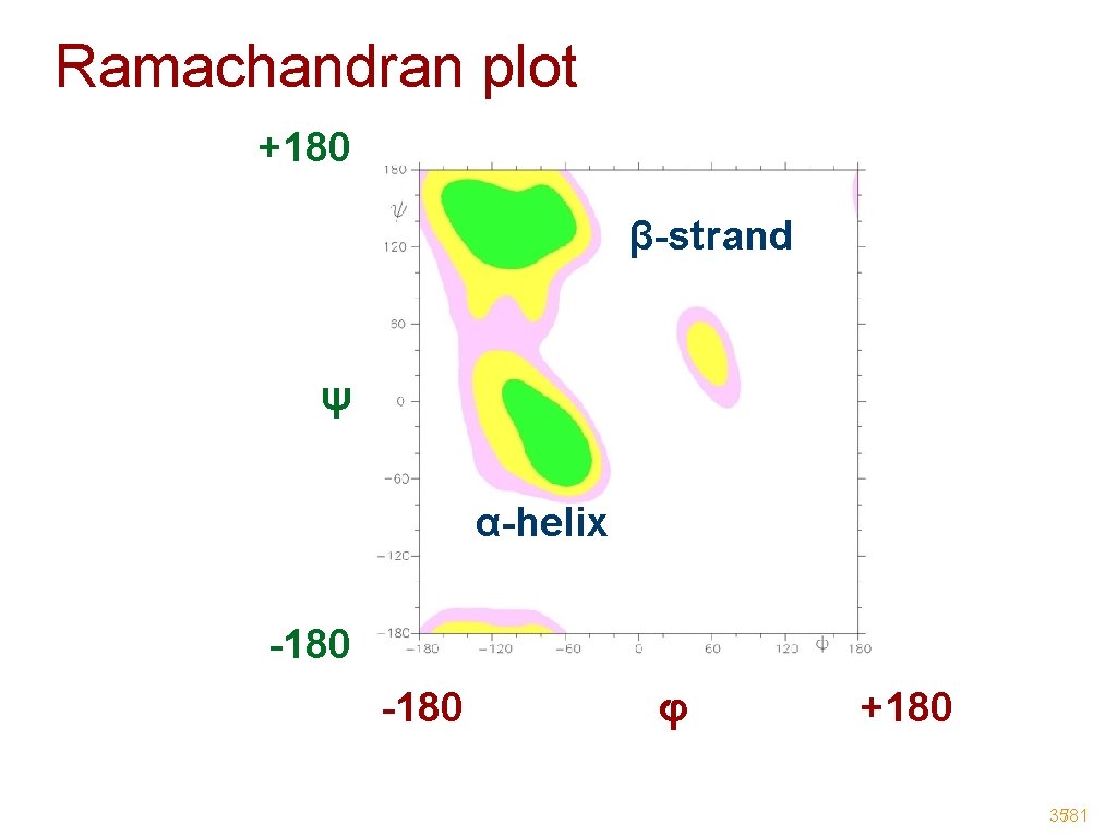
RESTRAINTS: dihedral angles ω ~ 180º C C C N φ C ψ O N ω C C O 30/81
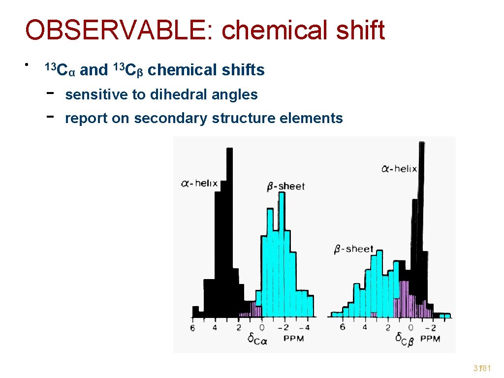
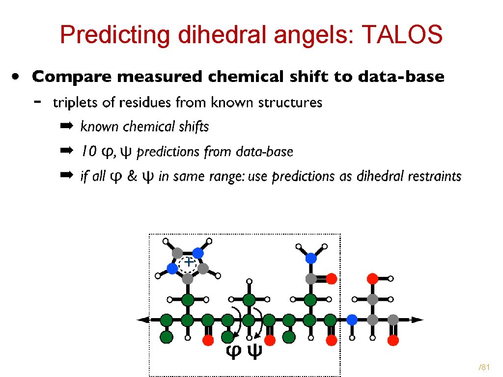
OBSERVABLE: chemical shift • 13 C - α and 13 Cβ chemical shifts sensitive to dihedral angles report on secondary structure elements 31/81
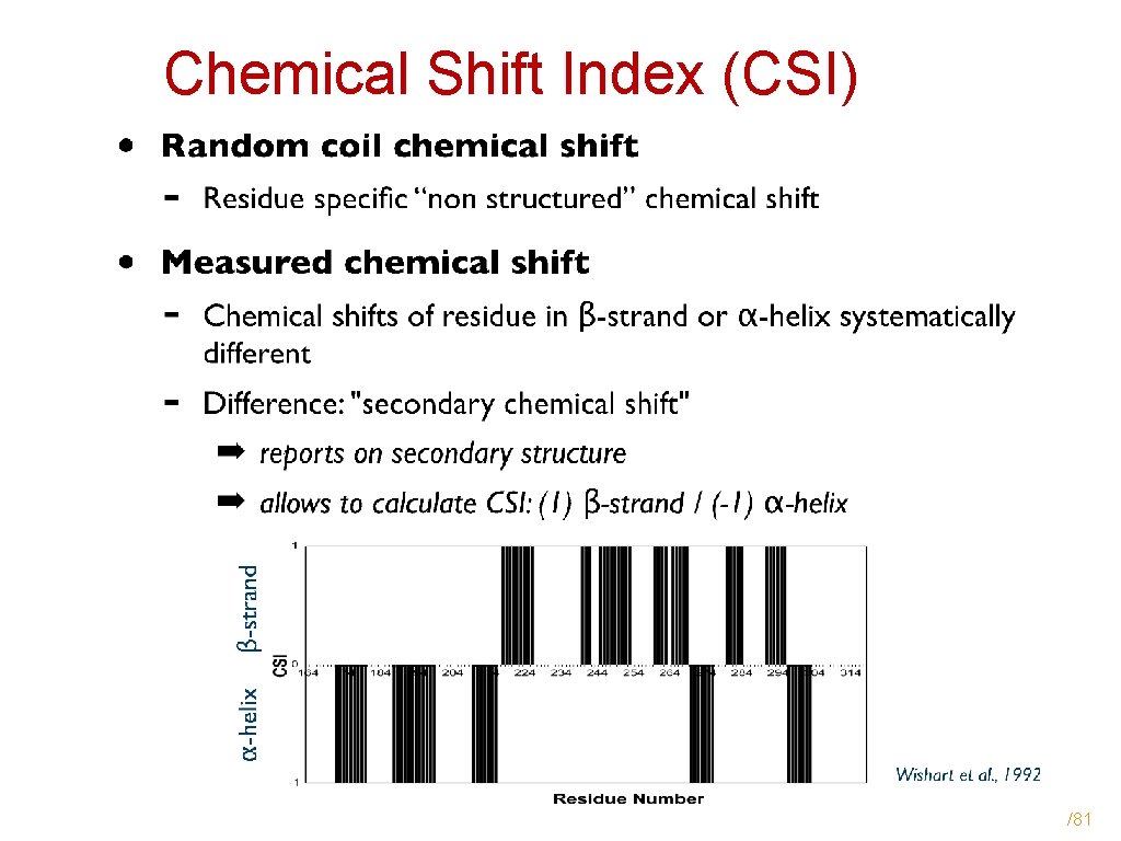
Chemical Shift Index (CSI) /81
Predicting dihedral angels: TALOS /81
RESTRAINTS: dihedral angles anti-parallel β-strand α-helix ψ φψ φ β-strand α-helix φ -130 -60 ψ 125 -45 34/81
Ramachandran plot +180 β-strand ψ α-helix -180 φ +180 35/81
OBSERVABLE: NOE • 1 H-1 H - r = NOEs r = signal intensity proportional to 1/r 6 reports on distance between protons ➡ distance restraints (up to 5 -6 Å) Longe range Sequential Intra-residue (used for identifying spin-systems) A B C D • • Z Medium range Sequential & medium range NOEs - SECONDARY STRUCTURE 36/81
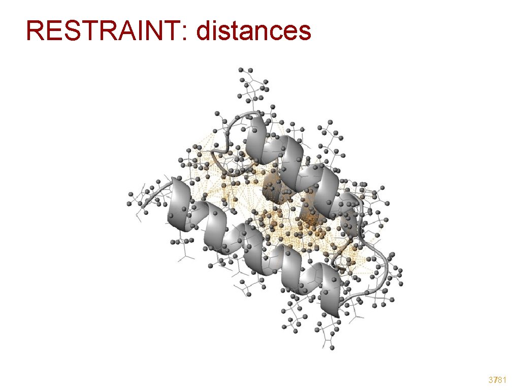
RESTRAINT: distances 37/81
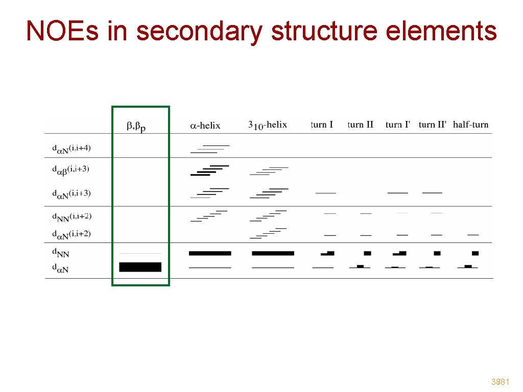
NOEs in secondary structure elements 38/81
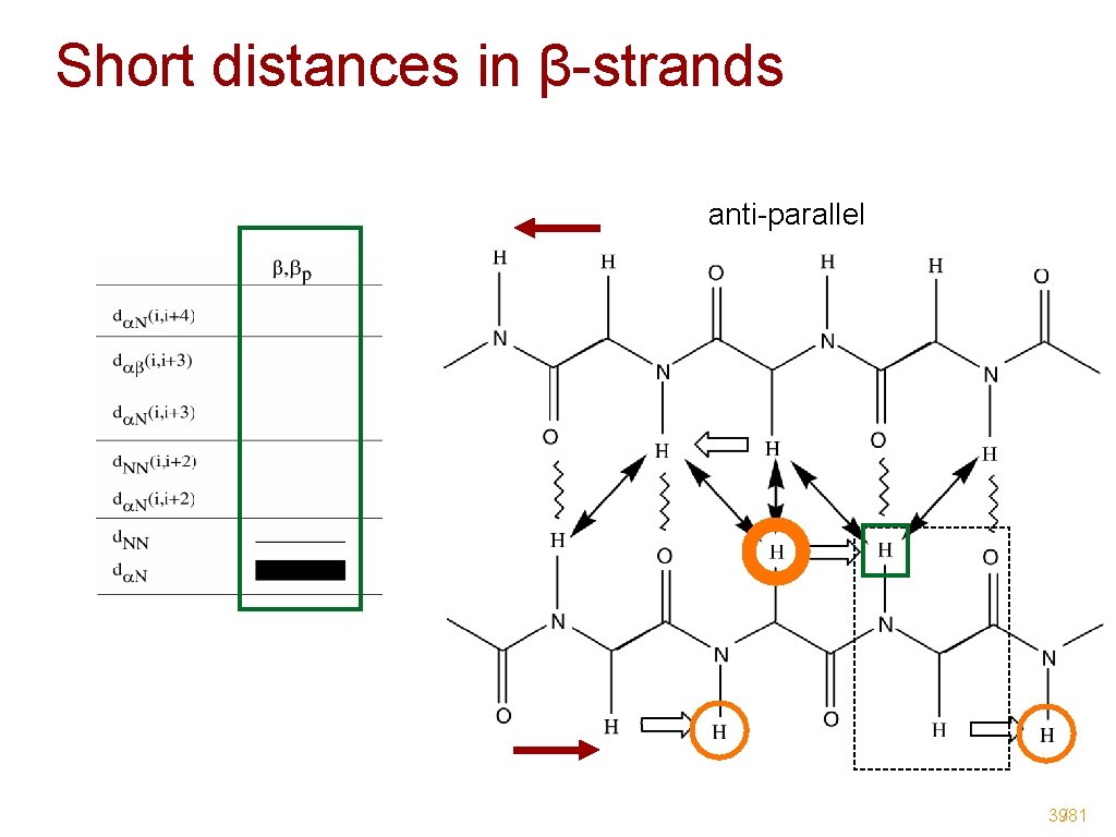
Short distances in β-strands anti-parallel 39/81
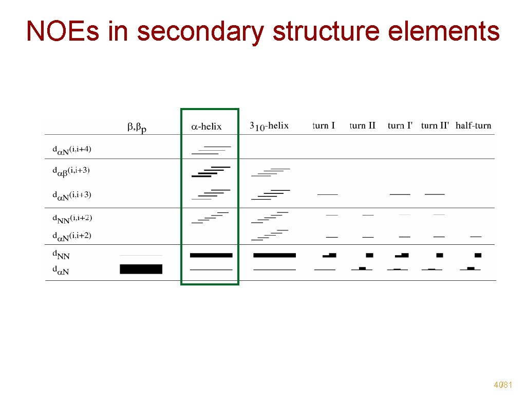
NOEs in secondary structure elements 40/81
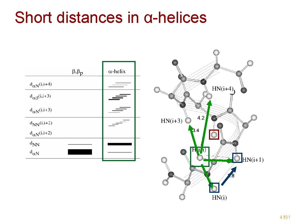
Short distances in α-helices 41/81
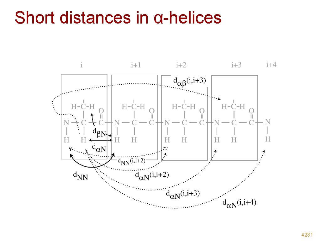
Short distances in α-helices 42/81
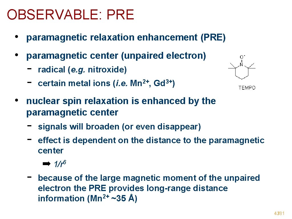
OBSERVABLE: PRE • paramagnetic relaxation enhancement (PRE) • paramagnetic center (unpaired electron) - radical (e. g. nitroxide) certain metal ions (i. e. Mn 2+, Gd 3+) • nuclear spin relaxation is enhanced by the paramagnetic center - signals will broaden (or even disappear) effect is dependent on the distance to the paramagnetic center ➡ 1/r 6 because of the large magnetic moment of the unpaired electron the PRE provides long-range distance information (Mn 2+ ~35 Å) 43/81
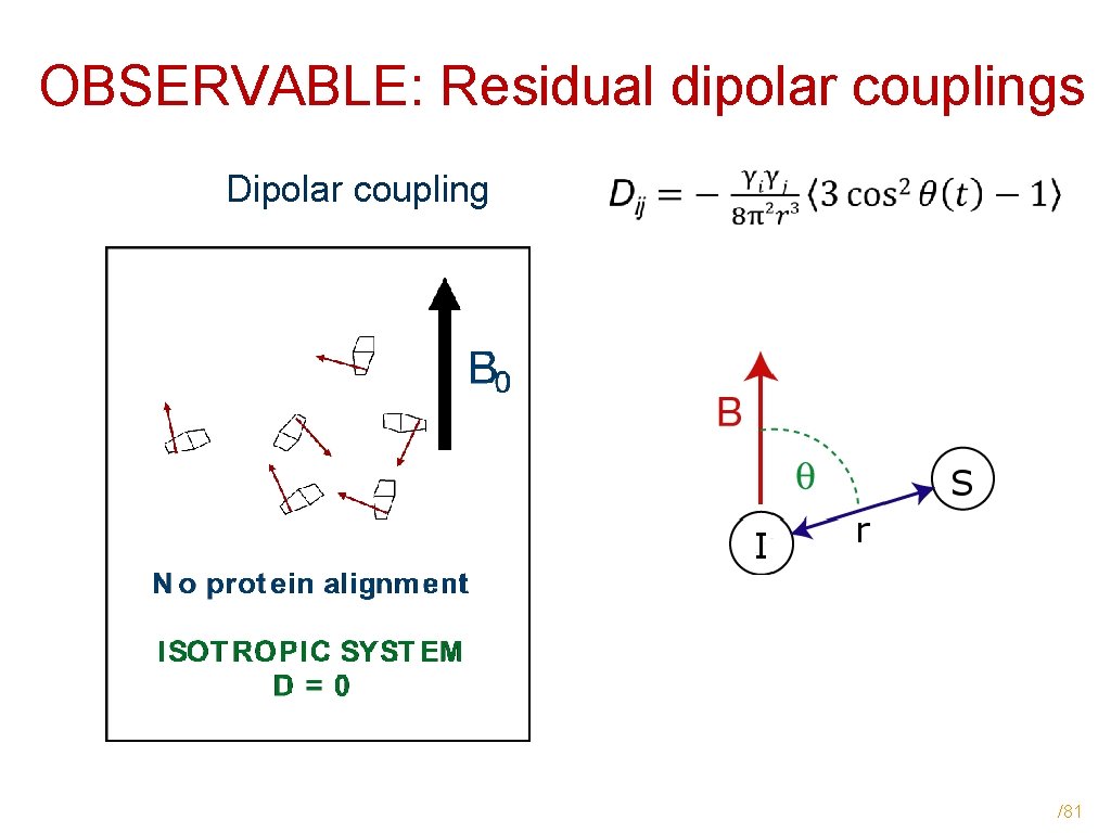
OBSERVABLE: Residual dipolar couplings Dipolar coupling /81
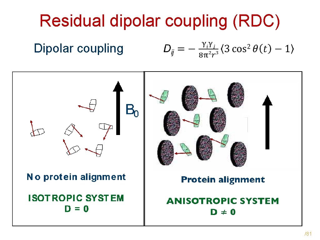
Residual dipolar coupling (RDC) Dipolar coupling /81
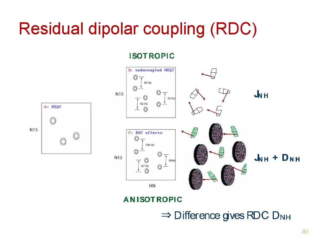
Residual dipolar coupling (RDC) /81
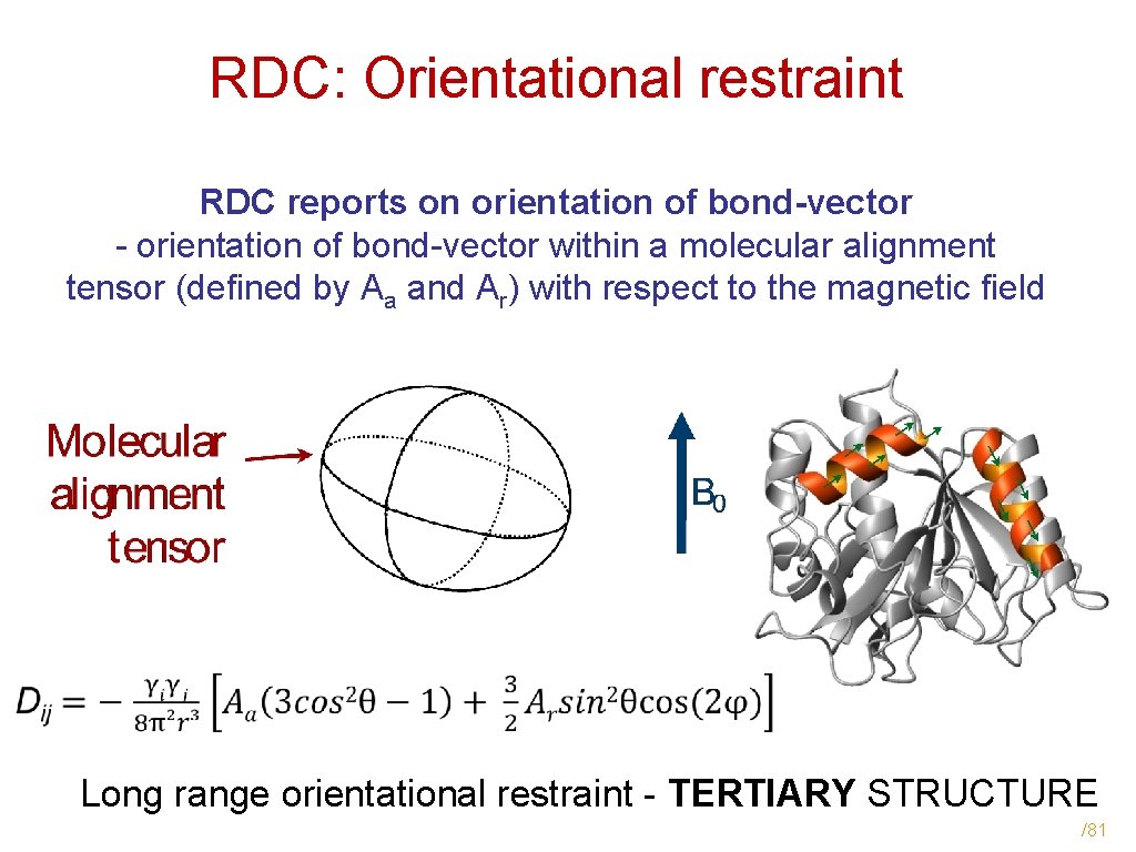
RDC: Orientational restraint RDC reports on orientation of bond-vector - orientation of bond-vector within a molecular alignment tensor (defined by Aa and Ar) with respect to the magnetic field Long range orientational restraint - TERTIARY STRUCTURE /81
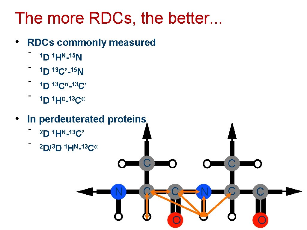
The more RDCs, the better. . . • RDCs commonly measured - 1 D 1 HN-15 N 1 D 13 C’-15 N 1 D 13 Cα-13 C’ 1 D 1 Hα-13 Cα • In perdeuterated proteins - 2 D 1 HN-13 C’ 2 D/3 D 1 HN-13 Cα C N C C C O N C C O 48/81
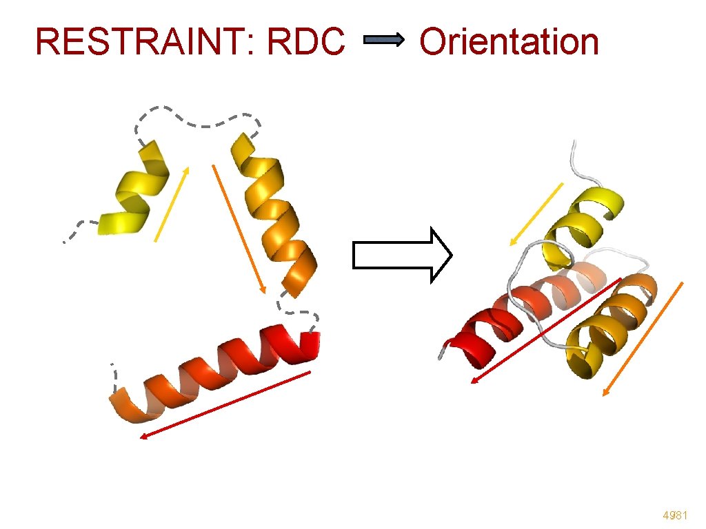
RESTRAINT: RDC Orientation 49/81
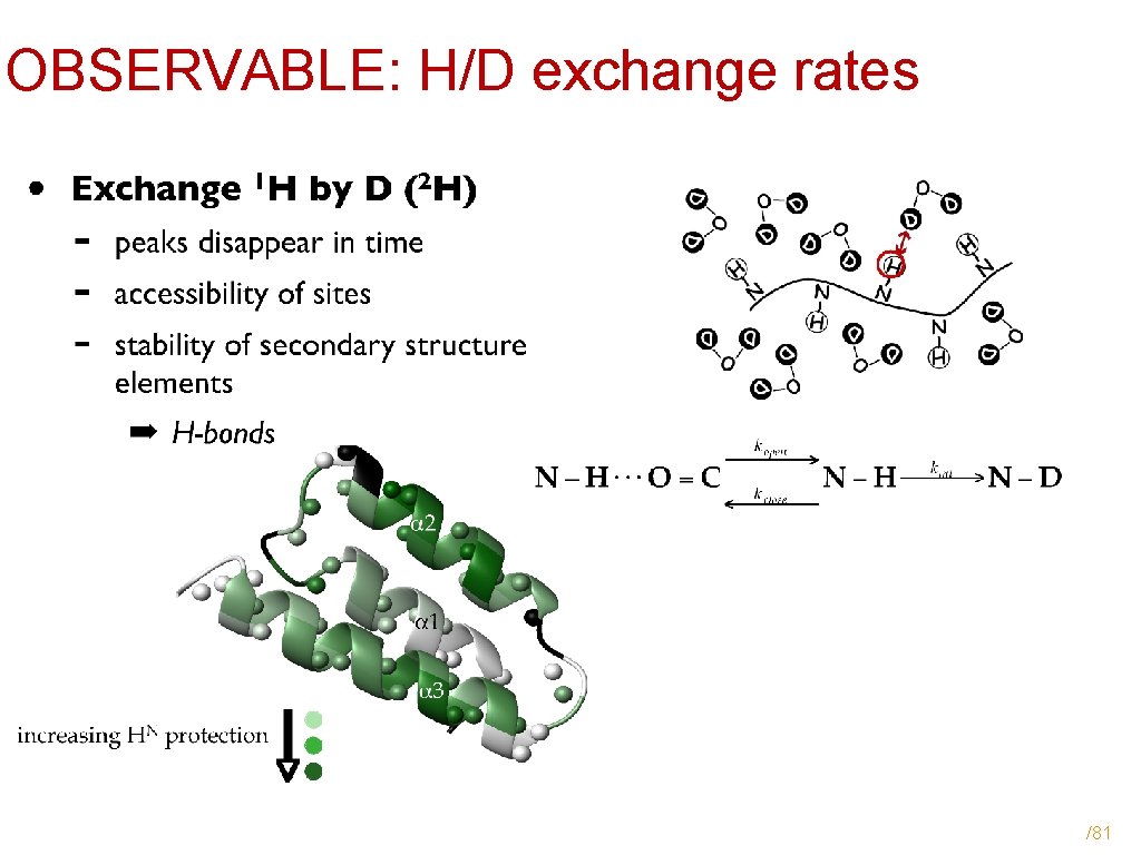
OBSERVABLE: H/D exchange rates /81
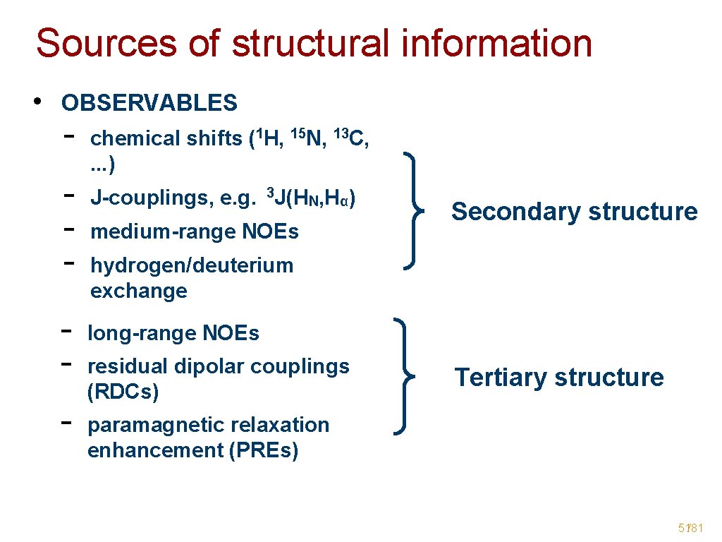
Sources of structural information • OBSERVABLES - - chemical shifts (1 H, 15 N, 13 C, . . . ) J-couplings, e. g. 3 J(HN, Hα) medium-range NOEs Secondary structure hydrogen/deuterium exchange long-range NOEs residual dipolar couplings (RDCs) Tertiary structure paramagnetic relaxation enhancement (PREs) 51/81
- Slides: 51