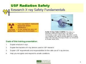
Research Xray Safety Fundamentals Office of Research Safety


![Analytical X-rays Diffraction [XRD] X-ray scattering from crystalline materials. “fingerprint” of crystalline atomic structure. Analytical X-rays Diffraction [XRD] X-ray scattering from crystalline materials. “fingerprint” of crystalline atomic structure.](https://slidetodoc.com/presentation_image_h2/ac4dc1f4ce8a7836657ef243da74f33d/image-17.jpg)


- Slides: 77

Research X-ray Safety Fundamentals Office of Research Safety 391 College Ave. , Suite 104

This Training will introduce you to • • • X-ray regulations Properties of x-rays Biological effects of x-ray and hazards associated with x-ray devices used in research Radiation dose; dose units Levels of radiation for analytical and industrial x-ray, as well as environmental radiation Basic principles of radiation protection Dose measurement and personnel dose monitoring Safety devices, postings and labels Requirements and responsibilities of the x-ray users

X-Ray Regulations In South Carolina Analytical and Industrial X-ray use is regulated by the Department of Health and Environmental Control (DHEC). These regulations may be found in R. 61 -64, X-Rays (Title B): Part III. Standards for Protection Against Radiation Part VII. Radiation Safety Requirements for Analytical X-Ray Equipment Part VIII. Radiation Safety Requirements for Industrial Uses of Radiographic Sources Part IX. Definitions This training provides overview of these regulations.

X-Ray Regulations • X-ray equipment may only be purchased from a vendor registered with the SC DHEC as a Class I Vendor • List of Vendors registered in South Carolina • All X-ray devices, including electron microscopes and handheld devices, must be registered with the SC DHEC • Each x-ray system MUST meet state x-ray safety requirements. • Please notify Radiation Safety Officer as soon as possible when you do or plan to purchase, dispose of, modify or relocate any X-ray equipment.

X-Ray Project Approval Any use of analytical or industrial x-ray (“x-ray project”) in the Clemson University must be reviewed and approved by the Radiation Safety Committee. X-ray project includes: • • • Responsible Investigator Approved x-ray equipment Locations of use List of Personnel Research protocol Special conditions (ex. , shielding, personnel monitoring)

What is Ionizing Radiation? Radiation in the form of electromagnetic waves or particles that has high enough energy to ionize atoms is called IONIZING RADIATION Ionizing radiation includes x-rays, gamma-rays, beta particles, alpha particles, and neutrons. Without the use of monitoring equipment, radiation is undetectable. Humans are not able to see, feel, taste, smell, or hear ionizing radiation.

What are X-rays? X-rays are a form of electromagnetic radiation produced when electrons are deflected from their original paths (“bremsstrahlung” or “breaking” x-rays) or change their orbital levels around the atomic nucleus (“characteristic” xray). X-rays can travel long distances through air and most other materials. Gamma- and x-rays usually require thicker and/or denser shielding to reduce their intensity than do beta or alpha particles. X- and gamma-rays only differ in their origin: • x-rays originate in the electronic shell • gamma rays originate in the nucleus

Discovery of X-rays were discovered in 1895 when Wilhelm Conrad Roentgen observed that a screen coated with a barium salt fluoresced when placed at significant distance from a “cathode ray” tube. Roentgen concluded that a form of penetrating radiation was being emitted by the cathode ray tube and called the unknown radiation: X-rays

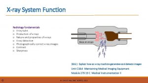
X-ray Tube When potential difference applied, these electrons start to Some metal alloys, when heated, release electrons, accelerate towards anode made of heavy material, ex. , tangsten, which create “electron cloud”… strike it, and create bremsstrahlung x-rays Cathode (-) Anode (+)

X-ray Interactions In passing through matter, energy is transferred from the xray photon to electrons and nuclei in the target material. An electron can be ejected from the atom with the subsequent creation of an ion. The amount of energy lost to the electron is dependent on the energy of the incident photon and the type of material through which it travels. There are three basic methods in which x-rays interact with matter: • photoelectric effect • Compton scattering • pair production

X-ray Effects The biological effects of x-ray exposure depend upon: • Duration of exposure to the radiation. • Energy of the x-ray photon defines depth of penetration • • Low Energy (<50 ke. V) - damage only to skin or outer body tissues • High Energy - damage to internal organs Dose – measure of the total amount of energy deposited in the body

Units of Radiation Dose The units of Roentgen (R), rad and rem are used to define radiation dose in different situations and for different types of ionizing radiation. For X-ray radiation all this units may be considered equivalent. Radiation dose rate measuring equipment is usually calibrated in units of R/h or m. R/h, while doses from the personal dosimeter are usually reported in units of mrem.

Mechanisms of Biological Damage Ionizing radiation causes atoms and molecules to become ionized or excited. These excitations and ionizations can: • Produce chemically active free radicals • Break chemical bonds • Produce new chemical bonds and cross-linkage between macromolecules • Damage molecules that regulate vital cell processes (e. g. DNA, RNA, proteins) The cell can repair certain levels of radiation-induced cell damage. At higher levels, cell death or mutation (cancer) results. At extremely high doses, cells cannot be replaced quickly enough, and tissues fail to function.

Tissues Affected by X-Ray Exposure With analytical XRD and XRF equipment, the x-ray energies are low. This means that the energy is deposited solely into the skin rather than deeper into the body. The effects are localized and limited to the irradiated site. There is no concern about genetic effects or fetal exposure for pregnant women because the x-ray diffraction radiation is not penetrating enough. Pregnant women may operate XRDs without taking any special precautions. X-rays from industrial radiographic applications will affect internal organs as well as skin due to their higher penetrating ability (higher xray photon energy).

Environmental Sources of Radiation Exposure Any human being, on average, is exposed to • Natural (“background”) radiation ~ 311 mrem/year • • • Medical procedures ~ 300 mrem/year • • • Radon-222 ~ 220 mrem Radionuclides inside human body ~ 29 mrem/year Chest x-ray ~ 10 -20 mrem Chest CT ~ 500 -700 mrem Consumer products ~ 13 mrem/year • • • Building materials ~ 3. 5 mrem/year Air travel ~ 1 mrem/hour Smoking 30 cigarettes per day ~ 16 rem/year

Occupational Radiation Exposure Occupational dose limits: • Whole body: 5 rem/year • Skin and extremities: 50 rem/year • Eyes: 15 rem/year • Embryo/fetus: 0. 5 rem/year Average US occupational doses ~110 mrem • • • Aviation (flight crew) ~ 310 mrem/year Nuclear power ~ 190 mrem/year Industry, medical, research, military ~ 60 -80 mrem/year
![Analytical Xrays Diffraction XRD Xray scattering from crystalline materials fingerprint of crystalline atomic structure Analytical X-rays Diffraction [XRD] X-ray scattering from crystalline materials. “fingerprint” of crystalline atomic structure.](https://slidetodoc.com/presentation_image_h2/ac4dc1f4ce8a7836657ef243da74f33d/image-17.jpg)
Analytical X-rays Diffraction [XRD] X-ray scattering from crystalline materials. “fingerprint” of crystalline atomic structure. Check known library vs. unknown sample. Fluorescence [XRF] Analytical method for determining the elemental composition of a substance.

Hazards of Analytical X-ray Equipment The primary beam: The primary beam is most hazardous because of the extremely high exposure rates. Exposure rates of 4 x 10⁵ R/min at the port have been reported for ordinary diffraction tubes. Leakage or scatter of the primary beam through cracks in ill fitting or defective equipment: The leakage or scatter of the primary beam through apertures in ill fitting or defective equipment can produce very high intensity beams of possibly small and irregular cross section. Penetration of the primary beam through the tube housing, shutters or diffraction apparatus: The hazard resulting from penetration of the useful beam through shutters or the x-ray tube housing is slight in well designed equipment. Adequate shielding is easily attained at the energies commonly used for diffraction and florescence analysis. Diffracted rays: Diffracted beams also tend to be small and irregular in shape. They may be directed at almost any angle with respect to the main beam, and occasionally involve exposure rates of the order of 80 R/h for short periods.

Improper Handling of X-ray Equipment • • • Putting fingers in X-ray beam to change sample Aligning X-ray beam visually Modification of shielding Failure to realize X-rays are emitted from several ports Failure to read & follow manufacturers X-ray operating instructions Any of these actions could cause unnecessary, potentially dangerous exposure!

Skin Damage from X-Ray Exposure When the skin receives a high dose of radiation within short period of time (acute exposure), the primary damage occurs to hair follicles, basal (dividing) cells of the outer skin layer, and small blood vessels. Effect Threshold Dose Erythema (skin reddening) 300 - 500 rem Temporary hair loss 300 - 500 rem Permanent hair loss 700 rem Transepidermal injury (skin burns) 1000 rem Dermal radio-necrosis (tissue death) 2000 - 3000 rem

Skin Burns from Analytical X-Ray X-ray burns can take many months to fully develop. These injuries are generally not life-threatening but take long time to heal, are extremely painful and have a huge impact on a person’s quality of life. Contact the Radiation Safety Officer immediately if an overexposure is suspected!

Safety Interlocks Safety interlocks may consist of switches or other devices that prevent operation of the x-ray device with shielding removed or altered. X-ray devices should NEVER be operated with the safety interlocks bypassed. A written procedure must be approved by the Radiation Safety Officer PRIOR to performing any work with safety interlocks defeated.

Warning Lights Regulations require every x-ray machine to have a failsafe “XRay On” light on the control panel near the switch that energizes the x-ray tube. X-Ray On light

Shutter Status Light There must also be a shutter status light that tells you whether the shutter is open or closed, though the shutter light is not required to be failsafe. Before you approach the beam path, always check the shutter status light, even if you are certain that you turned off the high voltage. X-Ray On light Shutter status lights

Safety Systems Test DHEC Regulations require that X-ray equipment safety systems (interlocks, shutters and warning lights) be checked at least annually. Records of these checks have to be maintained and available for inspection by DHEC.

Area Postings Each area or room containing analytical x-ray equipment must be posted with a radiation symbol and words “CAUTION – X-RAY EQUIPMENT” or similar

Labels All analytical x-ray equipment must be labeled with the radiation symbol and words: Placed near any switch which energizes x-ray tube Placed in the area immediately adjacent to each tube head, clearly visible to any person operating, aligning the unit or changing sample

Open Beam Devices This an example of an OLD open beam x-ray diffraction device. New diffraction x-ray devices for research should be contained in a fully shielded and interlocked cabinet.

XRD Devices The x-ray tube, detector and sample are contained in housing that provides shielding to the user and others in lab. The access doors are interlocked and will shut off x-rays when opened. The large viewing area is made possible by using leaded glass or Plexiglas.

XRD Devices cont. A small compact “totally enclosed” research X-ray device.

Industrial X-rays are used for non-destructive testing which has applications in a wide range of industries. Non-destructive testing (NDT) means the X-ray beam inspects the integrity of industrial products or processes without damaging the items under observation. The NDT field that uses radiation is called Industrial radiography. Industrial X-ray machines usually operate at higher tube voltages and, therefore, have higher penetrating ability and require more shielding. Industrial X-ray may be used inside a cabinet, in a shielded room or in the field. Each of these techniques involves specific requirements.

Industrial X-rays Security Industrial radiography X-ray equipment must be kept locked at all times except when under direct surveillance of an operator or Radiation Safety Officer. When in storage, radiographic x-ray equipment must be secured to prevent tampering or removal by unauthorized individuals.

Industrial X-rays Operating Procedures Industrial radiography projects must have written specific operating and emergency procedures besides operating and safety procedures outlined in the Clemson X-ray Safety Manual. These include, but are not limited to: • Methods for controlling access to radiographic areas • Methods for securing industrial x-ray equipment • Personnel monitoring • Proper actions in the event of an accident These procedures must be reviewed and approved by the Clemson Radiation Safety Committee.

Basic Principles of Radiation Protection: Time The dose of radiation a worker receives is directly proportional to the amount of time spent in a radiation field. Thus, reducing the time by one-half will reduce the radiation dose received by one-half. Operators should always work quickly and spend as little time as possible next to x-ray equipment while it is operating.

Basic Principles of Radiation Protection: Distance Radiation exposure from a point source, such as an X-ray tube, can be calculated by using the INVERSE SQUARE LAW: Amount of radiation at a given distance from a point source varies inversely with the square of the distance. For example, doubling the distance from an x-ray tube will reduce the dose to one-fourth of its original value. Maintaining a safe distance, therefore, represents one of the simplest and most effective methods for reducing radiation exposure to workers. Using the principle of distance is especially important when working around open beam analytical x-ray equipment.

Basic Principles of Radiation Protection: Shielding Radiation exposure can also be reduced by placing an attenuating material between a worker and the x-ray tube. The energy of the incident x-ray photon is reduced by Compton and photoelectric interactions in the shielding material. Substances such as lead, that are very dense and have a high atomic number, are very good shielding materials because of the abundance of atoms and electrons that can interact with the x-ray photon. Shielding is often incorporated into the equipment, such as the metal lining surrounding the x-ray tube. It may also consist of permanent barriers such as concrete and lead walls, leaded glass, and plastic movable screens in the case of analytical x-ray equipment.

Radiation Detection Instruments Radiation cannot be detected with any of the human senses. To detect and measure radiation dose or dose rate you need to uses special survey instruments. Survey instruments have to be calibrated periodically (usually, once a year) Ion Chamber – Best for precise measurements of dose and dose rate Geiger-Mueller probe – very good for detecting radiation, but usually overestimates dose rate measurement

Survey Instrument Checks Every day before use: 1. Check calibration expiration date. Contact radiation safety office when calibration expiration date is close or passed. Only use instrument with expired calibration in emergency and if all operational checks are OK.

Meter Dial m. R/hr Survey Instrument Checks Every day before use: 1. Check calibration expiration date. 2. Check battery. Set switch to “BAT” position. Needle should be within sector “BAT TEST” (or “BAT OK”, or similar). Replace batteries if it is not. It is a good idea to always have extra batteries of proper size for your instrument. On-Off Switch / Range Selector Battery Check Battery Compartment

Meter Dial m. R/hr Survey Instrument Checks Every day before use: 1. Check calibration expiration date. 2. Check battery. 3. Set switch to the proper range. Usually, radiation levels in research lab should be within the lowest range. If meter pegs, switch to higher scale and press Reset button. Highest scale usually has its own dial Reset Button On-Off Switch / Range Selector x 100 Dial

Meter Dial m. R/hr Survey Instrument Checks Every day before use: 1. 2. 3. 4. Check calibration expiration date. Check battery. Set switch to the proper range. Check background radiation level in a clean area away from radiation sources. You may want to set “Fast/Slow” switch to “S” to have stable reading. Fast/Slow Response

Meter Dial m. R/hr Survey Instrument Checks Every day before use: 1. 2. 3. 4. 5. Check calibration expiration date. Check battery. Set switch to the proper range. Check background radiation level. Check instrument response using dedicated check source or any other available known source of radiation (stock vial, sample, etc. )

Meter Dial m. R/hr Survey Instrument Checks Every day before use: 1. 2. 3. 4. 5. 6. Check calibration expiration date. Check battery. Set switch to the proper range. Audio Check background radiation level. On-Off Check instrument response When performing survey, move probe slowly (~2 in/sec) over Fast/Slow survey area. Set “Fast/Slow” Response switch to “F” for immediate response and Audio to ON.

Personnel Dose Monitoring The use of personnel monitors is required for: • • Anyone who may receive 10% of annual radiation dose limit; • Anyone who enters a high or very high radiation area • Anybody working with industrial x-ray • Your badge must remain at work. It is intended for measuring occupational dose only. • Only wear your issued dosimeter. • Store dosimeters away from radiation sources.

Personnel Monitoring Most analytical x-ray devices with fully enclosed beam do not require users to be issued personnel monitoring devices. Medical, veterinary and industrial x-ray use usually requires use of whole-body dosimeter. Open-beam application may require use of ring dosimeters in addition to the whole-body dosimeter.

Dose Report Clemson University uses Mirion’s instadose®+ dosimeters to monitor personnel dose. These dosimeters can measure dose for both low(analytical) and high-energy (radiography) x-rays. These dosimeters are Bluetoothequipped, which allows dose readings at any time by any Bluetoothcompatible device (laptop, tablet, smartphone) or with a special readers (“hot-spots”).

Ring Dosimeters Ring dosimeters are required for persons working with open beam x-ray systems.

Responsibilities of X-ray Owners & Users • Read, understand follow Clemson X-Ray Safety Manual • Operate x-ray device only as described in manufacturer’s operating instructions. • Notify Radiation Safety Officer of any repairs, modifications, disposal, or relocation of x-ray device. • X-ray users should address any radiation safety concerns to the Radiation Safety Officer @ 650 -3516.

In Case of Emergency In a case of known or suspected x-ray equipment malfunction or personnel overexposure: • • • Shut down equipment, if doing so is safe and does not increase personnel dose; Notify personnel and evacuate all potentially dangerous areas; Lock and/or post the room or otherwise prevent inadvertent use of the malfunctioning x-ray equipment; Notify Konstantin Povod (RSO) at 656 -3516 or kpovod@clemson. edu and the project’s Responsible Investigator Some cases of personnel overexposure may require medical evaluation and intervention.

This completes the training presentation. The following slides contain short quiz to evaluate how much knowledge you retained. At the end of this quiz you will be able to register by submitting email using the link provided.


Question 1 Occupational dose limits for a) whole body; b) eyes; c) skin; d) embryo: q a) 5 mrem; b) 15 mrem; c) 50 mrem; d) 0. 5 mrem q a) 5 rem; b) 15 rem; c) 50 rem; d) 5 rem q a) 5000 mrem; b) 15 rem; c) 50 rem; d) 500 mrem q There are no dose limits for analytical X-ray q Any radiation dose above 2 x background must be eliminated Right! Wrong! Please proceed review to the and next try question again


Question 2 Personal dosimeter: q Provides adequate protection from the ionizing radiation q Required for anybody likely to receive dose >0. 5 rem/year and for persons entering High and Very High Radiation Areas q Should be used for the monitoring of medical exposures q Only measures high-energy radiography x-rays q Must be worn by radiation worker 24/7 Right! Wrong! Please proceed review to the and next try question again


Question 3 All new X-ray projects at Clemson must be approved by: q Radiation Safety Officer (RSO) q Nuclear Regulatory Commission (NRC) q SC Dep’t of Health and Environmental Control (DHEC) q Vice President for Research and RSO q Clemson University Radiation Safety Committee (RSC) Right! Wrong! Please proceed review to the and next try question again


Question 4 Safety devices (interlocks, warning lights): q Must be checked at least annually per DHEC regulations q Only required for high-energy (>50 ke. V) x-ray equipment q Must be checked before each use per DHEC regulations q May be substituted by the ambient dose monitoring q Required for industrial and are optional for analytical x-ray Right! Wrong! Please proceed review to the and next try question again


Question 5 Basic principles of radiation protection: q Training and SOP’s q Warning lights and interlocks q Room postings and equipment labels q Time, Distance and Shielding q DHEC 61 -64 (RHB) X-Ray regulations Right! Wrong! Please proceed review to the and next try question again

Occupational Radiation Exposure Occupational dose limits: • Whole body: 5 rem/year • Skin and extremities: 50 rem/year • Eyes: 15 rem/year • Embryo/fetus: 0. 5 rem/year Average US occupational doses ~110 mrem • • • Aviation (flight crew) ~ 310 mrem/year Nuclear power ~ 190 mrem/year Industry, medical, research, military ~ 60 -80 mrem/year

Environmental Sources of Radiation Exposure Any human being, on average, is exposed to • Natural (“background”) radiation ~ 311 mrem/year • • • Medical procedures ~ 300 mrem/year • • • Radon-222 ~ 220 mrem Radionuclides inside human body ~ 29 mrem/year Chest x-ray ~ 10 -20 mrem Chest CT ~ 500 -700 mrem Consumer products ~ 13 mrem/year • • • Building materials ~ 3. 5 mrem/year Air travel ~ 1 mrem/hour Smoking 30 cigarettes per day ~ 16 rem/year

Personnel Dose Monitoring The use of personnel monitors is required for: • • Anyone who may receive 10% of annual radiation dose limit; • Anyone who enters a high or very high radiation area • Anybody working with industrial x-ray • Your badge must remain at work. It is intended for measuring occupational dose only. • Only wear your issued dosimeter. • Store dosimeters away from radiation sources.

Personnel Monitoring Most analytical x-ray devices with fully enclosed beam do not require users to be issued personnel monitoring devices. Medical, veterinary and industrial x-ray use usually requires use of whole-body dosimeter. Open-beam application may require use of ring dosimeters in addition to the whole-body dosimeter.

Dose Report Clemson University uses Mirion’s instadose®+ dosimeters to monitor personnel dose. These dosimeters can measure dose for both low(analytical) and high-energy (radiography) x-rays. These dosimeters are Bluetoothequipped, which allows dose readings at any time by any Bluetooth-compatible device (laptop, tablet, smartphone) or with a special readers (“hot-spots”).

Ring Dosimeters Ring dosimeters are required for persons working with open beam x-ray systems.

X-Ray Regulations In South Carolina Analytical and Industrial X-ray use is regulated by the Department of Health and Environmental Control (DHEC). These regulations may be found in R. 61 -64, X-Rays (Title B): Part III. Standards for Protection Against Radiation Part VII. Radiation Safety Requirements for Analytical X-Ray Equipment Part VIII. Radiation Safety Requirements for Industrial Uses of Radiographic Sources Part IX. Definitions This training provides overview of these regulations.

X-Ray Regulations • All X-ray devices, including electron microscopes and handheld devices, must be registered with the SC DHEC • Each x-ray system MUST meet state x-ray safety requirements. • Please notify Radiation Safety Officer as soon as possible when you do or plan to purchase, dispose of, modify or relocate any X-ray equipment.

X-Ray Project Approval Any use of analytical or industrial x-ray (“x-ray project”) in the Clemson University must be reviewed and approved by the Radiation Safety Committee. X-ray project includes: • • • Responsible Investigator Approved x-ray equipment Locations of use List of Personnel Research protocol Special conditions (ex. , shielding, personnel monitoring)

Safety Interlocks Safety interlocks may consist of switches or other devices that prevent operation of the x-ray device with shielding removed or altered. X-ray devices should NEVER be operated with the safety interlocks bypassed. A written procedure must be approved by the Radiation Safety Officer PRIOR to performing any work with safety interlocks defeated.

Warning Lights Regulations require every x-ray machine to have a failsafe “XRay On” light on the control panel near the switch that energizes the x-ray tube. X-Ray On light

Shutter Status Light There must also be a shutter status light that tells you whether the shutter is open or closed, though the shutter light is not required to be failsafe. Before you approach the beam path, always check the shutter status light, even if you are certain that you turned off the high voltage. X-Ray On light Shutter status lights

Safety Systems Test DHEC Regulations require that X-ray equipment safety systems (interlocks, shutters and warning lights) be checked at least annually. Records of these checks have to be maintained and available for inspection by DHEC.

Basic Principles of Radiation Protection: Time The dose of radiation a worker receives is directly proportional to the amount of time spent in a radiation field. Thus, reducing the time by one-half will reduce the radiation dose received by one-half. Operators should always work quickly and spend as little time as possible next to x-ray equipment while it is operating.

Basic Principles of Radiation Protection: Distance Radiation exposure from a point source, such as an X-ray tube, can be calculated by using the INVERSE SQUARE LAW: Amount of radiation at a given distance from a point source varies inversely with the square of the distance. For example, doubling the distance from an x-ray tube will reduce the dose to one-fourth of its original value. Maintaining a safe distance, therefore, represents one of the simplest and most effective methods for reducing radiation exposure to workers. Using the principle of distance is especially important when working around open beam analytical x-ray equipment.

Basic Principles of Radiation Protection: Shielding Radiation exposure can also be reduced by placing an attenuating material between a worker and the x-ray tube. The energy of the incident x-ray photon is reduced by Compton and photoelectric interactions in the shielding material. Substances such as lead, that are very dense and have a high atomic number, are very good shielding materials because of the abundance of atoms and electrons that can interact with the x-ray photon. Shielding is often incorporated into the equipment, such as the metal lining surrounding the x-ray tube. It may also consist of permanent barriers such as concrete and lead walls, leaded glass, and plastic movable screens in the case of analytical x-ray equipment.

REGISTRATION Good job! You successfully completed required Research X-Ray Safety Training Please register your achievement by clicking the button below. Enter your name and Clemson email address in the body of the email : SUBMIT (*) "Submit" button uses your default email app. If, after clicking on "Submit" button, unexpected email service (ex. , Mail for Windows) opens, please do the following (instructions are for Windows 10): 1. Go to your computer's Settings (gear icon) 2. Select "Apps" 3. Select "Default Apps" from the menu on the left 4. Set the app you usually use for email (ex. , Outlook, Gmail, Yahoo, etc. )
 Shell 3 golden rules
 Epsc process safety fundamentals
 X ray mastoid towne's view cholesteatoma
 Dental xray films
 Kub xray
 Xray xml editor
 Sza xray
 Jfk jr plane crash photos
 Pink tof vs blue tof
 Rickets xray
 Cvs x ray
 Rhese method rheese orbits positioning
 Occipito frontal
 Xray technique chart
 Faulty
 What is a falling load generator
 Lara xray
 Double exposure artifact
 Spectrum xray
 5 cyanotic congenital heart disease
 Lightbulb xray
 Hemothorax xray
 Rpo ribs
 Xray file cabinet
 Gimp xray
 Dark room radiographic darkroom layout
 Hampton hump sign คือ
 First xray ever taken
 Xray neck lateral view
 Xray laser
 Xray laser
 Triboluminescence x ray
 Xray laser
 Xray spectrometer
 Xray laser
 Xray laser
 Pancreatic calcification
 Xray laser
 Xray laser
 Xray laser
 Bga xray
 Acanthion anatomy
 Peter mueller mit
 68 xray
 Rigler sign
 Supine chest xray
 Facial bone xray
 Picker xray
 Ellis curve on chest x ray
 Atom xray
 Silicon valley xray
 Xray photoelectron spectroscopy
 Who discovered x rays
 X ray production
 Rds case study
 In capacitor/condenser discharge mobile units,
 Xray uranus
 Michigan xray
 Mmc xray
 Bacterial tracheitis xray
 Gladstone park xray
 Fadhl alakwaa
 Steeple sign
 Talipes equinovarus xray
 Bravais lattices
 Yxlon xray
 Xray searches
 Xray training
 Diffraction
 Xray telescope
 Xray gladstone
 Xray lithography
 Inertial confinement fusion lasers
 L
L Market research fundamentals
 The fundamentals of political science research 2nd edition
 Factory office plan
 Hazards in office


