Reproductive System Chapter 22 Continued Female Reproductive System


 Tubes • • Extend laterally from uterus Open end is funnel shaped")


 under influence of")


- Slides: 68
Reproductive System Chapter 22 Continued
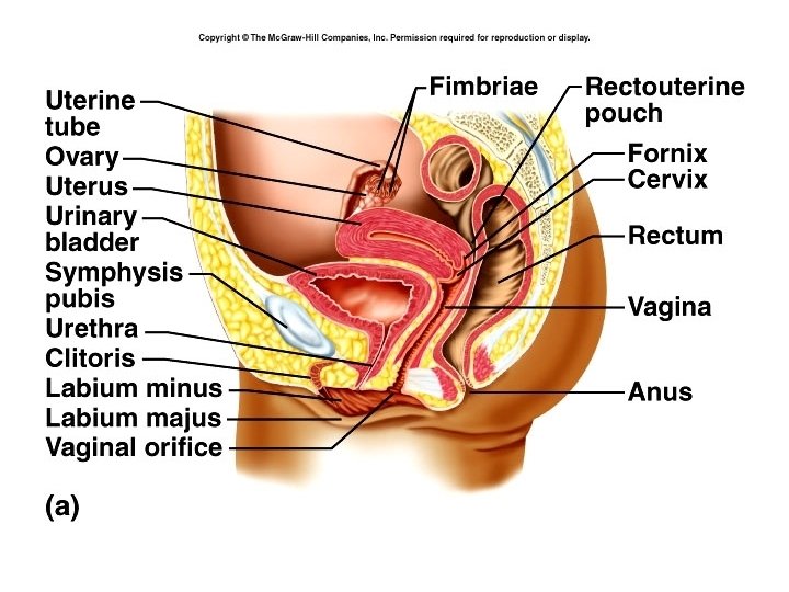
Female Reproductive System • Ovaries – gonads – produce oocytes and hormones • Uterine (Fallopian) tubes or oviducts • Uterus • Vagina • Vulva (female external reproductive organs)
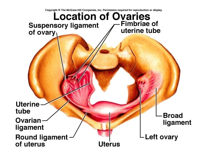
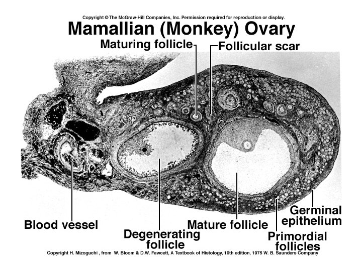
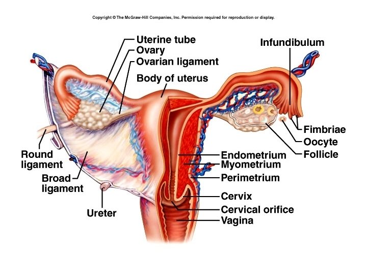
Ovaries • Located in upper pelvic cavity, on either side of uterus • Held in place by a number of ligaments • Descend from abdominal cavity to pelvic brim
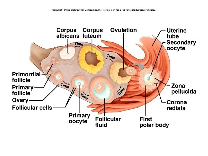
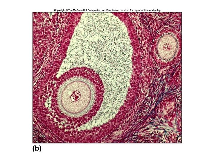
• Each ovary has a hilus – blood vessels and nerves enter • Several layers: – Germinal epithelium – simple squamous cells – Tunica albuginea- white capsule of C. T. – Stroma – connective tissue, can be divided into: • Medulla – loose connective tissue, blood vessels, nerves • Cortex - contains ovarian follicles – consist of oocytes at various stages of development
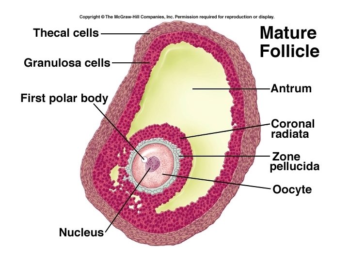
• Single layer – follicular cells • Several layers – granulosa cells • Mature or Graafian follicle is a large, fluid-filled follicle that will rupture and release a secondary oocyte in process called ovulation. • Corpus luteum is the remnant of a ruptured follicle – produces estrogen, progesterone and relaxin until degenerates into the corpus albicans.
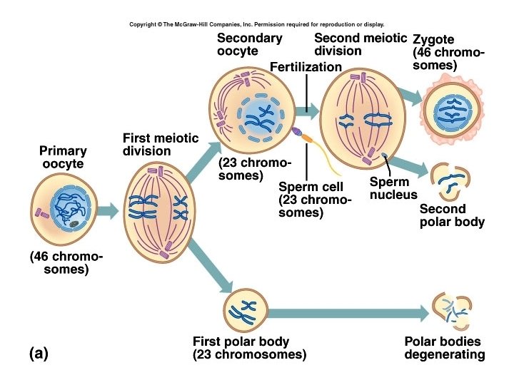
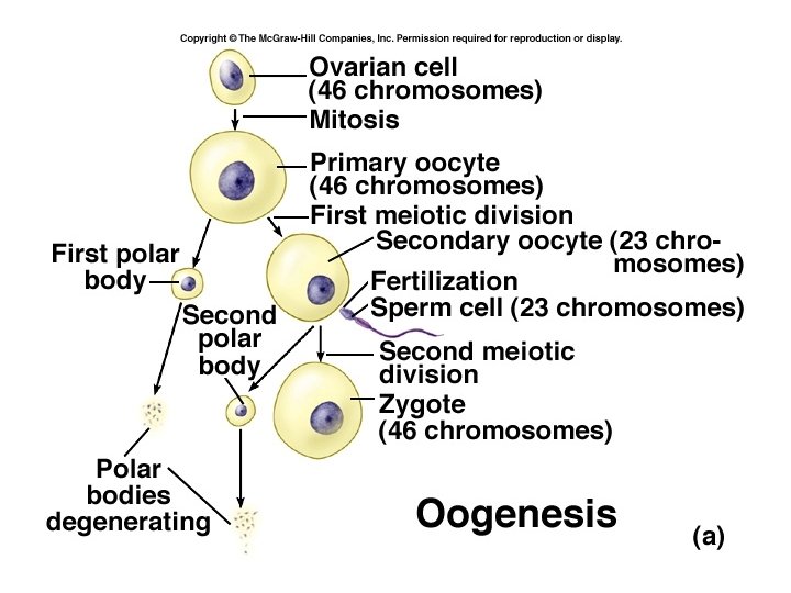
Oogenesis • During fetal development, germ cells differentiate into oogonia millions of germ cells. • Many degenerate, but a few develop into primary oocytes that enter Prophase I of meiosis before birth – stop there. • At birth, about 1 million oogonia and primary oocytes in each ovary • About 400 mature over a woman’s lifetime.
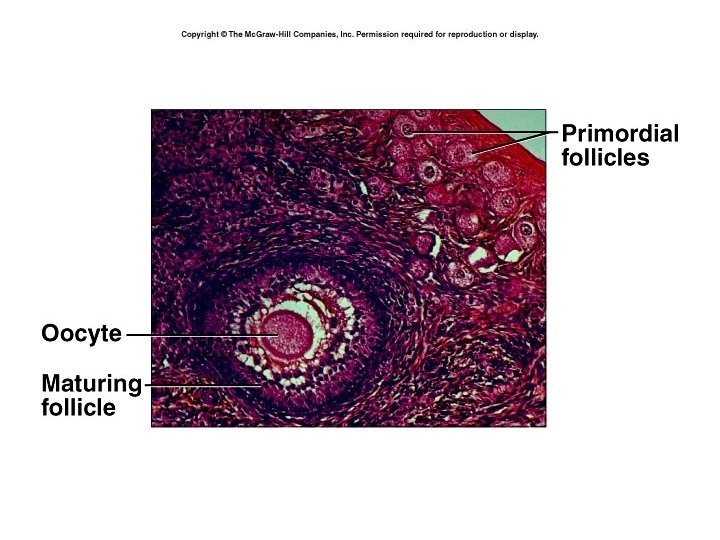
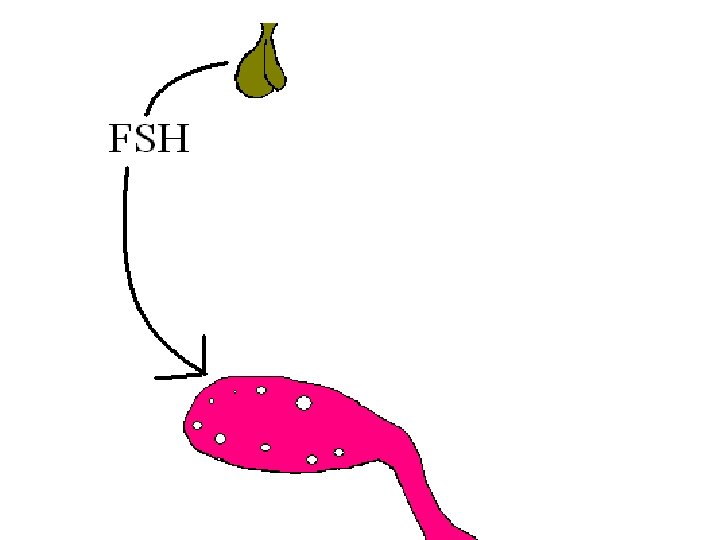
• Primordial follicle – primary oocyte and single layer of follicular cells • Under influence of FSH, become primary follicles which have several layers of cells. • Layer of glycoprotein, the zona pellucida, separates oocyte from the granulosa cells. • Ovarian cells outside follicle form two layers: – Inner vascular layer (theca interna) that secretes hormones – Outer fibrous layer (theca externa) – connective tissue.
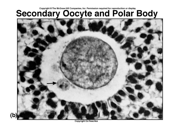
• Granulosa cells begin to secrete fluid, forms a cavity called the antrum. • Now called a secondary follicle. • After puberty, each month one secondary follicle resumes meiosis. • Meiosis I results in two unequal cells – secondary oocyte and a polar body. • Begins to divide again but stops at metaphase II.
• At ovulation, secondary oocyte and polar body are released. • If not fertilized, degenerates. • If penetrated by sperm, meiosis resumes, forming ovum and another polar body. • Nuclei of ovum and sperm unite to form a zygote. (2 n or diploid) • So, meiosis results in ONE OVUM and three polar bodies (which degenerate)
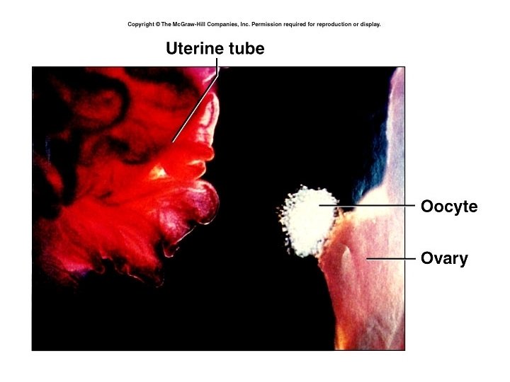
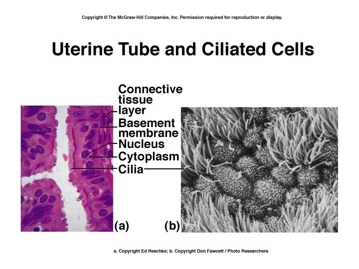
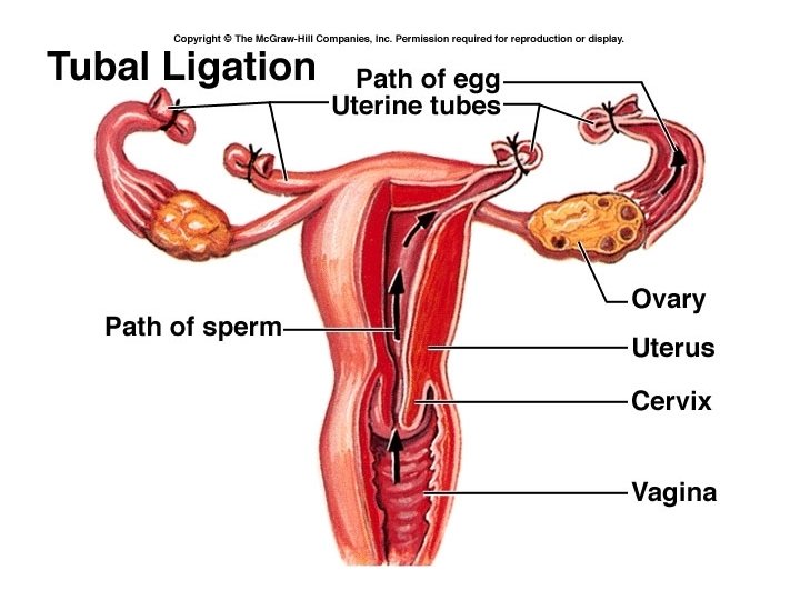
Uterine (Fallopian) Tubes • • Extend laterally from uterus Open end is funnel shaped – infundibulum Many finger-like projections – fimbriae Local currents produced by movement of fimbriae draws ovum into uterine tube. • Uterine tube is lined with ciliated columnar epithelium, which move ovum along. • Peristaltic movements also propel ovum toward uterus.
• The secondary oocyte is usually fertilized in the outer one-third of the uterine tube, although fertilization may occur in the abdominopelvic cavity. • Fertilization can occur up to about 24 hours after ovulation. • If oocyte is fertilized, it will reach the uterus in about 7 days.
The Uterus • Is an organ about the size and shape of an inverted pear. • Functions in menstruation, implantation of zygote, development of the fetus, and labor. • Also part of the pathway for sperm to reach ovum. • Normally held in position by ligaments
• Uterus can be divided into the fundus, body and cervix. • Secretory cells of the cervix produce about 20 – 60 ml of mucus / day. • At time of ovulation, this mucus becomes thinner and more alkaline. • Mucus provides for the energy needs of the sperm, protects sperm from environment of the vagina, and protects them from phagocytes. • At other times, mucus becomes thick and can form a cervical plug which impedes passage.
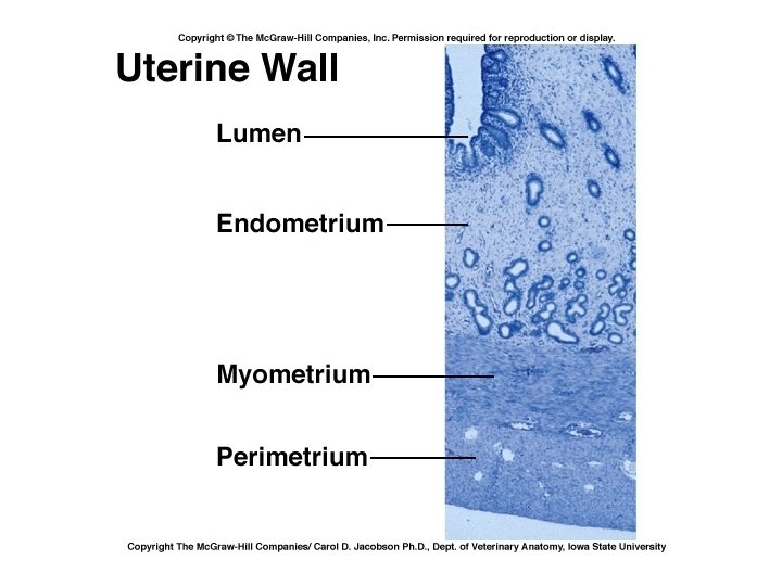
• Layers of uterus : perimetrium, myometrium and endometrium • Perimetrium – a part of visceral peritoneum • Myometrium – bulk of uterus – three layers of muscle that contract under influence of oxytocin during labor. • Endometrium – highly vascular mucosa – Stratum functionalis – shed during menstruation – Stratum basalis – deeper, permanent layer, gives rise to new stratum functionalis after each cycle.
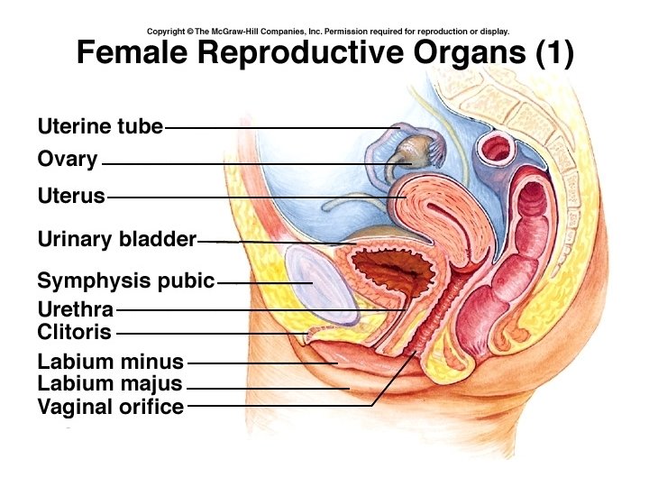
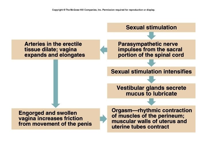
The Vagina • • • Passageway for sperm and menstrual flow Receptacle for penis during intercourse Inferior portion of birth canal Capable of considerable distention (stretching) Mucosa is continuous with that of uterus and consists of nonkeratinized stratified squamous epithelium. • Contains large stores of glycogen which decomposes to organic acids – lower p. H = less susceptible to infection & less hospitable to sperm
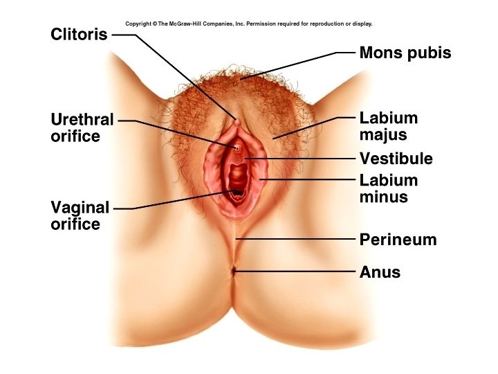
The Vulva • External genitals of female • Mons pubis – pad of adipose tissue which cushions pubic symphysis • Labia majora (labium majus) – folds of skin analogous to the scrotum – Adipose tissue, sebaceous glands, sweat glands • Labia minora (labium minus) – folds of skin with many blood vessels and sebaceous (oil) glands
• Clitoris – homologous to the penis, also composed of two corpora cavernosa and a glans. • Vestibule – between the labia minora – External urethral orifice – Openings for ducts of paraurethral glands : secrete mucus and are homologous to prostate – Beside vaginal orifice are the vestibular (Bartholin’s) glands – produce mucus during arousal – homologous to bulbourethral glands
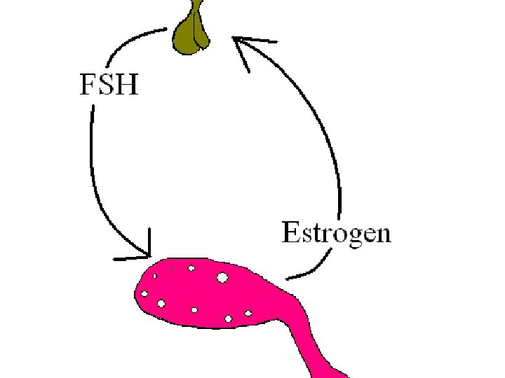
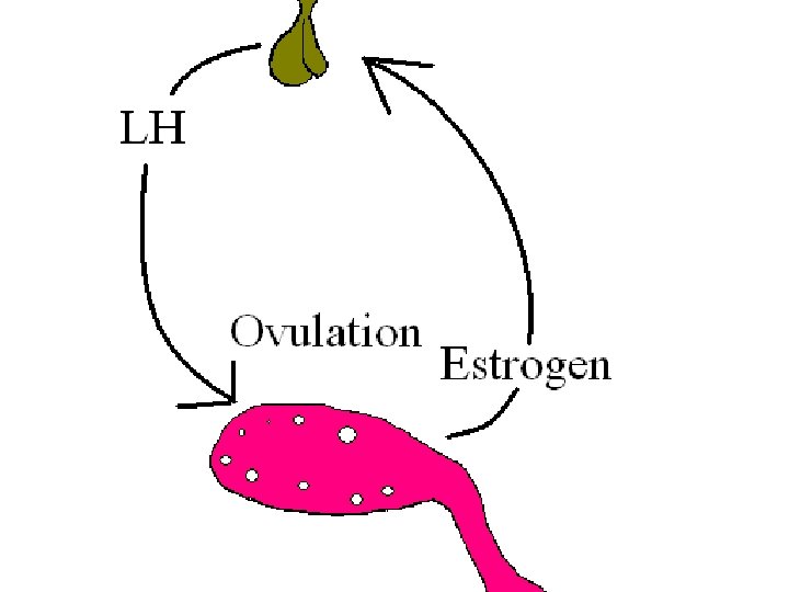
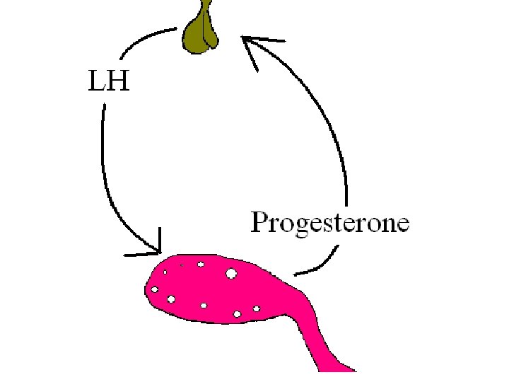
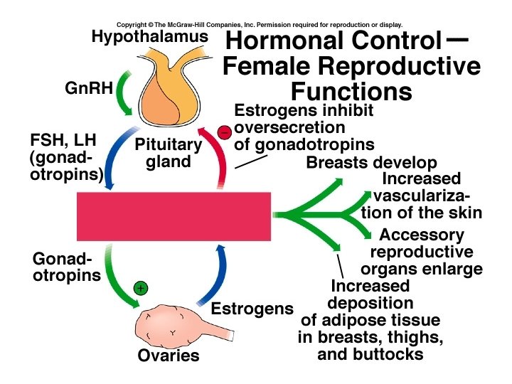
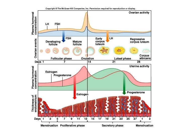
Female Reproductive Cycle • Two cycles: – Ovarian cycle – leading to maturation and release of oocyte – Uterine or menstrual cycle – prepares uterus to receive the fertilized ovum. • Both controlled by Gn. RH from the hypothalamus, which stimulates release of FSH and LH. • Estrogen and progesterone are produced by the ovaries.
Estrogens 1. Stimulate the growth, development, and maintenance of female reproductive structures, secondary sex characteristics and the breast. 2. They help regulate fluid and electrolyte balance. 3. The stimulate protein synthesis 4. They lower blood cholesterol levels
• Moderate levels of estrogen in the blood inhibit the release of Gn. RH, LH and FSH. • This is the basis for the birth control pill.
Progesterone • Is secreted mainly by the corpus luteum and works with estrogen to prepare the endometrium for implantation and mammary glands for lactation. • High levels of progesterone also inhibit Gn. RH, LH and FSH.
Relaxin • Produced by the corpus luteum • Inhibits uterine contractions which aids implantation. • During pregnancy, it is produced by the placenta, and continues to relax uterine smooth muscle. • Also relaxes the pubic symphysis and helps dilate the uterine cervix for delivery.
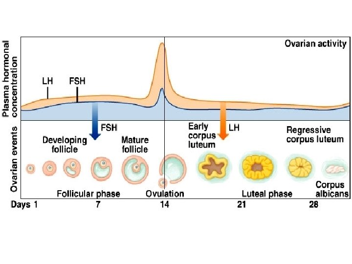
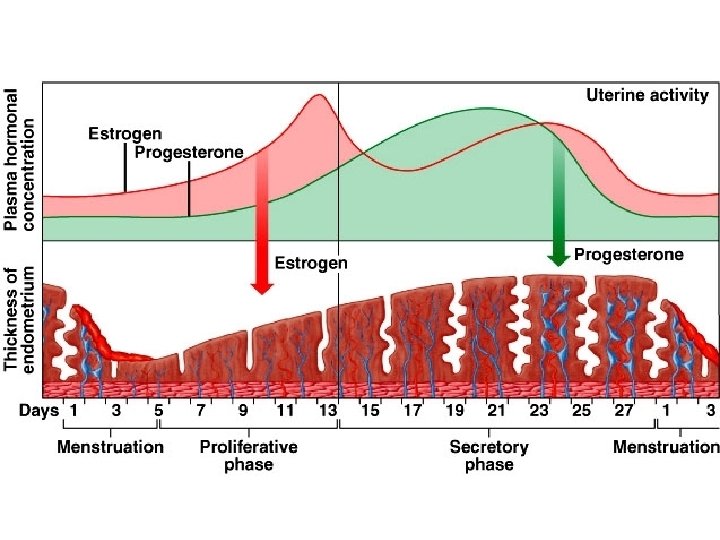
Phases of the Female Reproductive Cycle • Type cycle runs 24 -35 days, 28 days ave. • Three phases: – Menstrual phase – Preovulatory phase – Postovulatory phase
Menstrual phase • Days 1 – 5 • FSH release stimulates the maturation of follicles. 20 or so follicles, some in each ovary begin to enlarge. • In the uterus, the stratum functionalis is shed, discharging 50 -150 ml of blood, tissue fluid, mucus and epithelial cells. • Declines of estrogen and progesterone cause the spiral arteries to constrict, cells become ischemic and die, and are sloughed off.
Preovulatory phase • Most variable in length, us. Day 6 – 13 • Follicles continue to grow, granulosa cells produce increasing amounts of estrogen and some progesterone. This causes LH to be secreted, but stored in the pituitary. • Dominant follicle secretes inhibin, inhibiting growth of other follicles, and continues to develop into a mature follicle. • Called follicular phase in ovary.
• In the uterus, the estrogens produced by the developing follicles cause the cells of the stratum basalis to produce a new stratum functionalis, and thickness of endometrium doubles. • Proliferative phase in uterus.
Ovulation • About day 14, when estrogen levels are high enough, cause anterior pituitary to release a surge of LH that lasts about 36 hours. • This is what ovulation tests measure. • LH weakens wall of follicle, and ovulation occurs. • Secondary oocyte is released into pelvic cavity, taking the zona pellucida and first layer of granulosa cells.
Postovulatory phase • Mature follicle forms the corpus luteum (yellow body) under influence of Luteinizing Hormone (LH) and secretes progesterone, estrogen and relaxin. • Slight rise in progesterone just before ovulation causes an increase in basal temperature of about 0. 4 -0. 6 o. F – next 24 hours most likely time for conception. • Also see changes in cervical mucus • Mittelschmerz – pain around ovaries
• Postovulatory phase most constant in duration at 14 days, days 15 – 28. • In ovaries called the luteal phase. • If fertilization does not occur, at end of two weeks the secretions of corpus luteum decline, and it degenerates into corpus albicans (white body) • The decline in estrogens and progesterone bring on menstruation, and promote the release of Gn. RH and FSH and the cycle repeats.
• In the uterus, progesterone promotes : – growth of endometrial glands which secrete glycogen – Vacularization of stratum functionalis – Increase in amount of tissue fluid • These changes reach a peak about one week after ovulation, about the time the fertilized ovum would arrive. • Called secretory phase in uterus.
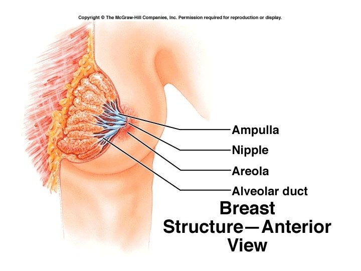
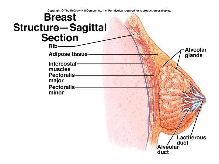
Mammary glands • Accessory organs of the female reproductive system • Modified sweat glands • Function is to synthesize, secrete and eject milk (lactation) • Lie over the pectoralis major muscles • Are attached to fascia by the suspensory ligaments (of Cooper) • Breast size is determined more by fat than by glandular tissue.
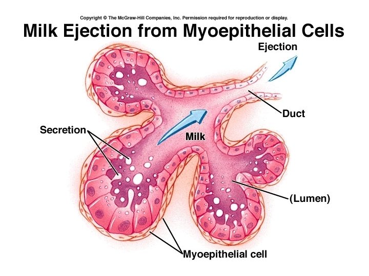
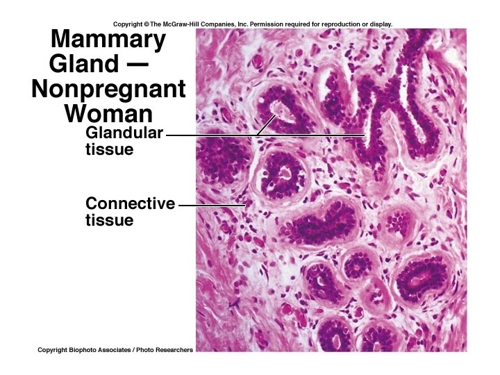
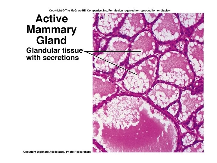
• Each breast has 15 – 20 lobes made up of several lobules. • Lobules are made of milk-secreting cells arranged in alveoli. • Alveoli are surrounded by myoepithelial cells which contract to move milk out of alveoli. • Milk passes from alveoli to secondary tubules into mammary ducts
• Close to nipple, ducts expand to form lactiferous sinuses where some milk may be stored. • Milk then passes through lactiferous ducts which open onto the nipple. • Colored area around the nipple is the areola. • Milk production is stimulated mostly by prolactin with some help from estrogen and progesterone. • Ejection or milk let down occurs under the • Influence of oxytocin.