Reproductive System Animals reproductive systems can be divided

Reproductive System Animals’ reproductive systems can be divided into the internal reproductive organs and the external genitalia. The gonads are the actual organs that produce the gametes. In the male, testes (singular = testis) produce sperm, and in the female, ovaries make eggs.

In most animals, individuals are either definite males or definite females. However, in some species, individual organisms are both male and female. Hermaphroditism is when one organism has both sexes.

Male Reproductive Tract Anatomy

Learning Objectives • To be able to describe the male reproductive system in the bull. – Structure – Function

Testis Factory • 1 -25 X 109 sperm/day • “Plant” must be air conditioned Penis Delivery System • Erection • Protrusion • Ejaculation Epididymis Finishing School • Fluid Absorption • 8 -25 X 109 sperm – membrane changes – nuclear & flagellar stabilization – motility, fertility – cytoplasmic droplet translocation Tail of Epididymis Warehouse and Delivery Accessory Sex Glands Alterations & Packaging • Metabolic substrates • Surface coatings • Transport for sperm • Storage 10 -50 X 109 sperm • Sperm for 5 -10 ejaculates • Smooth muscle contractions upon sexual stimulation

The male reproductive system is illustrated to the next slide. Sperm are produced in the testes located in the scrotum. Normal body temperature is too hot thus is lethal to sperm so the testes are outside of the abdominal cavity where the temperature is about 2° C (3. 6° F) lower.

From the testes, sperm are transferred to the epididymis, coiled tubules found within the scrotum, that store sperm and are the site of their final maturation. In ejaculation, sperm are forced up into the vas deferens (plural = vasa deferentia).

From the epididymis, the vas deferens goes up, around the front of, over the top of, and behind the bladder. The ends of the vasa deferentia, behind and slightly under the bladder, are called the ejaculatory ducts. The seminal vesicles are also located behind the bladder.

Their secretions are about 60% of the total volume of the semen (= sperm and associated fluid) and contain mucus, amino acids, fructose as the main energy source for the sperm, and prostaglandins to stimulate female uterine contractions to move the semen up into the uterus.

The seminal vesicles empty into the ejaculatory ducts. The ejaculatory ducts then empty into the urethra (which, in males, also empties the urinary bladder).
.")
The initial segment of the urethra is surrounded by the prostate gland (note spelling!). The prostate is the largest of the accessory glands and puts its secretions directly into the urethra. These secretions are alkaline to buffer any residual urine, which tends to be acidic, and the acidity of the woman’s vagina.

The prostate needs a lot of zinc to function properly, and insufficient dietary zinc (as well as other causes) can lead to enlargement which potentially can constrict the urethra to the point of interferring with urination.

Mild cases of prostate hypertrophy can often be treated by adding supplemental zinc to the man’s diet, but severe cases require surgical removal of portions of the prostate. This surgery, if not done very carefully can lead to problems with urination or sexual performance.

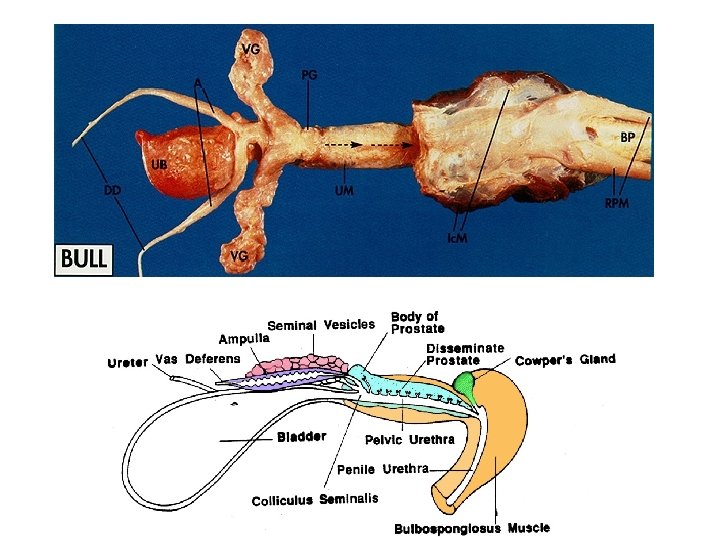
Bull Reproductive Tract

Reproductive Organs of the Bull Rectum Prostate Cowpers Gland Seminal Vesicles Ampulla Bladder Retractor Penis Muscle Vas Deferens Sigmoid Flexure Glans Penis Caput Epididymis Scrotum Testis Gubernaculum Cauda Epididymis

Reproductive Organs of the Bull

Reproductive Organs of the Bull Testis

Bull Testis Tunica Vaginalis • parietal • visceral Tunica Albuginea

Bull Tract • • • Thermosensor Radiator Protective sac Scrotum

Bull Tract Longitudinal Section Cross Section

Scrotal Cross Section testicular parenchyma mediastinum

Testis Testicular Parenchyma Mediastinum
")
Structure of the Testis Seminiferous Tubule Tunica Albuginea Rete Testis (within the mediastinum)

Mediastinum Rete Testis Seminiferous Tubule The Testis
")
Structure of the Testis Seminiferous Tubule Tunica Albuginea Rete Testis (within the mediastinum)

Structure of the Testis Caput Epididymis Spermatic Cord Vas Deferens • Vasectomy Vas Efferentia 6 -12 tubules Corpus Epididymis Cauda Epididymis

Structure of the Testis Caput Epididymis Spermatic Cord Vas Deferens • Vasectomy Vas Efferentia 6 -12 tubules Seminiferous Tubule Tunica Albuginea Corpus Epididymis Rete Testis (within the mediastinum) Cauda Epididymis

Spermatic Chord • Vascular, lymphatic and nerves Pampiniform Plexus • Heat exchanger • Houses cremaster muscle Testicular Artery Pampiniform Plexus • Counter Current Exchange Ø 39°C - 34°C Ø 70 ng T/ml - 4. 8 ng T/ml

Testis Arterial Pulse

Spermatic Chord Vas Deferens Cremaster Muscle Pampiniform Plexus Parietal Tunica Vaginalis

Bull Tract Tunica Dartos

Contraction of the Tunica Dartos Testis Tunica Dartos • Smooth muscle • Androgen dependent (Testosterone)

Bull Tract Gubernaculum

Bull Tract Pelvic Genitalia

Pelvic Genitalia of the Bull Pelvic Urethra Muscle

Bulb of the Penis

Cross Section of Bull Penis

Penis Cross Section Corpus Cavernosum Urethra Corpus Spongiosum

Arterial Supply

Glans Penis

Glans Penis
- Slides: 42