Reproduction and Development The Reproductive System Human Development


- Slides: 62
Reproduction and Development The Reproductive System Human Development
The Reproductive System Sexual Development The Male Reproductive system The Female Reproductive System The Menstrual Cycle Fertilization
The Reproductive System • Reproductive system=group of specialized organs that carries out the function of reproduction – Single most important system to the continuation of a species • Organism produces the next generation
The Reproductive System • Produces, stores, nourishes, and releases specialized sex cells known as gametes – Released in ways that make possible the fusion of egg and sperm • Zygote=fertilized egg cell
Sexual Development • Human male and female embryo are identical in the first six weeks after fertilization – Major changes occur in seventh week • Testes begin to produce steroid hormones known as androgens – Tissues of embryo respond by developing into male reproductive organs
Sexual Development • If the embryo is female – Ovaries produce steroid hormones known as estrogens • Tissues of embryo develop in a pattern that produces female reproductive organs
Sexual Development • After birth, testes and ovaries continue to produce small amounts of androgens and estrogens – Cont to influence the development of the reproductive organs • Neither testes nor ovaries can produce active reproductive cells
Sexual Development • Puberty – Period of rapid growth and sexual maturation • Reproductive system becomes fully functional – Completion • Gonads are fully developed – Onset varies • Anytime between 9 to 15 – Earlier in females
Sexual Development • Puberty – Begins with a change in the hypothalamus • Part of brain that regulates the secretions of the pituitary gland • Causes pituitary gland to produce increased levels of FSH and LH
The Male Reproductive System • Reproductive organs – Testes • Just before birth the testes descent through a canal into an external sac called the scrotum – 1 to 3 below the internal body temp • Clusters of hundreds of tiny tubules called seminiferous tubules – Seminiferous=seed bearing » Sperm are produced – Tightly coiled and twisted together to form a compact organ • As FSH and LH are released, they stimulate the testes to make testosterone
The Male Reproductive System • Testosterone – Cells that can respond are found all over the body – Produces a number of secondary sex characteristics • Appear in males at puberty – – Voice deeper Facial hair Chest broadens Easier to develop muscles
The Male Reproductive System • Sperm – FSH and testosterone stimulate the development of sperm – Puberty is complete when large numbers of sperm have been produced in the testes – Reproductive system now function • Can produce and release active sperm
Sperm Development • Derived from special cells within the testes that go through the process of meiosis to for the haploid nuclei found in mature sperm • Consists of – Head-contains the highly condensed nucleus – Midpiece-packed with energy-releasing mitochondria – Tail-propels the cell forward – Cap- enzyme vital to the process of fertilization
Sperm Development • Developed sperm travel from the seminiferous tubules into the epididymis – Fully mature and stored – Forced into vas deferens • Passes into the abdominal cavity – 3 glands produce seminal fluid » Semen – Merges with the urethra • Leads to the outside of body through the penis
Sperm Development • 100 to 200 million sperm are present in 1 milliliter of semen – 5 million sperm per drop
Sperm Release • When male is sexually excited, autonomic nervous system prepares the male organs to deliver sperm – Sperm are ejected from the penis by the contractions of smooth muscles lining the vas deferens • Ejaculation – Not completely voluntary » ~2 to 3 milliliters of sperm
Female Reproductive System • Primary reproductive organs – Ovaries • Located in abdominal cavity • Produce one egg (ovum) per month – Prepare to nourish a developing embryo
Female Reproductive System • Puberty – Starts with changes in the hypothalamus • Cause release of FSH and LH from the pituitary gland – Same hormones found in males » Target cells and effects different • FSH-stimulates cells within the ovaries to produce hormones known as estrogens – Cause reproductive system to complete its development – Produce secondary sex characteristics » Breast development » Enlargement of reproductive organs » Widening of hips » Hair growth under arms and in pubic areas
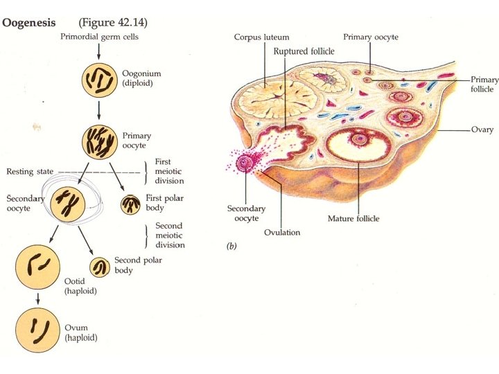
Ova Development • Each ovary contains about 400, 000 primary follicles – Clusters of cells surrounding a single ovum – Prepare a single ovum for release • mature within their follicles – Female born with about 400, 000 immature ova in her ovaries » Fewer than 500 will actually be released » FSH influences ovum to complete meiosis and increase in size • As nutrients are added to its cytoplasm
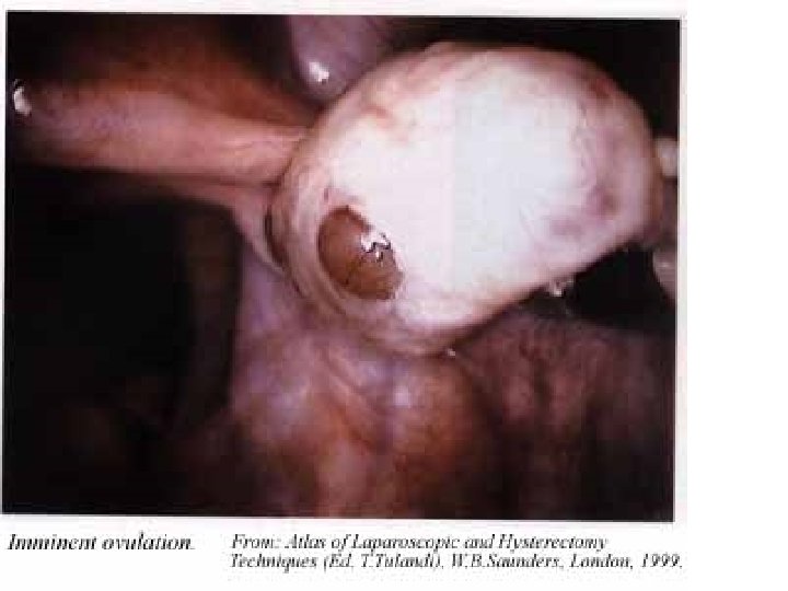
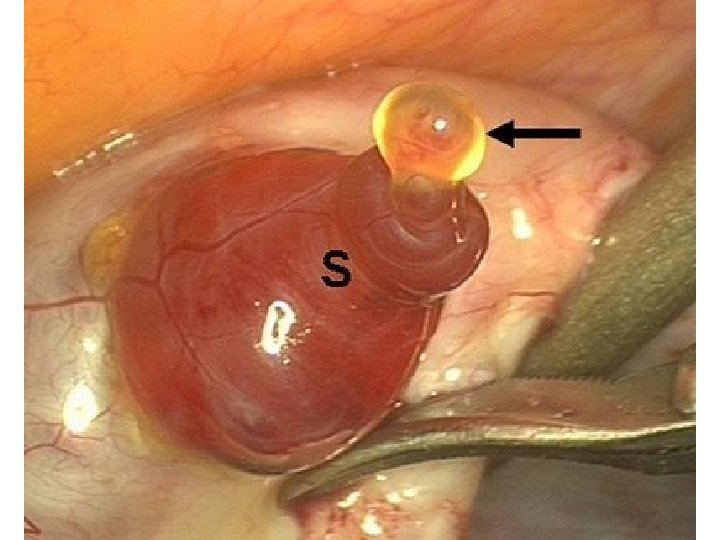
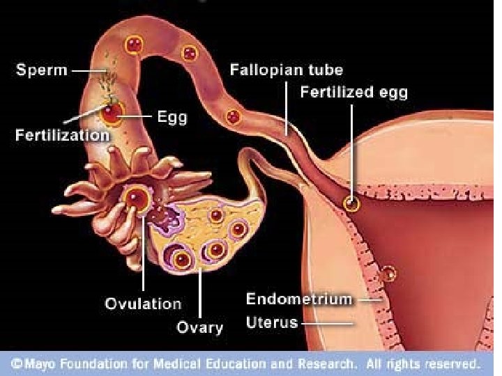
Ovulation • When follicle has completely matured, the ovum is released – Ovulation • Follicle literally ruptures – Ovum is swept from the surface of the ovary into the opening of one of the two Fallopian tubes – Ovum moves through the fluid-filled Fallopian tubes, pushed along by cilia » Egg can be fertilized during this journey » After a few days, ovum arrives at the uterus » Uterus opens into vagina which leads to the outside of the body
Ovulation • Begins at puberty and continues till late 40’s – Menopause occurs • Follicle development no longer occurs and female is no longer capable of bearing a child
The Menstrual Cycle • The interaction of the reproductive system and the endocrine system • Complex series of periodic events • In females • About 28 days • Latin, mensis=month
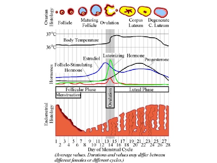
The Menstrual Cycle • Controlled by hormones • The development and the release of an egg for fertilization and the preparation of the uterus to receive a fertilized egg – Egg not fertilized…it is discharged along with the lining of the uterus • 4 phases – Follicle phase – Ovulation – Luteal phase – menstruation
Follicle Phase • Begins when estrogen levels are low • Hypothalamus reacts by producing a releasing hormone that acts on the pituitary gland – Stimulates FSH and LH into the blood – Travel through the circulatory system to the ovaries where they cause a follicle to develop to maturity • As follicle develops, cells around ovum enlarge and produce more estrogen – Estrogen level rise dramatically • Causes lining of uterus to thicken • 10 days
Ovulation Shortest of menstrual cycle Occurs at 14 days Lasts 3 -4 days Hypothalamus send a large amount of releasing hormone to the pituitary gland • Pituitary gland produces a sudden rush of FSH and LH • • – Dramatic effect on the follicle • It ruptures • Mature ovum is released
Luteal Phase • 10 -14 days after the completion of the last menstrual cycle • Ruptured follicle is now known as corpus luteum – Continues to release estrogen – Begins to release progesterone • Adds finishing touches to uterus lining – Blood supply is increased – Tissue matures – Lining is fully prepared to accept a fertilized ovum • First 2 days of luteal phase, chances of getting pregnant is greatest!
Luteal Phase • Ovum is fertilized by a sperm – Resulting zygote starts to divide • After several divisions…ball of cells will implant itself in the lining of the uterus • Within a few days of implantation…the uterus and growing embryo will release hormones that keep the corpus luteum functioning for several weeks – Allows the lining of the uterus to nourish and protect the developing embryo
Menstruation • Fertilization does not occur • Within 2 -3 days of ovulation – Ovum passes through the uterus without implantation – Corpus luteum begins to disintegrate – As old follicle breaks down, it releases les and less estrogen and progesterone • Hormone level in blood decreases
Menstruation • When estrogen levels fall below a certain point, lining of uterus begins to detach from the uterine wall – This tissue along with blood from the unfertilized ovum are discharged through the vagina in the last phase of the menstrual cycle=menstruation • 3 -7 days, average=4 – At end, new cycle begins
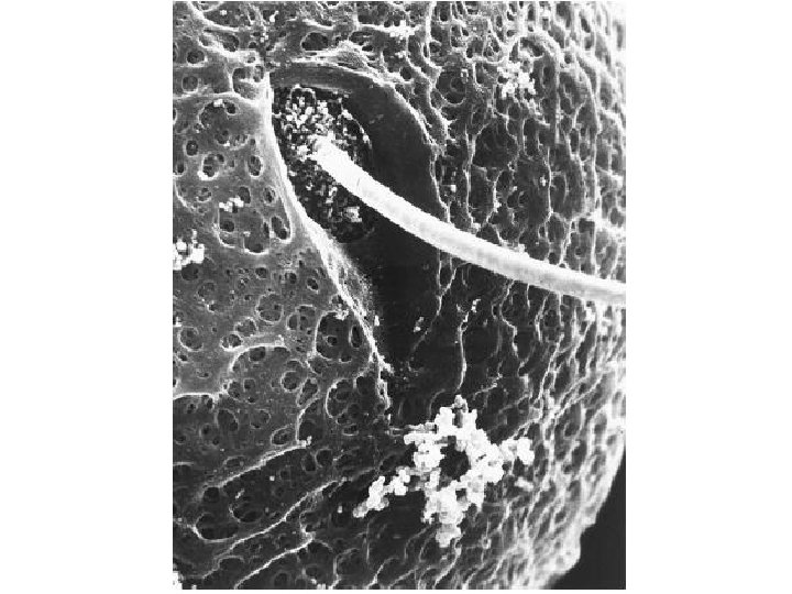
Fertilization • Sperm must be present in the female reproductive tract – Fallopian tube • Sperm are released during sexual intercourse – Semen is ejaculated through the penis into the vagina • To cervix – Sperm swim actively through the uterus and up into the Fallopian tubes • Only a few of the hundreds of millions of sperm will reach the ovum – Only a single sperm will fertilize the ovum
Fertilization • Ovum is surrounded by a dense protective layer that contains receptor sites to which sperm bind – Binding causes a vesicle in the sperm head to rupture and release enzymes that break down the protective layer and form a pathway through which the sperm nucleus can reach the ovum • Cell membrane of the ovum changes – Preventing other sperm from entering the cell
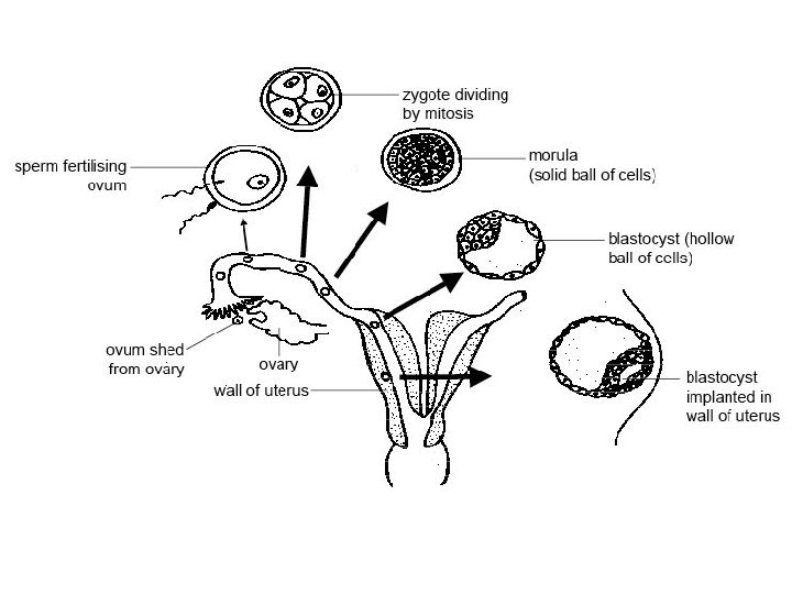
Fertilization • Zygote=fertilized egg – After two haploid nuclei fuse to form a single diploid nucleus • Goes through several rounds of cell division – Ball of cells will attach itself to the wall of the uterus and begin to grow into an embryo
Human Development Early Development Later Development Childbirth After Childbirth
Human Development • Begins when an ovum is fertilized – Cell the size of this. Will undergo cell divisions that will result in the formation of a new human being
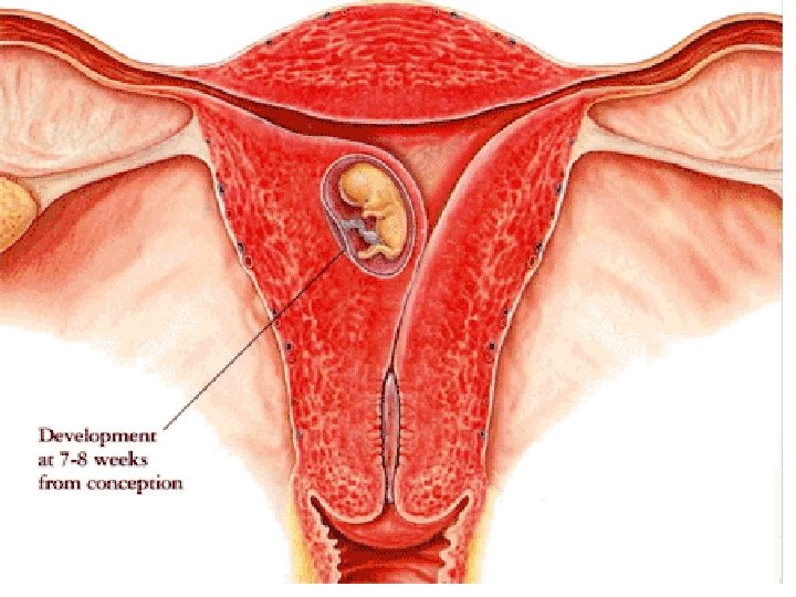
Early Development • Mitotic cell divisions of the zygote=cleavages – Take place while the zygote is still in a Fallopian tube – 4 days after fertilization • Embryo is solid ball of about 50 cells – Morula • As embryo grows, a fluid-filled cavity forms in the center, transforming it into a hollow structure known as a blastocyst – 6 -7 days after fertilization • Blastocyst attaches itself to the wall of the uterus and begins to grow inward – implantation
Early Development • Cluster of cells forms within the cavity of the blastocyst – Sorts itself into 2 layers • Then produce a 3 rd layer – Gastrulation » Result is the formation of three cell layers • Ectoderm • Mesoderm • Endoderm • Primary germ layers • Organs and tissues will be formed from these
Early Development • During implantation – Outer layer of cells of the blastocyst produces two membranes that surround, protect, and nourish developing embryo • Amnion • chorion
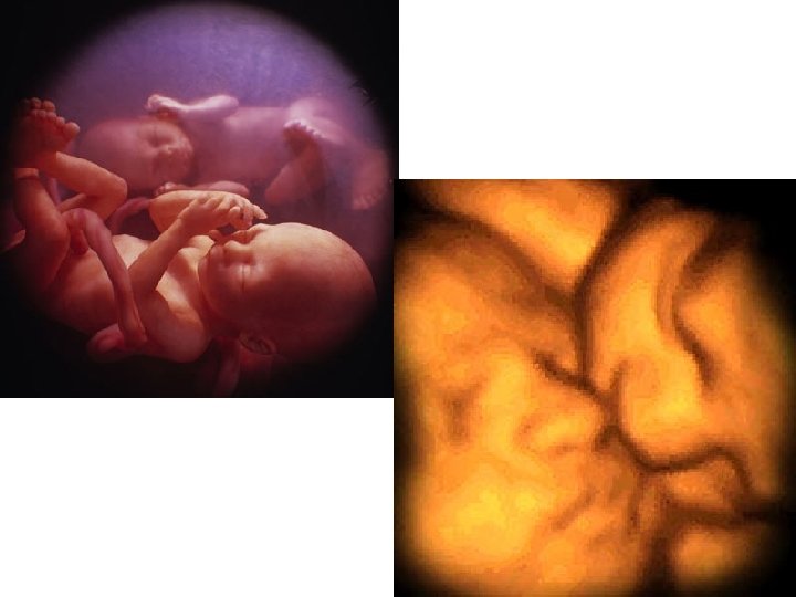
Early Development • End of 3 rd week of development – Nervous system and digestive system have begun to form – Chorion has grown into the uterine tissue • Forms placenta – Connection between the mother and developing embryo » Needs a supply of nutrients and oxygen » Means of eliminating carbon dioxide and metabolic wastes – Blood of mother and embryo flow past each other, but do not mix » Thin membrane for diffusion of gases, food, waste
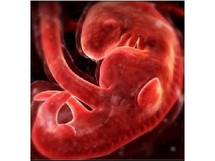
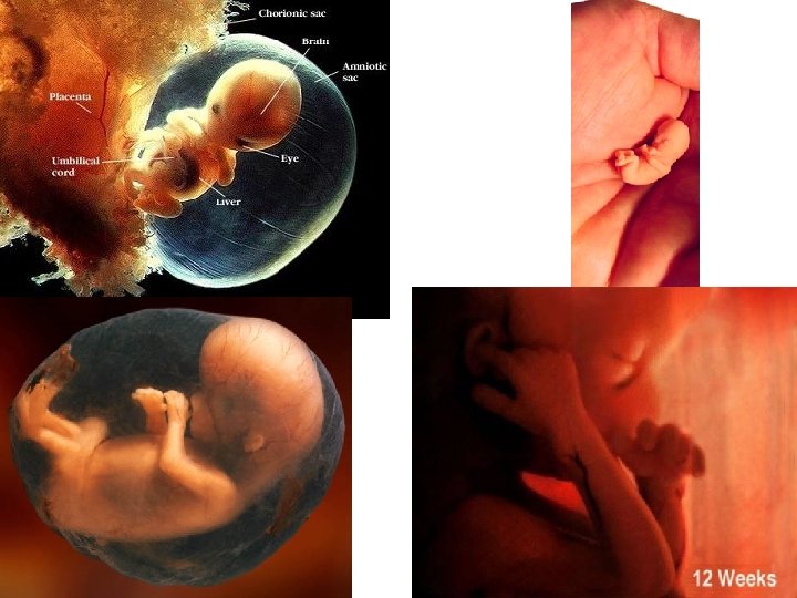
Early Development • 8 weeks – Embryo is known as a fetus • 3 months – Most of major organs and tissues of the fetus are fully formed – Umbilical cord forms • Contains two arteries, one vein • Connects the fetus to the placenta – Muscular system well developed • Fetus may begin to move • Show signs of reflexes – Fetus about 9 cm long – Mass of about 15 grams – Amnion developed into a fluid-filled sac • Amniotic sac – Cushions and protects the developing fetus
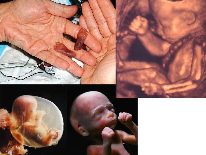
Later Development • During 4 th, 5 th, and 6 th months – Tissues of fetus continue to become more complex and specialized – Skeleton begins to form – Fetal heart beat becomes strong enough to be heard by a stethoscope – Layer of soft hair grows over fetus’s skin – As fetus increases in sizes so does the mothers abdomen • Fetus is about 35 cm in length with a mass of about 700 grams by the end of the 6 th month
Later Development • Fetus is capable of leading a completely independent existence during the final 3 months – Months are important • Fetus doubles in mass • Lungs and other organs undergo a series of changes that prepare them for life outside the mother – Premature babies (before 8 months) have difficult time breathing because lungs are not fully developed
Childbirth
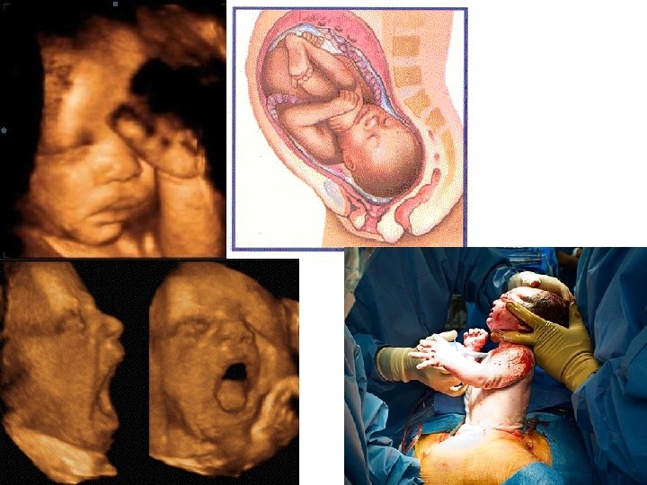
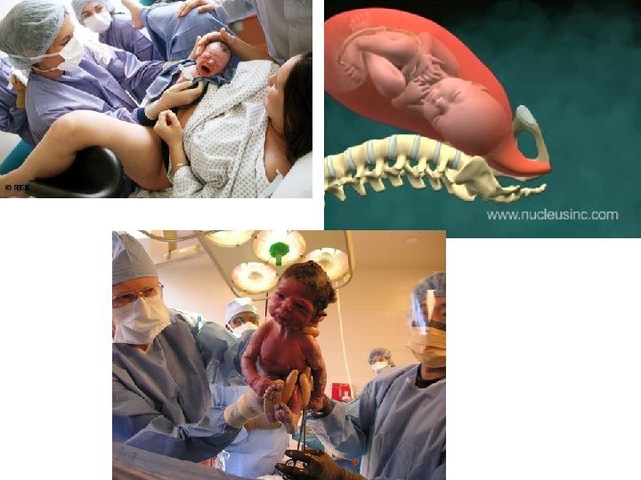
Childbirth • About 9 months after fertilization – End of full-term pregnancy • Fetus is ready for birth • How process is triggered is unknown • Oxytocin is released from the pituitary gland – Effects group of large involuntary muscles that surround the uterus • Muscles are stimulated, begin a series of rhythmic contractions known as labor that expand the opening of the cervix (about 10 cm) to allow the baby to pass through it
Childbirth • As contractions continue they become more powerful and more frequent – Once every 1 -2 min • Last 2 -16 hours • baby is forced toward the vagina • Amniotic sac breaks – Fluid rushes out vagina • Baby forced out of the uterus and the vagina – Usually head first still attached to its mother by the umbilical cord
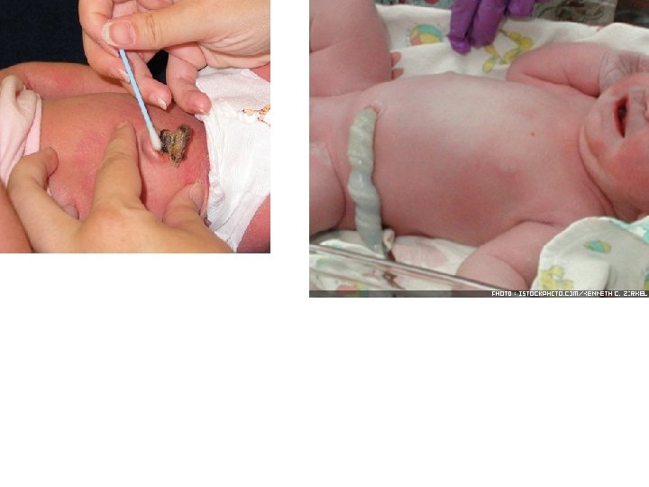
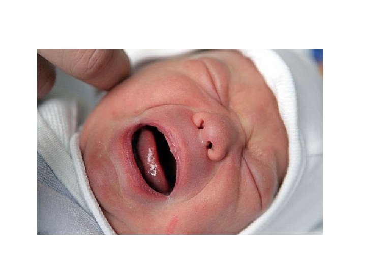
Childbirth • As baby meets outside world – May cough or cry to rid lungs of fluid • Breathing starts almost immediately • Blood supply to placenta begins to dry up • Umbilical cord clamped and cut – Scar=navel • Placenta and amniotic sac are expelled with more contractions – afterbirth
After Childbirth • Within a few hours – Pituitary hormone, prolactin stimulates milk production in the breast tissue – If mom breastfeeds • Milk is always ready when needed • Stops whenever breastfeeding stops – Stimulation of nerve cells in the breast transmit impulses to the hypothalamus • Causes pituitary gland to release nearly 10 times the normal amount of prolactin – Enables milk production