REPAIR WOUND HEALING Repair Wound healing by primary

REPAIR & WOUND HEALING

Repair Wound healing by primary & secondary union Factors influencing wound healing

Healing is the body response to injury in an attempt to restore normal structure and function. Healing involves 2 distinct processes: q Regeneration when healing takes place by proliferation of parenchymal cells and usually results in complete restoration of the original tissues. q Repair when healing takes place by proliferation of connective tissue elements resulting in fibrosis and scarring.

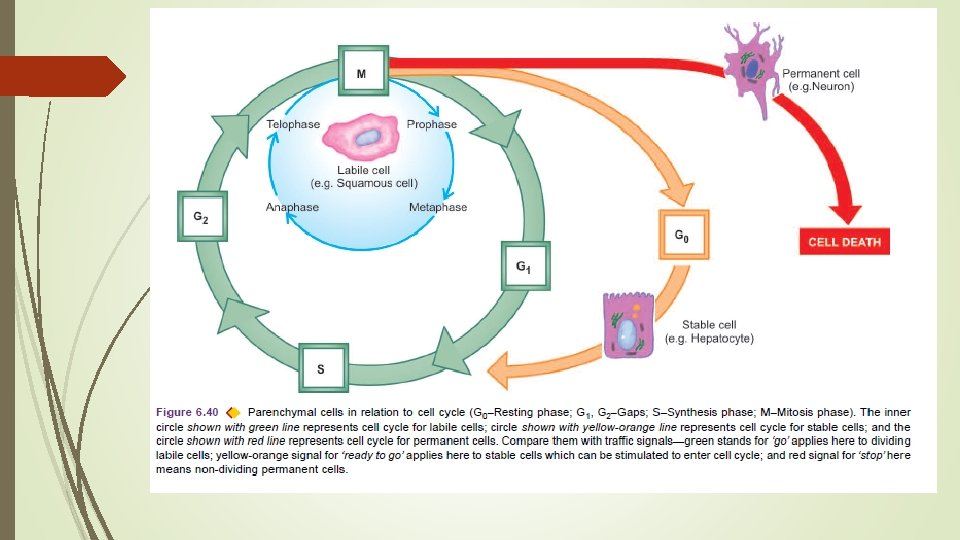
CELL CYCLE Cell cycle is defined as the period between two successive cell divisions and is divided into 4 unequal phases M (mitosis) phase: Phase of mitosis. G 1 (gap 1) phase: The daughter cell enters G 1 phase after mitosis. S (synthesis) phase: During this phase, the synthesis of nuclear DNA takes place. G 2 (gap 2) phase: After completion of nuclear DNA duplication, the cell enters G 2 phase. G 0 (gap 0) phase: This is the quiescent or resting phase of the cell after an M phase.

Not all cells of the body divide at the same pace. Some mature cells do not divide at all while others complete a cell cycle every 1624 hours. The main difference between slowly dividing and rapidly-dividing cells is the duration of G 1 phase.

Depending upon their capacity to divide, the cells of the body can be divided into 3 groups: labile cells, stable cells, and permanent cells. 1. Labile cells. These cells continue to multiply throughout life under normal physiologic conditions. These include: surface epithelial cells of the epidermis, alimentary tract, respiratory tract, urinary tract, vagina, cervix, uterine endometrium, haematopoietic cells of bone marrow and cells of lymph nodes and spleen.

2. Stable cells. These cells decrease or lose their ability to proliferate after adolescence but retain the capacity to multiply in response to stimuli throughout adult life. These include: parenchymal cells of organs like liver, pancreas, kidneys, adrenal and thyroid; mesenchymal cells like smooth muscle cells, fibroblasts, vascular endothelium, bone and cartilage cells. 3. Permanent cells. These cells lose their ability to proliferate around the time of birth. These include: neurons of nervous system, skeletal muscle and cardiac muscle cells.

RELATIONSHIP OF PARENCHYMAL CELLS WITH CELL CYCLE. 1. Labile cells which are continuously dividing cells remain in the cell cycle from one mitosis to the next. 2. Stable cells are in the resting phase (G 0) but can be stimulated to enter the cell cycle. 3. Permanent cells are non-dividing cells which have left the cell cycle and die after injury.
")
Regeneration of any type of parenchymal cells involves the following 2 processes: i) Proliferation of original cells from the margin of injury with migration so as to cover the gap. ii) Proliferation of migrated cells with subsequent differentiation and maturation so as to reconstitute the original tissue.

REPAIR Repair is the replacement of injured tissue by fibrous tissue. Two processes are involved in repair: 1. Granulation tissue formation; and 2. Contraction of wounds. Repair response takes place by participation of mesenchymal cells (consisting of connective tissue stem cells, fibrocytes and histiocytes), endothelial cells, macrophages, platelets, and the parenchymal cells of the injured organ.

1. Granulation Tissue Formation The term granulation tissue derives its name from slightly granular and pink appearance of the tissue. Each granule corresponds histologically to proliferation of new small blood vessels which are slightly lifted on the surface by thin covering of fibroblasts and young collagen.
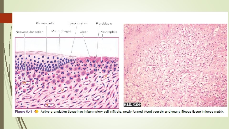

3 phases in the formation of granulation tissue : 1. PHASE OF INFLAMMATION. Following trauma, blood clots at the site of injury. There is acute inflammatory response with exudation of plasma, neutrophils and some monocytes within 24 hours. 2. PHASE OF CLEARANCE. Combination of proteolytic enzymes liberated from neutrophils, autolytic enzymes from dead tissues cells, and phagocytic activity of macrophages clear off the necrotic tissue, debris and red blood cells.

3. PHASE OF INGROWTH OF GRANULATION TISSUE. consists of 2 main processes: Ø angiogenesis or neovascularisation, and Ø fibrogenesis.
 Angiogenesis (neovascularisation). Formation of new blood vessels at the site of injury takes")
i) Angiogenesis (neovascularisation). Formation of new blood vessels at the site of injury takes place by proliferation of endothelial cells from the margins of severed blood vessels. Initially, the proliferated endothelial cells are solid buds but within a few hours develop a lumen and start carrying blood. The newly formed blood vessels are more leaky, accounting for the oedematous appearance of new granulation tissue. Soon, these blood vessels differentiate into muscular arterioles, thin-walled venules and true capillaries.
 Vascular endothelial growth")
Angiogenesis takes place under the influence of following factors: a) Vascular endothelial growth factor (VEGF) b) Platelet-derived growth factor (PDGF), transforming growth factor-β (TGF-β), basic fibroblast growth factor (b. FGF) and surface integrins
 Fibrogenesis. The newly formed blood vessels are present in an amorphous ground substance")
ii) Fibrogenesis. The newly formed blood vessels are present in an amorphous ground substance or matrix. The new fibroblasts originate from fibrocytes as well as by mitotic division of fibroblasts. Collagen fibrils begin to appear by about 6 th day. As maturation proceeds, more and more of collagen is formed while the number of active fibroblasts and new blood vessels decreases. This results in formation of inactive looking scar known as cicatrisation.

2. Contraction of Wounds The wound starts contracting after 2 -3 days and the process is completed by the 14 th day. During this period, the wound is reduced by approximately 80% of its original size. Contracted wound results in rapid healing since lesser surface area of the injured tissue has to be replaced.

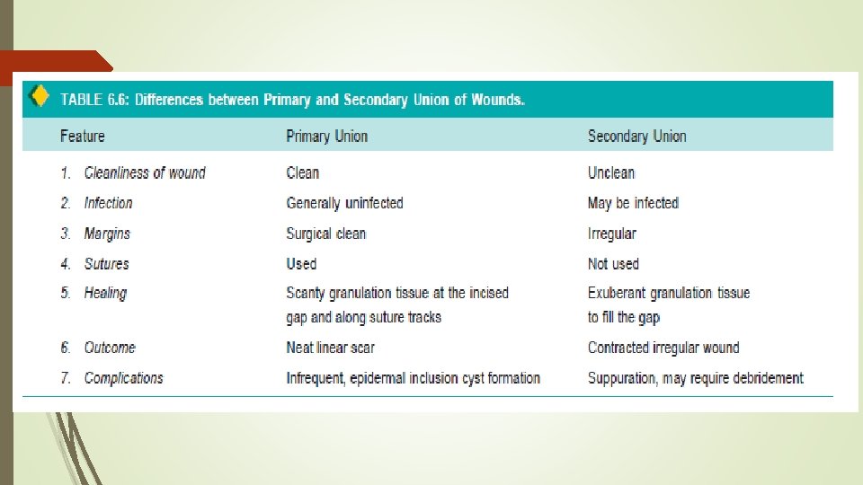
WOUND HEALING Healing of skin wounds provides a classical example of combination of regeneration and repair. Wound healing can be accomplished in one of the following two ways: Ø Healing by first intention (primary union) Ø Healing by second intention (secondary union).
 This is defined as healing of a wound")
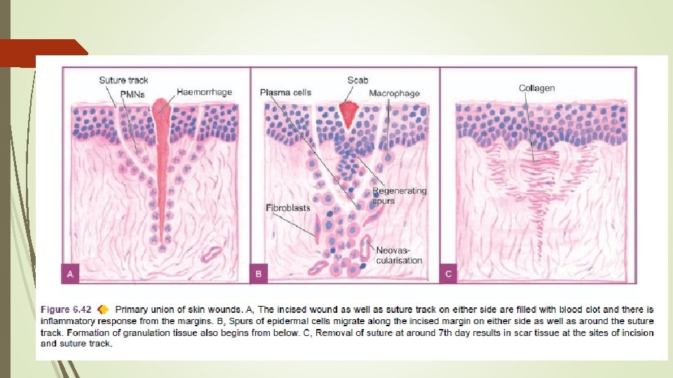
Healing by First Intention (Primary Union) This is defined as healing of a wound which has the following characteristics: i) clean and uninfected; ii) surgically incised; iii) without much loss of cells and tissue; and iv) edges of wound are approximated by surgical sutures.

Sequence of events in primary union : 1. Initial haemorrhage. Immediately after injury, the space between the approximated surfaces of incised wound is filled with blood which then clots and seals the wound against dehydration and infection. 2. Acute inflammatory response. This occurs within 24 hours with appearance of polymorphs from the margins of incision. By 3 rd day, polymorphs are replaced by macrophages.

3. Epithelial changes. The basal cells of epidermis from both the cut margins start proliferating and migrating towards incisional space in the form of epithelial spurs. A well approximated wound is covered by a layer of epithelium in 48 hours. The migrated epidermal cells separate the underlying viable dermis from the overlying necrotic material and clot, forming scab which is cast off. The basal cells from the margins continue to divide. By 5 th day, a multilayered new epidermis is formed which is differentiated into superficial and deeper layers.

4. Organisation. By 3 rd day, fibroblasts also invade the wound area. By 5 th day, new collagen fibrils start forming which dominate till healing is completed. In 4 weeks, the scar tissue with scanty cellular and vascular elements, a few inflammatory cells and epithelialised surface is formed.

5. Suture tracks. When sutures are removed around 7 th day, much of epithelialised suture track is avulsed and the remaining epithelial tissue in the track is absorbed. Sometimes the suture track gets infected (stitch abscess), or the epithelial cells may persist in the track (implantation or epidermal cysts).
 This is defined as healing of a wound")
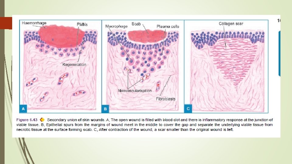
Healing by Second Intention (Secondary Union) This is defined as healing of a wound having the following characteristics: i) open with a large tissue defect, at times infected; ii) having extensive loss of cells and tissues; and iii) the wound is not approximated by surgical sutures but is left open.

The basic events in secondary union are similar to primary union but differ in having a larger tissue defect which has to be bridged. Healing takes place from the base upwards as well as from the margins inwards. The healing by second intention is slow and results in a large, at times ugly, scar as compared to rapid healing and neat scar of primary union.

Sequence of events in secondary union: 1. Initial haemorrhage. As a result of injury, the wound space is filled with blood and fibrin clot which dries. 2. Inflammatory phase. There is an initial acute inflammatory response followed by appearance of macrophages which clear off the debris as in primary union.

3. Epithelial changes. As in primary healing, the epidermal cells from both the margins of wound proliferate and migrate into the wound in the form of epithelial spurs till they meet in the middle and re-epithelialise the gap completely. Proliferating epithelial cells do not cover the surface fully until granulation tissue from base has started filling the wound space. In this way, pre-existing viable connective tissue is separated from necrotic material and clot on the surface, forming scab which is cast off. In time, the regenerated epidermis becomes stratified and keratinised.

4. Granulation tissue. Main bulk of secondary healing is by granulations. The newly-formed granulation tissue is deep red, granular and very fragile. With time, the scar on maturation becomes pale and white due to increase in collagen and decrease in vascularity.

5. Wound contraction. Contraction of wound is an important feature of secondary healing. Due to the action of myofibroblasts present in granulation tissue, the wound contracts to one-third to one-fourth of its original size. Wound contraction occurs at a time when active granulation tissue is being formed.

6. Presence of infection. Bacterial contamination of an open wound delays the process of healing due to release of bacterial toxins that provoke necrosis, suppuration and thrombosis. Surgical removal of dead and necrosed tissue, debridement, helps in preventing the bacterial infection of open wounds.

Complications of Wound Healing 1. Infection of wound due to entry of bacteria delays the healing. 2. Implantation (epidermal) cyst formation due to persistence of epithelial cells in the wound after healing. 3. Pigmentation. Healed wounds may at times have rust-like colour due to staining with haemosiderin. Some coloured particulate material left in the wound may persist and impart colour to the healed wound. 4. Deficient scar formation. due to inadequate formation of granulation tissue.

5. Incisional hernia. A weak scar, especially after a laparotomy, may be the site of bursting open of a wound (wound dehiscence) or an incisional hernia. 6. Hypertrophied scars and keloid formation. At times the scar formed is excessive, ugly and painful. Excessive formation of collagen in healing may result in keloid (claw-like) formation, seen more commonly in Blacks. Hypertrophied scars differ from keloid in that they are confined to the borders of the initial wound while keloids have tumour-like projection of connective tissue.

7. Excessive contraction. An exaggeration of wound contraction may result in formation of contractures or cicatrisation e. g. Dupuytren’s (palmar) contracture, plantar contracture and Peyronie’s disease (contraction of the cavernous tissues of penis). 8. Neoplasia. Rarely, scar may be the site for development of carcinoma later e. g. squamous cell carcinoma in Marjolin’s ulcer i. e. a scar following burns on the skin.

Factors Influencing Healing A. LOCAL FACTORS: 1. Infection delays the process of healing. 2. Poor blood supply to wound slows healing e. g. injuries to face heal quickly due to rich blood supply injury to leg with varicose ulcers having poor blood supply heals slowly.

3. Foreign bodies including sutures interfere with healing and cause intense inflammatory reaction and infection. 4. Movement delays wound healing. 5. Exposure to ionising radiation delays granulation tissue formation. 6. Exposure to ultraviolet light facilitates healing. 7. Type, size and location of injury determines whether healing takes place by resolution or organisation.

B. SYSTEMIC FACTORS: 1. Age. Wound healing is rapid in young and somewhat slow in aged and debilitated people due to poor blood supply to the injured area in the latter. 2. Nutrition. Deficiency of constituents like protein, vitamin C (scurvy) and zinc delays the wound healing. 3. Systemic infection delays wound healing. 4. Administration of glucocorticoids has anti-inflammatory effect. 5. Uncontrolled diabetics more prone to develop infections delay in healing. 6. Haematologic abnormalities like defect of neutrophil functions (chemotaxis and phagocytosis), and neutropenia and bleeding disorders slow the process of wound healing.

THANK YOU
- Slides: 42