
Renal Disorders nutritional therapy aspects A Norouzy Assistant

- Slides: 65
Renal Disorders: nutritional therapy aspects A Norouzy Assistant Professor in Clinical Nutrition Mashad Medical School
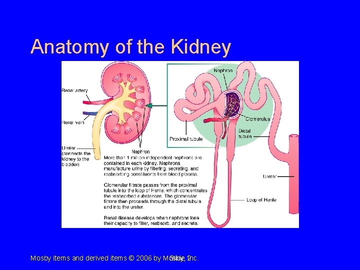
Anatomy of the Kidney Mosby items and derived items © 2006 by Mosby, Slide 2 Inc.
• • Creatinine BUN Urea Urine output Fluid load Urine analysis (U/A) GFR
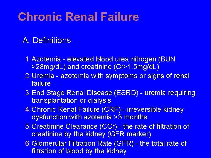
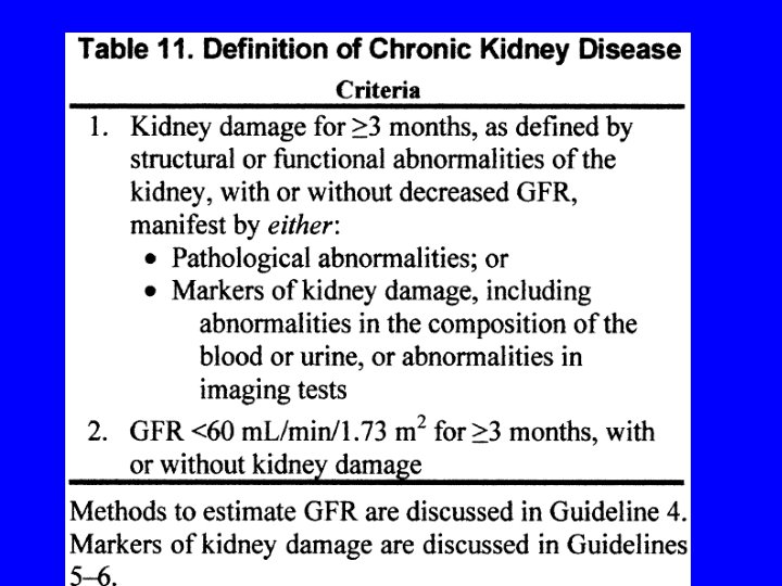
Chronic Renal Failure A. Definitions 1. Azotemia - elevated blood urea nitrogen (BUN >28 mg/d. L) and creatinine (Cr>1. 5 mg/d. L) 2. Uremia - azotemia with symptoms or signs of renal failure 3. End Stage Renal Disease (ESRD) - uremia requiring transplantation or dialysis 4. Chronic Renal Failure (CRF) - irreversible kidney dysfunction with azotemia >3 months 5. Creatinine Clearance (CCr) - the rate of filtration of creatinine by the kidney (GFR marker) 6. Glomerular Filtration Rate (GFR) - the total rate of filtration of blood by the kidney
Acute Renal Failure
Acute Renal Failure Definition • Acute decrement in GFR/creatinine • May heal partially or completely or progress to more severe renal insufficiency, including end -stage renal disease
Acute Renal Failure Classification • Pre-renal (functional) • Renal (structural) • Post-renal (obstruction)
Acute Renal Failure Pre-renal Causes • Intravascular volume depletion – Hemorrhage – Sodium depletion • Redistribution of ECF – “Third space” accumulation – Edematous disorders • Drugs
Acute Tubular Necrosis Classification • Ischemic • Nephrotoxic
Acute Renal Failure Nephrotoxic ATN • Endogenous Toxins – Heme pigments (myoglobin, hemoglobin) – Myeloma light chains • Exogenous Toxins – – Antibiotics (e. g. , aminoglycosides, amphotericin B) Radiocontrast agents Heavy metals (e. g. , cis-platinum, mercury) Poisons (e. g. , ethylene glycol)
Acute Renal Failure Post-renal Causes • Intra-renal Obstruction – Acute uric acid nephropathy – Drugs (e. g. , acyclovir) • Extra-renal Obstruction – Renal pelvis or ureter (e. g. , stones, clots, tumors, papillary necrosis, retroperitoneal fibrosis) – Bladder (e. g. , BPH, neuropathic bladder) – Urethra (e. g. , stricture)
Acute Renal Failure Urine Volume (1) • Anuria (< 100 ml/24 h) – Acute bilateral arterial or venous occlusion – Bilateral cortical necrosis – Acute necrotizing glomerulonephritis – Obstruction (complete) – ATN (very rare)
Acute Renal Failure Urine Volume (2) • Oliguria (100 -500 ml/24 h) – Pre-renal azotemia – ATN • Non-Oliguria (> 500 ml/24 h) – ATN – Obstruction (partial)
CHRONIC RENAL FAILURE
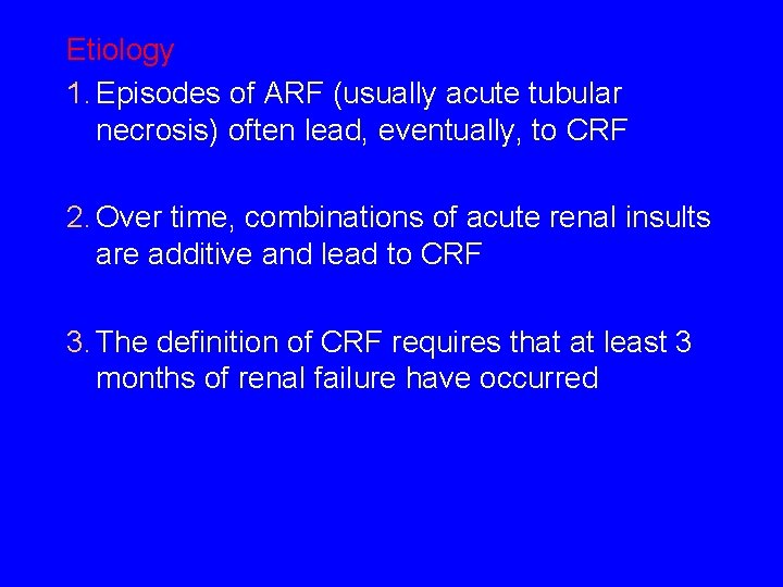
Etiology 1. Episodes of ARF (usually acute tubular necrosis) often lead, eventually, to CRF 2. Over time, combinations of acute renal insults are additive and lead to CRF 3. The definition of CRF requires that at least 3 months of renal failure have occurred
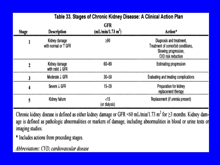
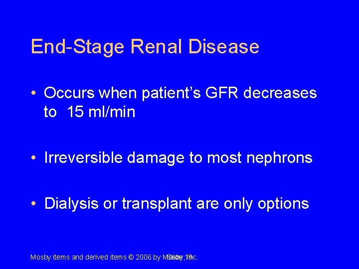
End-Stage Renal Disease • Occurs when patient’s GFR decreases to 15 ml/min • Irreversible damage to most nephrons • Dialysis or transplant are only options Mosby items and derived items © 2006 by Mosby, Slide 19 Inc.
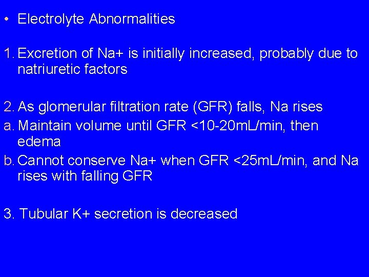
• Electrolyte Abnormalities 1. Excretion of Na+ is initially increased, probably due to natriuretic factors 2. As glomerular filtration rate (GFR) falls, Na rises a. Maintain volume until GFR <10 -20 m. L/min, then edema b. Cannot conserve Na+ when GFR <25 m. L/min, and Na rises with falling GFR 3. Tubular K+ secretion is decreased
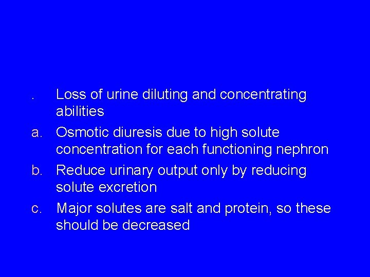
. Loss of urine diluting and concentrating abilities a. Osmotic diuresis due to high solute concentration for each functioning nephron b. Reduce urinary output only by reducing solute excretion c. Major solutes are salt and protein, so these should be decreased
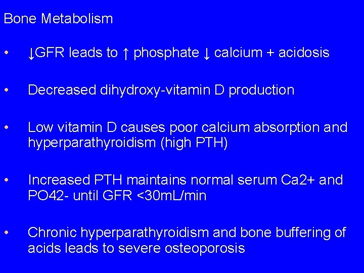
Bone Metabolism • ↓GFR leads to ↑ phosphate ↓ calcium + acidosis • Decreased dihydroxy-vitamin D production • Low vitamin D causes poor calcium absorption and hyperparathyroidism (high PTH) • Increased PTH maintains normal serum Ca 2+ and PO 42 - until GFR <30 m. L/min • Chronic hyperparathyroidism and bone buffering of acids leads to severe osteoporosis
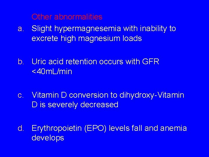
Other abnormalities a. Slight hypermagnesemia with inability to excrete high magnesium loads b. Uric acid retention occurs with GFR <40 m. L/min c. Vitamin D conversion to dihydroxy-Vitamin D is severely decreased d. Erythropoietin (EPO) levels fall and anemia develops
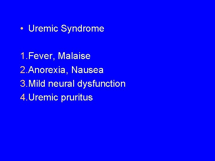
• Uremic Syndrome 1. Fever, Malaise 2. Anorexia, Nausea 3. Mild neural dysfunction 4. Uremic pruritus
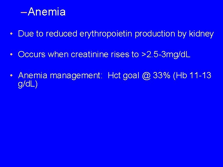
– Anemia • Due to reduced erythropoietin production by kidney • Occurs when creatinine rises to >2. 5 -3 mg/d. L • Anemia management: Hct goal @ 33% (Hb 11 -13 g/d. L)
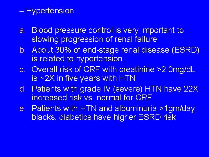
– Hypertension a. Blood pressure control is very important to slowing progression of renal failure b. About 30% of end-stage renal disease (ESRD) is related to hypertension c. Overall risk of CRF with creatinine >2. 0 mg/d. L is ~2 X in five years with HTN d. Patients with grade IV (severe) HTN have 22 X increased risk vs. normal for CRF e. Patients with HTN and albuminuria >1 gm/day, blacks, diabetics have higher ESRD risk
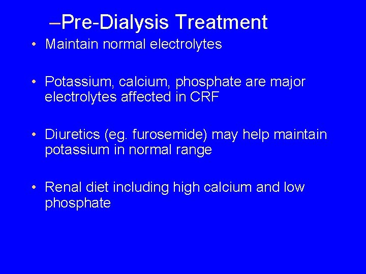
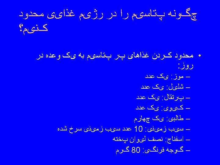
–Pre-Dialysis Treatment • Maintain normal electrolytes • Potassium, calcium, phosphate are major electrolytes affected in CRF • Diuretics (eg. furosemide) may help maintain potassium in normal range • Renal diet including high calcium and low phosphate
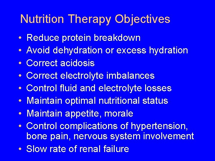
Nutrition Therapy Objectives • • Reduce protein breakdown Avoid dehydration or excess hydration Correct acidosis Correct electrolyte imbalances Control fluid and electrolyte losses Maintain optimal nutritional status Maintain appetite, morale Control complications of hypertension, bone pain, nervous system involvement • Slow rate of renal failure
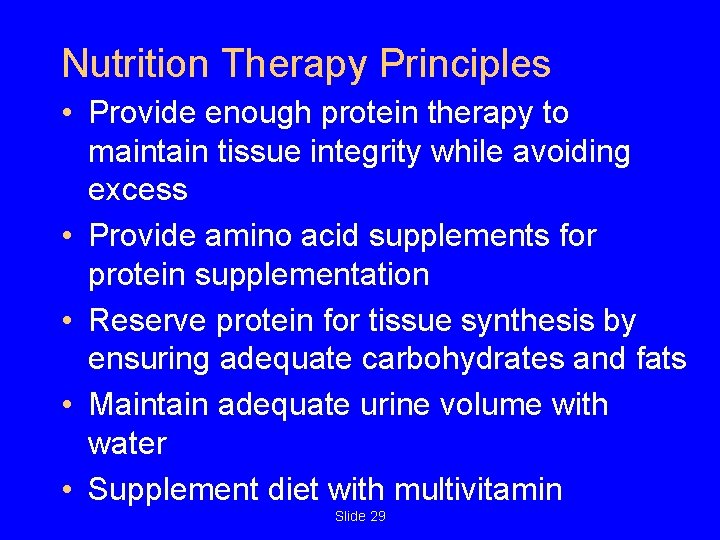
Nutrition Therapy Principles • Provide enough protein therapy to maintain tissue integrity while avoiding excess • Provide amino acid supplements for protein supplementation • Reserve protein for tissue synthesis by ensuring adequate carbohydrates and fats • Maintain adequate urine volume with water • Supplement diet with multivitamin Slide 29
Hemodialysis Patient: Objectives for Diet • Maintain protein and energy balance • Prevent dehydration or fluid overload • Maintain normal serum potassium and sodium levels • Maintain acceptable phosphate and calcium levels Mosby items and derived items © 2006 by Mosby, Slide 30 Inc.
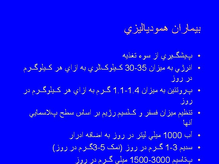
Hemodialysis Patient: Other Dietary Concerns • Avoid protein-energy malnutrition via careful calculation of protein allowance • Maintain body mass index within upper 50 th percentile via generous amounts of carbohydrates and some fats • Fluid intake: 1000 ml/day, plus amount equal to urine output • Limit sodium: 1000 -3000 mg/day • Limit potassium: 1500 -3000 mg/day • Supplement of water soluble vitamins
Peritoneal Dialysis • Performed at home • Patient introduces dialysate solution directly into peritoneal cavity 4 -5 times/day • Surgical insertion of permanent catheter is required • Disposable bag containing dialysate solution is attached to catheter • Diet is more liberal than with hemodialysis
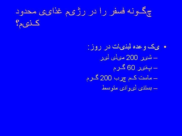
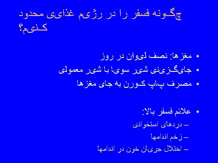
Peritoneal Dialysis: Nutritional Therapy • Increase protein intake to 1. 2 -1. 5 g/kg body weight • Limit phosphorus to 1200 -1500 mg/day • Increase potassium via wide variety of fruits and vegetables • Encourage liberal fluid intake • Avoid sweets and fats • Maintain lean body weight Slide 33
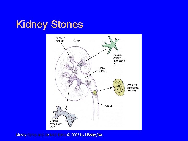
Kidney Stones Mosby items and derived items © 2006 by Mosby, Slide 54 Inc.
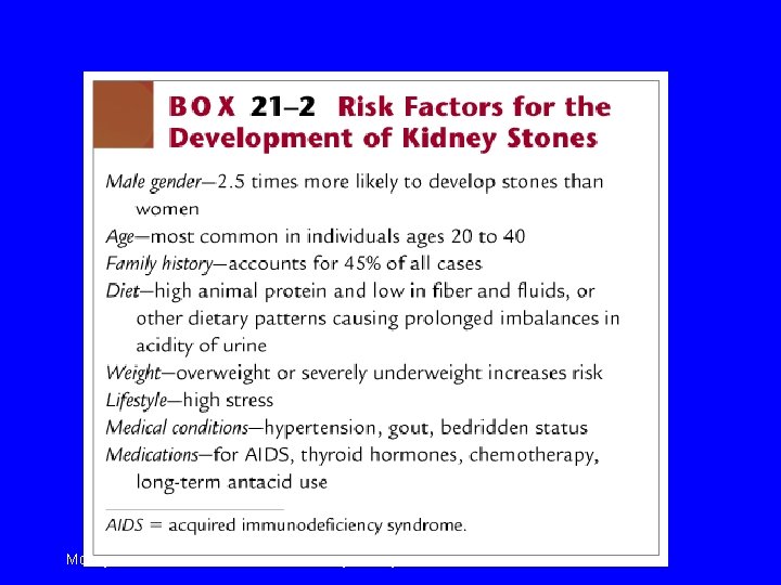
Mosby items and derived items © 2006 by Mosby, Slide 55 Inc.
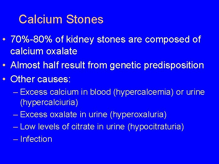
Calcium Stones • 70%-80% of kidney stones are composed of calcium oxalate • Almost half result from genetic predisposition • Other causes: – Excess calcium in blood (hypercalcemia) or urine (hypercalciuria) – Excess oxalate in urine (hyperoxaluria) – Low levels of citrate in urine (hypocitraturia) – Infection
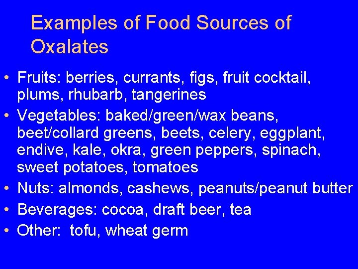
Examples of Food Sources of Oxalates • Fruits: berries, currants, figs, fruit cocktail, plums, rhubarb, tangerines • Vegetables: baked/green/wax beans, beet/collard greens, beets, celery, eggplant, endive, kale, okra, green peppers, spinach, sweet potatoes, tomatoes • Nuts: almonds, cashews, peanuts/peanut butter • Beverages: cocoa, draft beer, tea • Other: tofu, wheat germ
Struvite Stones • Composed of magnesium ammonium phosphate • Mainly caused by urinary tract infections rather than specific nutrient • No diet therapy is involved • Usually removed surgically
Other Stones • Cystine stones – Caused by genetic metabolic defect – Occur rarely • Xanthine stones – Associated with treatment for gout and family history of gout – Occur rarely
Kidney Stones: Symptoms and Treatment • Clinical symptoms: severe pain, other urinary symptoms, general weakness, and fever • Several considerations for treatment – Fluid intake to prevent accumulation of materials – Dietary control of stone constituents – Achievement of desired p. H of urine via medication – Use of binding agents to prevent absorption of stone elements – Drug therapy in combination with diet therapy
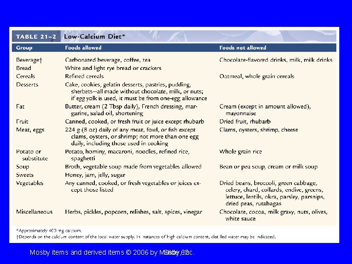
Nutrition Therapy: Calcium Stones • Low-calcium diet (approx. 400 mg/day) recommended for those with supersaturation of calcium in the urine and who are not at risk for bone loss • If stone is calcium phosphate, sources of phosphorus (meats, legumes, nuts) are controlled • Fluid intake increased • Sodium intake decreased • Fiber foods high in phytates increased
Low-Calcium Diet Mosby items and derived items © 2006 by Mosby, Slide 62 Inc.
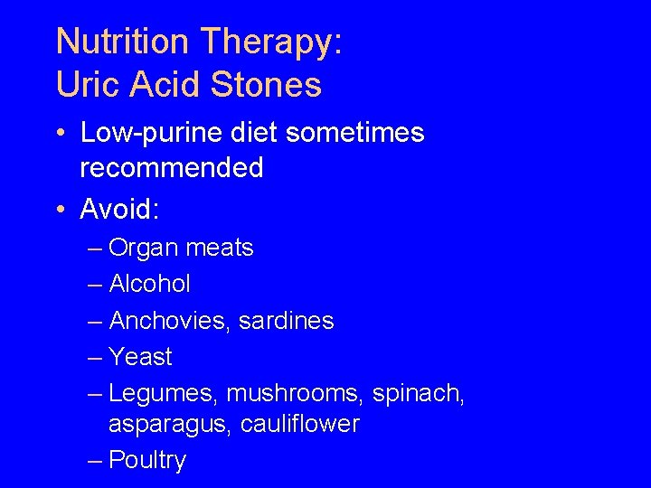
Nutrition Therapy: Uric Acid Stones • Low-purine diet sometimes recommended • Avoid: – Organ meats – Alcohol – Anchovies, sardines – Yeast – Legumes, mushrooms, spinach, asparagus, cauliflower – Poultry
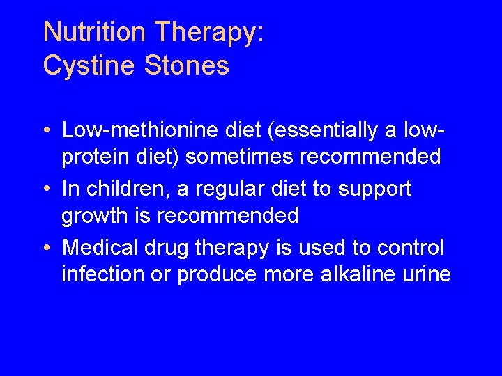
Nutrition Therapy: Cystine Stones • Low-methionine diet (essentially a lowprotein diet) sometimes recommended • In children, a regular diet to support growth is recommended • Medical drug therapy is used to control infection or produce more alkaline urine
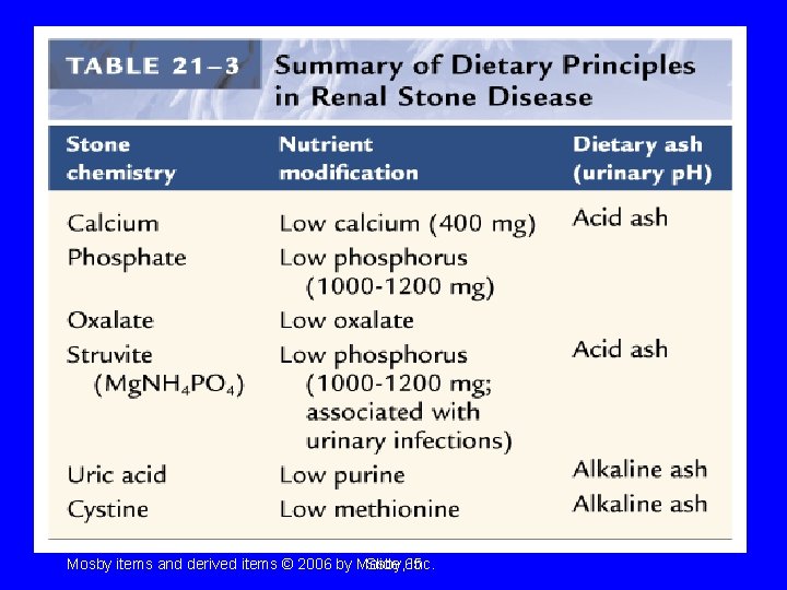
General Dietary Principles: Kidney Stones Mosby items and derived items © 2006 by Mosby, Slide 65 Inc.