Rehabilitation of Neurologic Diseases Stroke Sudden focal sometimes

Rehabilitation of Neurologic Diseases
 neurologic deficit secondary to occlusion or rupture of")
Stroke • Sudden focal (sometimes global) neurologic deficit secondary to occlusion or rupture of blood vessels supplying the brain • Symptoms > 24 hours = stroke • Symptoms < 24 hours = transient ischemic attack (TIA) • Reversible ischemic neurologic deficit (RIND) = (this term is no longer used)

RISK FACTORS Nonmodifiable: • Age—single most important risk factor for stroke worldwide; after age 55, incidence increases for both males and females • Risk more than doubles each decade after age 55 • Sex ( male > female) • Race ( African Americans 2 x> whites > Asians) • Family history (Hx) of stroke
 risk factors: • Hypertension—probably the most important modifiable risk factor for both")
Modifiable (treatable) risk factors: • Hypertension—probably the most important modifiable risk factor for both ischemic and hemorrhagic stroke; increases risk by sevenfold • History of TIA/prior stroke (~ 5% of patients with TIA will develop a completed stroke within 1 month if untreated) • Heart disease (Dz. ) -Congestive heart failure (CHF) and coronary artery disease (CAD): increases risk by twofold – Valvular heart Dz. and arrhythmias atrial fibrillation (A. Fib. )—increases risk of embolic stroke - A. Fib. : fivefold increase risk • Diabetes—twofold increase in risk; unfortunately, good blood sugar control has not been shown to alter the risk of stroke • Cigarette smoking • Carotid stenosis (and carotid bruit); risk of stroke decreases with carotid endarterectomy (CEA) on selected symptomatic patients (> 70% stenosis)
—considerable increase in risk")
• ETOH abuse/cocaine use • High-dose estrogens (birth control pills)—considerable increase in risk when linked to cigarette smoking • Systemic diseases associated with hypercoagulable states – – – Elevated RBC count, hematocrit, fibrinogen – Protein S and C deficiency – Sickle-cell anemia – Cancer • Hyperlipidemia—several clinical trials have shown a reduction in stroke with use of cholesterol reducing agents (~ 30% reduction risk of stroke with use of HMG-Co. A reductase inhibitors) • Migraine headaches • Sleep apnea • Patent Foramen Ovale • [Obesity/sedentary life style (no clear relationship with increased risk of stroke)]

TYPES OF STROKE

Diagnosis • Clinical • MRI scan • CT scan

Treatment • IMMEDIATE MANAGEMENT – Respiratory support/ABCs of critical care – Airway obstruction can occur with paralysis of throat, tongue, or mouth muscles and pooling of saliva. Stroke patients with recurrent seizures are at increased risk of airway obstruction. Aspiration of vomiting is a concern in hemorrhagic strokes (increased association of vomiting at onset). Breathing abnormalities (central) occasionally seen in patients with severe strokes – Control of blood pressure – Obtain blood sugar levels immediately • Hypoglycemia → bolus 50% dextrose • Hyperglycemia: shown to potentiate severity of brain ischemia in animal studies. • Insulin if blood sugar > 300 mg/dl – Control of Intracranial Pressure (ICP) – Fever: potentially damaging to the ischemic brain. • Antipyretics (acetaminophen) should be given early while the source of fever is being ascertained • Intravenous Fluid: Normal Saline Solution (NSS) or Ringer’s lactate; avoid hypotonic solutions or excessive loading because they may worsen brain edema

Stroke rehabilitation • The primary goal of stroke rehabilitation is functional enhancement by maximizing the independence and life style of the patient. • This approach implies rehabilitative efforts from a physical, behavioral, cognitive, social, vocational, adaptive, and re-educational point of view. The multidimensional nature of stroke and its consequences make coordinated and combined interdisciplinary team care the most appropriate strategy to treat the stroke patient.

Recovery of impairments Recovery of movement: – In the UE, a flexor synergy pattern develops (with shoulder, elbow, wrist and finger flexion) followed by development of an extensor synergy pattern. Voluntary movement in the lower limb also begins with flexor synergy (also proximal—hip) followed by extensor synergy pattern • With increase of voluntary movement, there is a decrease in the spasticity of the muscles involved • Tendon reflexes remain increased despite complete recovery of movement • At onset of hemiplegia, the arm is more involved than the leg, and eventual motor recovery in the leg occurs earlier, and is more complete, than in the arm • Most recovery takes place in the first three months and only minor additional recovery occurs after six months post onset

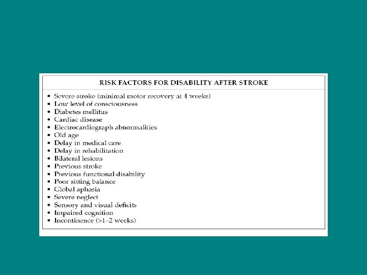
Predictors of motor recovery: • Severity of arm weakness at onset: – With complete arm paralysis at onset, there is a poor prognosis of recovery of useful hand function • Timing of return of hand movement: – If the patient shows some motor recovery of the hand by four weeks, there is up to 70% chance of making a full or good recovery – Poor prognosis with no measurable grasp strength by four weeks • Poor prognosis associated also with: – Severe proximal spasticity – Prolonged “flaccidity” period
 No “voluntary” movements on the")
Brunnstrom recovery stages 1. Flaccidity (immediately after the onset) No “voluntary” movements on the affected side can be initiated 2. Spasticity appears. Basic synergy patterns appear. Minimal voluntary movements may be present 3. Patient gains voluntary control over synergies. Increase in spasticity 4. Some movement patterns out of synergy are mastered (synergy patterns still predominate). Decrease in spasticity 5. If progress continues, more complex movement combinations are learned as the basic synergies lose their dominance over motor acts. Further decrease in spasticity 6. Disappearance of spasticity. Ιndividual joint movements become possible and coordination approaches normal 7. Normal function is restored

REHABILITATION METHODS FOR MOTOR DEFICITS • Traditional Therapy: – Traditional therapeutic exercise program consists of positioning, ROM exercises, strengthening, mobilization, compensatory techniques, endurance training (e. g. , aerobics). – Traditional approaches for improving motor control and coordination: emphasize need of repetition of specific movements for learning, the importance of sensation to the control of movement, and the need to develop basic movements and postures.
Neuromuscular Facilitation (PNF) – Uses spiral and diagonal components of")
• Proprioceptive (or peripheral)Neuromuscular Facilitation (PNF) – Uses spiral and diagonal components of movement rather than the traditional movements in cardinal planes of motion with the goal of facilitating movement patterns that will have more functional relevance than the traditional technique of strengthening individual group muscles
 – The goal of NDT is")
• Bobath approach / neurodevelopmental technique (NDT) – The goal of NDT is to normalize tone, to inhibit primitive patterns of movement, and to facilitate automatic, voluntary reactions and subsequent normal movement patterns.

• Brunstrom approach/Movement therapy: – Uses primitive synergistic patterns in training in attempting to improve motor control through central facilitation

UPPER EXTREMITY MANAGEMENT • Shoulder pain: 70%– 84% of stroke patients with hemiplegia have shoulder pain with varying degrees of severity • Of the patients with shoulder pain, the majority (85%) will develop it during the spastic phase of recovery • It is generally accepted that the most common causes of hemiplegic shoulder pain are the shoulder-hand syndrome/reflex sympathetic dystrophy (RSD) and soft-tissue lesions (including plexus lesions)
/Reflex Sympathetic Dystrophy (RSD)")
• Complex Regional Pain Syndrome Type I (CRPS, Type I)/Reflex Sympathetic Dystrophy (RSD) – Disorder characterized by sympathetic-maintained pain and related sensory abnormalities, abnormal blood flow, abnormalities in the motor system, and changes in both superficial and deep structures with trophic changes • Sholuder subluxation: – Characterized by the presence of a palpable gap between the acromion and the humeral head – Etiology is unknown, but may be due to changes in the mechanical integrity of the glenohumeral joint

• Brachial Plexus/Peripheral Nerve Injury – Etiology: “Traction” neuropathy • Bicipital Tendinitis • Rotator Cuff Tear, Impingement Syndrome, Adhesive Capsulitis
")
OTHER ASPECTS OF STROKE REHABILITATION • • • Spasticity Management Deep Vein Thrombosis (DVT) Bladder Dysfunction Bowel Dysfunction Dysphagia APHASIA Sexual Dysfunction Poststroke depression Seizures
 • DEFINITION OF CP – A group of motor syndromes resulting")
CEREBRAL PALSY (CP) • DEFINITION OF CP – A group of motor syndromes resulting from disorders of early brain development – Genetic, metabolic, ischemic, infectious, other

CLASSIFICATION OF CP • The modified neurologic classification system divides patients into the following categories: – 1. Spastic (pyramidal) cerebral palsy} 75% • • Hemiplegia Diplegia Quadriplegia monoplegia – 2. Dyskinetic (extrapyramidal) cerebral palsy – 3. Mixed types

Pathophysiology of CP • Caused by an insult to the immature brain at any time prior to birth up to 2 years of age. • Alteration in muscle tone, muscle stretch reflexes, primitive reflexes, postural reactions • MR, vision and hearing problems, seizure • Oromotor and dental problems

Etiology of CP • • Vascular Hypoxic-ischemic, Metabolic, Infectious, toxic, Teratogenic, Traumatic Genetic

• Prenatal: – – – • Neonatal: – – – • İntrauterine infections Congenital malformations Toxic or teratogenic agents Multipl births Abdominal trauma Maternal illness Prematurity Birth weight less than 2500 gr Growth retardation İntracranial hemorhage Trauma, infection Hyperbilirubinemia Postnatal: – Trauma, infection – IC hemorrhage – coagulopathy

Presentation • Failing to meet expected developmental milestones or failing to suppress primitive reflexes. • Abnormal muscle tone • May have an early period of hypotoni followed by hypertonia • Asymetric crawl or failure to crawl

Findings • Joint contractures secondary to spastic muscles • Hypotonic to spastic tone • Persistent primitive reflexes (asymmetric tonic neck, palmar grasp, moro reflex exc)

Typical Gait Abnormalities Include • Spastic diplegia – – Scissoring gait pattern Hips flexed and adducted Knees flexed with valgus Ankles in equinus • Spastic hemiplegia – – – Weak hip flexion and ankle dorsiflexion Overactive posterior tibialis Hip hiking or hip circumduction Supinated foot in stance phase Upper extremity posturing

THERAPEUTIC MANAGEMENT • • ROM exercises Stretching exercises PRE Postural and motor control training

Spasticity Management • The mainstay of treatment is through the application of modalities, primarily therapeutic exercise, range of motion, hot and cold application, as well as casting and splinting
 with essentially")
• Botulinum Toxin – Botulinum toxin affects the neuromuscular junction (NMJ) with essentially the same results – Botox acts by irreversibly blocking the NMJ presynaptictically preventing acetylcholine (ACh) release. Given as an IM injection, onset of effect occurs at 24– 72 hrs.

• Surgical Procedures – Note: surgery to improve ambulation remains problematic – Surgery may be indicated to improve function and appearance and to prevent or correct deformities
 • Knee Ankle Foot Orthoses (KAFO) • Hip")
• Ankle Foot Orthoses (AFO) • Knee Ankle Foot Orthoses (KAFO) • Hip Knee Ankle Foot Orthoses (HKAFO):
- Slides: 36