Regulation of Body Functions Hormonal System of Regulation

Regulation of Body Functions • Hormonal System of Regulation • Nervous System. •

The nervous system is composed of three • major parts: the sensory input portion • the central nervous system • and the motor output portion •

Sensory receptors detect the state of the body or • the state of the surroundings. For instance The eyes are sensory organs that give one a visual image of the surrounding area, . The central nervous system is composed of the • brain and spinal cord. The brain can store information, generate thoughts, create ambition, and determine reactions that the body performs in response to the sensations. Appropriate signals are then transmitted through the motor output portion of the nervous system to carry out one’s desires.

Hormonal System of Regulation. Located in • the body are eight major endocrine glands that secrete chemical substances called hormones. Hormones are transported in the extracellular fluid to all parts of the body to help regulate cellular function. For instance Insulin controls glucose metabolism and parathyroid hormone controls bone calcium and phosphate. Thus, the hormones are a system of regulation that complements the nervous system. The nervous system regulates mainly muscular and secretary activities of the body, whereas the hormonal system regulates many metabolic functions.

Membranous Structures of the Cell • Most organelles of the cell are covered by • membranes composed primarily of lipids and proteins. These membranes include the cell membrane, • nuclear membrane, membrane of the endoplasmic reticulum, and membranes of the mitochondria, lysosomes, and Golgi apparatus.
, which")
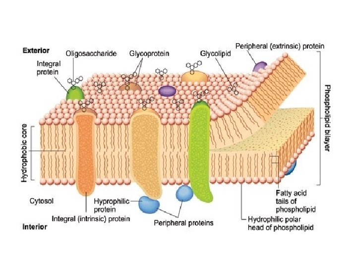
The Cell Membrane • The cell membrane (also called the plasma • membrane), which envelops the cell, is a thin, pliable, elastic structure only 7. 5 to 10 nanometers thick. It is composed almost entirely of proteins and lipids. The approximate composition is proteins, 55 %; phospholipids, 25 %; cholesterol, 13% other lipids, 4 %; and carbohydrates, 3 %. Cell membranes are selectively permeable semipermeable (some things can pass through and some can’t).

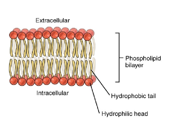
Lipid Barrier of the Cell Membrane • Its basic structure is a lipid bilayer, which is a thin, double-layered • film of lipids—each layer only one molecule thick that is continuous over the entire cell surface. Interspersed in this lipid film is large globular protein molecules. The basic lipid bilayer is composed of phospholipid molecules. One end of each phospholipid molecule is soluble in water; that is, it is hydrophilic. The other end is soluble only in fats; that is, it is hydrophobic. Because the hydrophobic portions of the phospholipid molecules are repelled by water, but are mutually attracted to one another, they have a natural tendency to attach to one another in the middle of the membrane, as shown in Figure. The hydrophilic phosphate portions then constitute the two surfaces of the complete cell membrane, in contact with intracellular water on the inside of the membrane and extracellular water on the outside surface.

The lipid layer in the middle of the membrane is • impermeable to the usual water-soluble substances, such as ions, glucose, and urea. Conversely, fatsoluble substances, such as oxygen, carbon dioxide, and alcohol, can penetrate this portion of the membrane with ease. The cholesterol molecules in the membrane are also lipid in nature because their steroid nucleus is highly fat soluble. These molecules, in a sense, are dissolved in the bilayer of the membrane. They mainly help determine the degree of permeability (or impermeability) of the bilayer to water-soluble constituents of body fluids. Cholesterol controls much of the fluidity of the membrane as well.

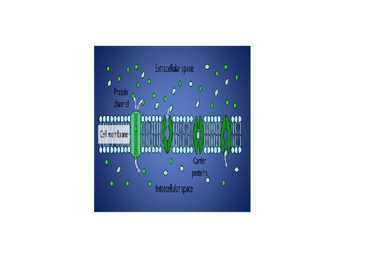
Cell Membrane Proteins • most of the cell membrane are glycoproteins. • Two types of proteins occur: integral proteins that protrude all the way through the membrane, and peripheral proteins that are attached only to one surface of the membrane and do not penetrate all the way through
 through which")
. Many of the integral proteins provide • structural channels (or pores) through which water molecules and water-soluble substances, especially ions, can diffuse between the extracellular and intracellular fluids Other integral proteins act as carrier proteins • for transporting substances that otherwise could not penetrate the lipid bilayer

Integral membrane proteins can also serve as • receptors for water-soluble chemicals, such as peptide hormones, that do not easily penetrate the cell membrane. Peripheral protein molecules are often attached • to the integral proteins. These peripheral proteins function almost entirely as enzymes or as controllers of transport of substances through the cell membrane “pores. ”

Membrane Carbohydrates—The Cell • “Glycocalyx. ” Membrane carbohydrates occur almost • invariably in combination with proteins or lipids in the form of glycoproteins or glycolipids. In fact, most of the integral proteins are glycoproteins, and about one tenth of the membrane lipid molecules are glycolipids. The “glyco” portions of these molecules almost invariably protrude to the outside of the cell, dangling outward from the cell surface.

The carbohydrate moieties attached to the outer surface of • the cell have several important functions: (1) Many of them have a negative electrical charge, which • gives most cells an overall negative surface charge that repels other negative objects. (2) The glycocalyx of some cells attaches to the glycocalyx • of other cells, thus attaching cells to one another. (3) Many of the carbohydrates act as receptor substances • for binding hormones, such as insulin; when bound, this combination activates attached internal proteins that, in turn, activate a cascade of intracellular enzymes. (4) Some carbohydrate moieties enter into immune • reactions.

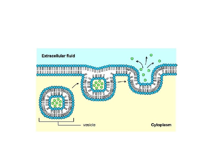
Mechanism of Solutes Transport • All cells need to import oxygen, sugars, amino acids, • and some small ions and to export carbon dioxide, metabolic wastes, and secretions. At the same time, specialized cells require mechanisms to transport molecules such as enzymes, hormones, and neurotransmitters. The movement of large molecules is carried out by Endocytosis and Exocytosis, the transfer of substances into or out of the cell, respectively, by vesicle formation and vesicle fusion with the plasma membrane. Cells also have mechanisms for the rapid movement of ions and solute molecules across the plasma membrane.
, which requires")
hese mechanisms are of two general types: • Passive movement (Passive Transport), which requires no direct expenditure of metabolic energy, and Active movement (Active transport), • which uses metabolic energy to drive solute transport.
 Endocytosis – is a")
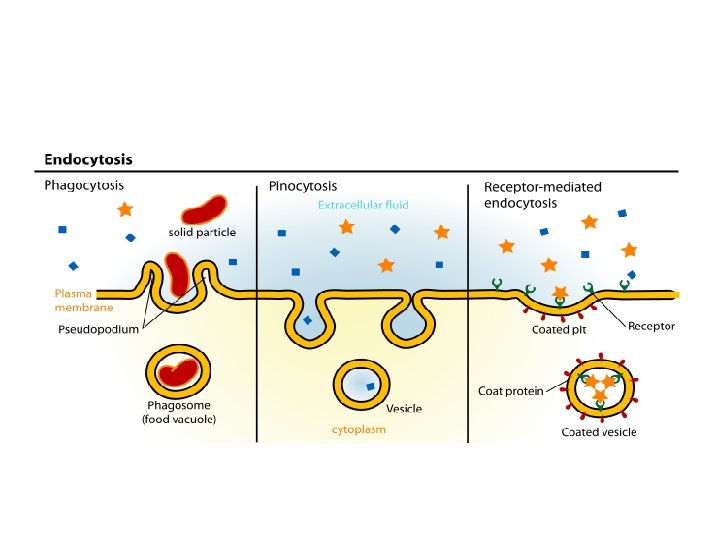
Macromolecules Cross the Plasm Membrane • by Vesicle Fusion A) Endocytosis – is a general term for the • process in which a region of the plasma membrane is pinched off to form an endocytic vesicle inside the cell. During vesicle formation, some fluid, dissolved solutes, and particulate material from the extracellular medium are trapped inside the vesicle and internalized by the cell

Three main types of endocytosis can be distinguished : - • 1 - Phagocytosis – is the ingestion of large particles or • microorganisms, usually occurring only in specialized cells such as macrophages. 2 - Pinocytosis: is the nonspecific uptake of the • extracellular fluid and all its dissolved solutes. The material is trapped inside the endocytic vesicle as it is pinched off inside the cell 3 - Receptor mediated Endocytosis is a more efficient • process that uses receptors on the cell surface to bind specific molecules. These receptors accumulate at specific depressions known as coated pits, so named because the cytosolic surface of the membrane at this site is covered with a coat of several proteins.

Receptor-mediated endocytosis is the • mechanism by which cells take up a variety of important molecules, including hormones; growth factors; and serum transport proteins, such as transferrin (an iron carrier). Foreign substances, such as diphtheria toxin and certain viruses, also enter cells by this pathway.
 Exocytosis • Many cells synthesize important macromolecules that are • destined for Exocytosis")
B) Exocytosis • Many cells synthesize important macromolecules that are • destined for Exocytosis or export from the cell. These molecules are synthesized in the endoplasmic reticulum, modified in the Golgi apparatus, and packed inside transport vesicles. The vesicles move to the cell surface, fuse with the cell membrane, and release their contents outside the cell. There are two exocytic pathways constitutive and regulated. Some proteins are secreted continuously by the cells that make them. Secretion of mucus by goblet cells in the small intestine is a specific example. In this case, exocytosis follows the constitutive pathway, which is present in all cells. In other cells, macromolecules are stored inside the cell in secretory vesicles. These vesicles fuse with the cell membrane and release their contents only when a specific extracellular stimulus arrives at the cell membrane. This pathway, known as the regulated pathway, is responsible for the rapid “on-demand” secretion of many specific hormones, neurotransmitters, and digestive enzymes.
- Slides: 24