REGIONAL CEREBRAL OXYGEN SATURATION MONITORING IN CARDIAC SURGERY

REGIONAL CEREBRAL OXYGEN SATURATION MONITORING IN CARDIAC SURGERY 연세대학교 의과대학 마취통증의학교실 송종욱

Cerebral oximetry • oxygen saturation of hemoglobin at the microvascular level (70 -75% of blood in brain is venous) • Balance of cerebral oxygen supply/demand • Sensitive index of cerebral ischemia • Continuous and non-invasive

Near-infrared spectroscopy • 700 -900 nm • Traverse up to 8 cm of tissue Fig. 1. (A) Light absorption spectra of different absorbers in tissue. Water, lipids, melanin and myoglobin show constant absorption effects, whereas the concentration of deoxyhemoglobin (HHb) and oxyhemoglobin (O 2 Hb) varies with oxygen status. (B) At 800 nm, the absorption spectra of O 2 Hb and HHb is equivalent (isobestic point), which is used to calculate hemoglobin concentration independent of oxygen saturation. Pellicer et al. Seminars in Fetal and Neonatal Medicine 16 (2011) 42 -49
 = α •")
A. Beer-Lambert law B. Modified Beer-Lambert law A = log(I 0/I) = α • c • d • Light absorption에 의한 attenuation (A)은 chromophore의 농도(c) 및 optical pathlength(d)에 비례함 A = log(I 0/I) = α • c • d • DPF + G • • 생체 내에서는 attenuation의 80% 정도가 scattering에 기인 함 (G) Scattering에 의해 photon들은 geometric pathlength보다 평균 적으로 먼 거리를 투과한다. (differential pathlength factor)

Types of spectrometer • Continuous wave spectroscopy üModified Beer-Lambert law를 이용 ü측정 중 Scattering loss 와 differential pathlength factor가 일 정하며 attenuation의 변화는 absoprtion의 변화만을 반영한 다고 가정함 ü따라서 chromophore 농도의 변화량을 측정할 수 있다.

Types of spectrometer a. Time-resolved spectroscopy • Ultashort pulsed laser를 사용 • 조직을 통과하는 light의 time difference 및 통 과하는데 걸린 mean time으로 부터 DP를 계 산 • Absolute optical absorption and scattering b. Frequency-resolved spectroscopy • Modulate a continuous laser source • 조직을 통과하면서 생기는 phase shift로 부터 DP 계산 • Absolute optical absorption and scattering c. Spatially resolved spectroscopy • Closely spaced multi-detector를 사용 • 거리에 따른 light attenuation의 차이로 부터 DPF 를 추산 • 측정한 절대값의 정확도는 각 기기들과 사용하는 algorithms에 따라 다를 수 있다. • 임상에서 사용되는 대부분의 cerebral oximeter는 continuous wave, spatially resolved spectroscopy
 Fore-Sight (CASMED) Equanox (Nonin) Cer. Ox (Ornim)")
Commercial NIRS cerebral oximeters INVOS (Covidien) Fore-Sight (CASMED) Equanox (Nonin) Cer. Ox (Ornim)
 • 2 wavelengths (730 and 810 nm) • 2")
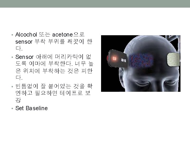
INVOS Fore-Sight Equanox INVOS (Covidien) • 2 wavelengths (730 and 810 nm) • 2 channels for bilateral brain monitoring • 2 light detectors at fixed distances from the light source (3 and 4 cm for adult sensor) • r. SO 2 normal value 67 ± 9% for adult cardiac surgical patients • FDA indications for use; adjunct trend monitoring of regional hemoglobin oxygen saturation of the brain • Baseline has to be established before induction

Validation • No gold standard or index test • Based on Sjv. O 2, Sv. O 2, ti. PO 2 or Sw. O 2 (weighted average of Sa. O 2 and Sjv. O 2) Reading Bias of 5 Cerebral Oximeters Mean bias ± SD Arms Fore-Sight 1. 76 ± 3. 92 4. 28 INVOS 5100 c 0. 05 ± 9. 72 9. 69 NIRO-200 NX -1. 13 ± 9. 64 9. 68 Equanox 7600 3 -wave 2. 48 ± 8. 12 8. 47 Equanox 7600 4 -wave 2. 84 ± 6. 27 6. 86 Arms; root mean square error Bickler et al. Anesth Analg 2013; 117: 813 -23

r. SO 2 vs Sw. O 2 r. SO 2 vs Sv. O 2

Validation • Limitations • No gold standard • Many assumptions (scattering loss, DPF, ratio of arterial/venous component…) • Extracranial contamination • Each device uses a unique algorithm, different light source, wavelengths, number of wavelengths • Hair, ambient light, hematoma, venous sinus, skin pigmentation, chromophore other than Hb (bilirubin)… • r. SO 2 values appear to reasonably reflect regional cerebral oxygen balance • Best used as a trend monitor • Claims of absolute thresholds for cerebral ischemia should be treated with caution

Clinical applications • r. SO 2 – venous weighted oxygen saturation, balance of oxygen supply/demand • Decline in r. SO 2 • Increase in CMRO 2 • Decrease in CBF • Decrease in Ca. O 2 (anemia or hypoxemia) u General Strategy - maintain bilateral CMRO 2 values ü within 75 -80% of baseline ü above 50% for patients with a baseline value of ≤ 50%

www. casecag. com

www. casecag. com

Denault et al. Semin Cardiothorac Vasc Anesth 2007; 11; 274 -281

Zheng et al. Anesth Analg 2013; 116: 663 -76

Zheng et al. Anesth Analg 2013; 116: 663 -76

Summary • r. SO 2 reflects balance of cerebral oxygen supply/demand • Easy to use, non-invasive, high temporal resolution • r. SO 2 monitoring may lead to improved outcome after cardiac surgery. • Full potential of r. SO 2 has yet to be determined • Further technological advances are necessary
- Slides: 19