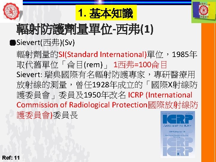
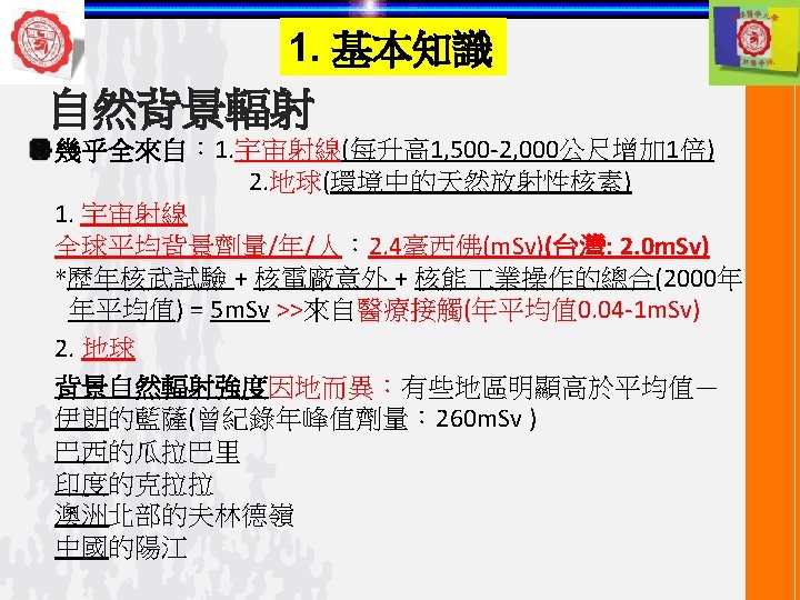
References 1 Harring JI Jansen L Dental radiography

 ON RADIATION • Breakage of one or")
 ON RADIATION base deletion or Single strand")

 Killing")


")


 結 Typical Doses during Radiographic Examinations Effective Dose (m. Sv) Dental 0")
 結 X-ray Exam (2 Effective dose (m. Sv) CT chest 8. 0")
 % Relative Dose % Improve in Radiation Relative Dosage for Intra-Oral")
- Slides: 46
References: 1. Harring JI & Jansen L: Dental radiography- principle & techniques 2 nd ed. , Chapters 2, 3, 5 -9, 22, 23 p. 11 -34, 35 -46, 63 -80, 81 -90, 91 -107, 108 -122, 123 -153, 342 -362, 363 -383 2. Eric Whaites: Essentials of dental radiography & radiology 3 rd ed. , Chapter 5, p. 33 -49 3. 何復辰: 牙科放射線學 1 st edition, p. 109 -13 4. White & Pharaoh: Oral radiology: principle & interpretation, 5 th ed. , Chapter 6, p. 94 -109 5. Kaohsiung Medical University, Oral Pathology Department 6. Manufacturer’s data sheet 7. Cha JY, Mah J, Sinclair P. Incidental findings in the maxillofacial area with 3 -dimensional cone-beam imaging. Am J Orthod Dentofacial Orthop 2007; 132: 7 -14 8. Web et al. Background radiation levels & medical exposure in Australia. Radiation Protection in Australia. 1999; 15: 25 -32 9. Farman AG. ALARA still applies. Oral Surg Oral Med Oral Pathol Oral Radiol 2005; 100: 395 -6 10. White & Pharoah: Oral radiology: principle & interpretation, 6 th ed. , Chapter 2, p. 19 -22 11. http: //www. icrp. org/ICRP publication 125 12. zh. wikipedia. org/zh-tw 13. big 5. ce. cn 14. tupian. baike. com 15. www. fly 168. com. tw/ 16. What you need to know about cancer. Sci Am 1996; 289: 28 -119 17. Oral Pathology for the Dental Hygienist. Olga AC Ibsen, Joan Anderson Phelan, 4 th ed. , 2004, p. 21645 18. Bushberg, et al. The Essential Physics of Medical Imaging, 2 nd ed. , p. 819 19. Bushberg, et al. The Essential Physics of Medical Imaging, 2 nd ed. , p. 816
1. 基本知識 CHANGES IN DEOXYRIBONUCLEIC ACID (DNA) ON RADIATION • Breakage of one or both DNA strands • Cross-linking of DNA strands within the helix to other DNA strands or to proteins A G • Change or loss of a base • Disruption of hydrogen bonds between DNA strands T C Codon Hydrogen Bond Ref. 17
1. 基本知識 CHANGES IN DEOXYRIBONUCLEIC ACID (DNA) ON RADIATION base deletion or Single strand break or LET : Linear Low LET Energy (x-ray) Transfer Ref. 18 High LET ( -particle) base substitution hydrogen bond disruption or Double strand break
1. 基本知識 CHROMOSOME ABERRATIONS ON RADIATION + DNA synthesis + X-ray A A Irradiation of cell after DNA synthesis results single-arm (chromatid) aberration + X-ray Cell cycle. A proliferating cell moves in the cycle from mitosis to Gap 1 (G 1) to the period of DNA synthesis (S) to Gap 2 (G 2) to the next mitosis Refs. 10, 16 + DNA synthesis B B Irradiation before DNA synthesis results double-arm (chromosome) aberration
1. 基本知識 DETERMINISTIC & STOCHASTIC EFFECTS OF RADIATION Radiation injury to organisms: (1) Killing of large numbers of cells (deterministic effects) or (2) Sublethal damage to individual cells results in cancer formation or heritable mutation (stochastic effects) Comparison of Deterministic & Stochastic Effects of Radiation Deterministic Effects Stochastic Effects Examples *Mucositis resulting from radiation therapy to oral cavity *Radiation-induced cataract formation *Radiation-induced cancer *Heritable effects Caused by *Killing of many cells *Sublethal damage to DNA Threshold dose? *Yes: sufficient cell killing required to cause a clinical response *No: even one photon could cause a change in DNA that leads to cancer or heritable effect Severity of clinical effects and dose *Severity of clinical effects is proportional to dose *The greater the dose the greater the effect *Severity of clinical effects is independent of dose *All-or-none response; an individual either has effect or does not Probability of having effect and dose *Probability of effect independent of dose *All individuals show effect when dose is above threshold *Frequency of effect proportional to dose *The greater the dose. the greater the chance of having the effect Ref. 10
1. 基本知識 RELATIVE RADIOSENSITIVITY OF VARIOUS CELLS HIGH INTERMEDIATE LOW Characteristics *Divide regularly *Long mitotic futures *Undergo no or little differentiation between mitoses *Divide occasionally in response to a demand for more cells *Highly differentiated *When mature are incapable of division Examples *Vascular endothelial cells *Fibroblasts *Acinar & ducal salivary glands cells *Parenchymal cells of liver, kidney, & thyroid *Neurons *Striated muscle cells *Squamous epithelial cells *Erythrocytes Ref. 10 *Spermatogenic & erythroblastic stem cells *Basal cells of oral mucous membrane
1. 基本知識 RELATIVE RADIOSENSITIVITY OF VARIOUS ORGANS HIGH INTERMEDIATE Lymphoid organs Fine vasculature Optic lens Bone marrow Growing cartilage Muscle Testes Growing bone Intestines Salivary glands Mucous membrane Lung Kidney Liver Ref. 10 LOW
1. 基本知識 INTERACTION OF RADIATION WITH TISSUE v v v Ionizing radiation energy deposited randomly and rapidly (< 10 -10 sec) via excitation, ionization & thermal heating Interactions producing biologic changes classified as either direct or indirect Direct v v Critical targets (e. g. , DNA, RNA or protein) directly ionized or excited Indirect v Radiation interacts within the medium (e. g. , cytoplasm) creating reactive chemical species (free radicals) which in turn interact with the a critical target macromolecule
1. 基本知識 INTERACTION OF RADIATION WITH TISSUE v Vast majority of interactions are indirect (body 70% 85% water) v v Results in an unstable ion pair, H 2 O+, H 2 ODissociate into another ion and a free radical (lifetime < 10 -5 sec) v v H 2 O+ H+ + OH • H 2 O- H • + OH- Combine with other free radicals to form molecules such as hydrogen peroxide (H 2 O 2) → highly toxic to cell Oxygen enhances free radical damage via production of reactive oxygen species (e. g. , H • + O 2 → HO 2 • )
1. 基本知識 INTERACTION OF RADIATION WITH TISSUE Indirect action Direct action Repair Rad iatio n Free radicals and Hydrogen peroxide Radiation H+ OHIonization & excitation H • ; OH- ; HO 2+ ; H 2 O 2 DNA Molecular Damage Mutation Genetic Biological Response Somatic Death 10 -17 to 10 -5 sec Ref. 19 Teratogenic min to 10 -5 decades
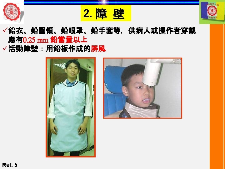
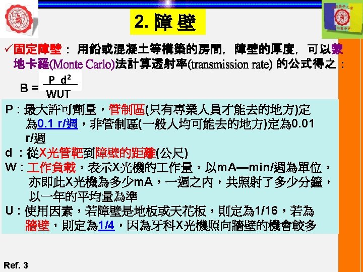
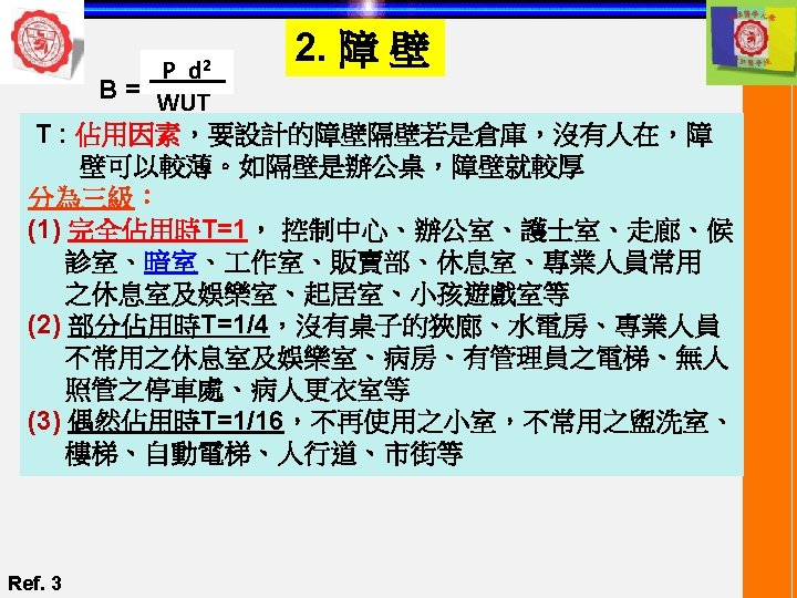
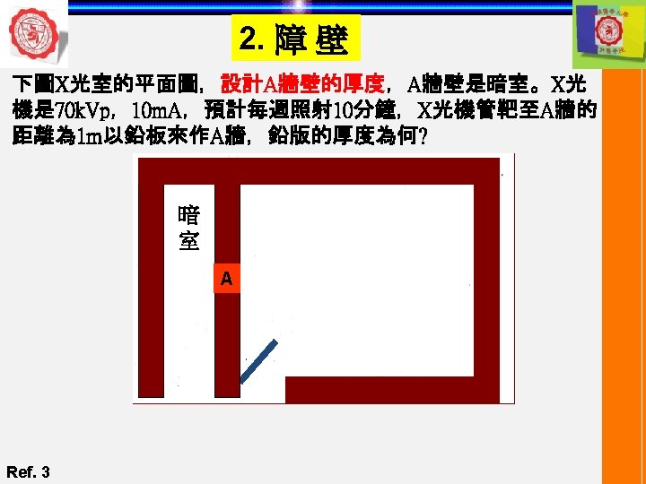
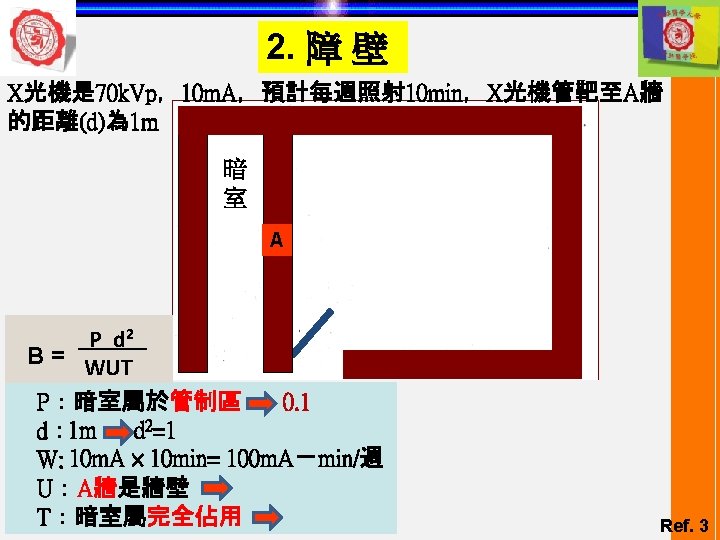
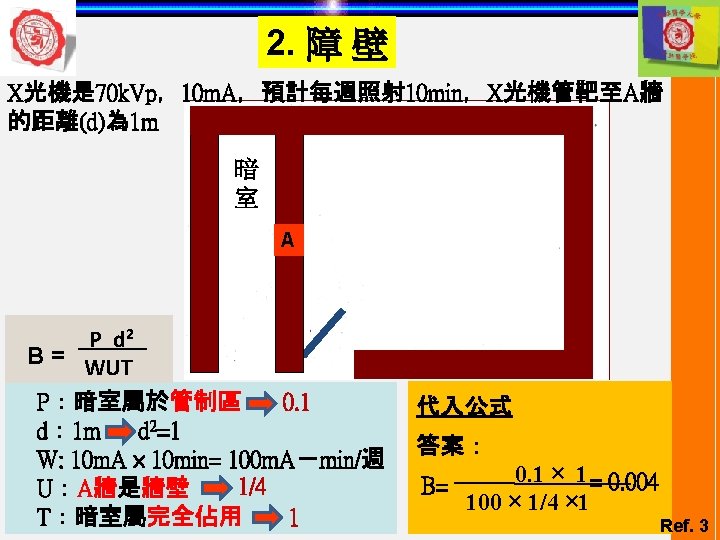
2. 障 壁 B= 0. 004 圖 1 Ref. 3 圖 2 0. 5 mm
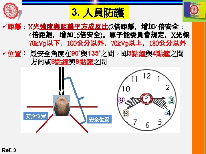
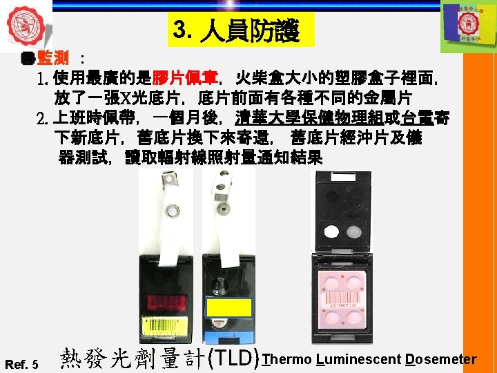
3. 人員防護 ü安全規定: 安全標準 Current Guidelines International Commission on Radiological Protection (ICRP, Pub. 26) 1. No practice unless a positive net benefit 2. As low as reasonably achievable (ALARA) 3. Not exceed recommended dose limits 1. Patients 2. Radiation workers 3. General public
3. 人員防護 1. Patients 2. Radiation workers 3. General public Classified workers Non-classified workers 1. Exam associated with illness 2. Periodic health checks 3. Exam for occupational, medico-legal, insurance 4. Medical research 1. No set dose limits Not receiving radiation dose as a patient or radiation worker 2. IRB approval 3. Child avoided Ref. 2 ICRP annual dose limits Classified workers Non-classified workers General public Whole body dose 50 m. Sv 15 m. Sv Individual organs and tissues 500 m. Sv 150 m. Sv Lens of the eye 150 m. Sv 45 m. Sv 15 m. Sv Abdomen of women of reproductive capacity 13 m. Sv In any consecutive 3 months Abdomen of 10 m. Sv During the declared term of pregnancy 1 m. Sv
3. 人員防護 Radiation absorbed dose Amount of energy absorbed from the radiation beam per unit mass (joules/kg) (1 c. Gy = 1 rad) 1 Gray (Gy) =100 rad Equivalent dose = radiation absorbed dose x quality factor (Sievert, Sv) X-rays, particles Q=1 Fast neutrons & protons Q=10 particles Q=20 Tissues Weighting factor Testes & ovaries 0. 25 Breast 0. 15 Red bone marrow 0. 12 Effective dose = equivalent dose x weighting factor (Sievert, Sv) Lung 0. 12 Thyroid 0. 03 Bone surfaces 0. 03 Remainder 0. 30 Ref. 2
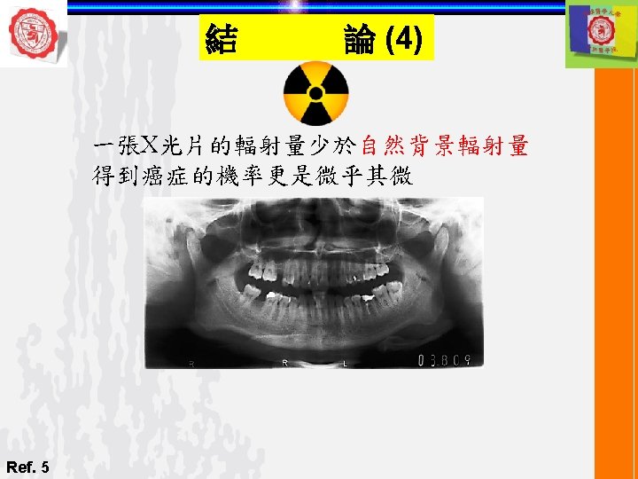
論 (1) 結 Typical Doses during Radiographic Examinations Effective Dose (m. Sv) Dental 0 2 4 6 8 10 Web et al. Background radiation levels and medical exposure in Australia, Radiation Protection in Australia, 1999; 15(2): 25 -32 Ref. 8 12
論 (2) 結 X-ray Exam (2 Effective dose (m. Sv) CT chest 8. 0 Barium enema 7. 7 Lumbar spine 2. 2 CT head 2. 0 Skull 0. 1 Chest 0. 04 Dental panoramic tomograph -Using rare-earth screens -Using calcium tungstate screens 0. 007 0. 014 2 dental intraoral films -using 70 k. V, 200 mm fsd (focal spot diameter), rectangular collimation & E speed film -using 50 k. V, 100 mm fsd, round collimation & D speed film Ref. 2 0. 002 0. 016
結 論 (3) % Relative Dose % Improve in Radiation Relative Dosage for Intra-Oral Radiography (1919 -2005) 100 80 60 40 20 0 1919 1925 1941 1955 1981 1991 2005 Time frame (Year) Since 1919 a reduction in the dosage of ionizing radiation needed to produce an intraoral x-ray image is 2 orders of magnitude Ref. 9