RED EYE differential diagnosis RED EYE l Red


 - onset")


")
 , nodular – young, female Presentation")


 injection Miosis due to sphincter spasm")
")


- Slides: 51
RED EYE – differential diagnosis
RED EYE l „Red eye“ is sign of pathology of anterior or posterior ocular segment, of orbit or of ocular adnexa.
Anamnesis l Systemic disease l Eye disease l Devolopment of difficulties l Character of diffuculties
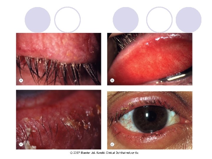
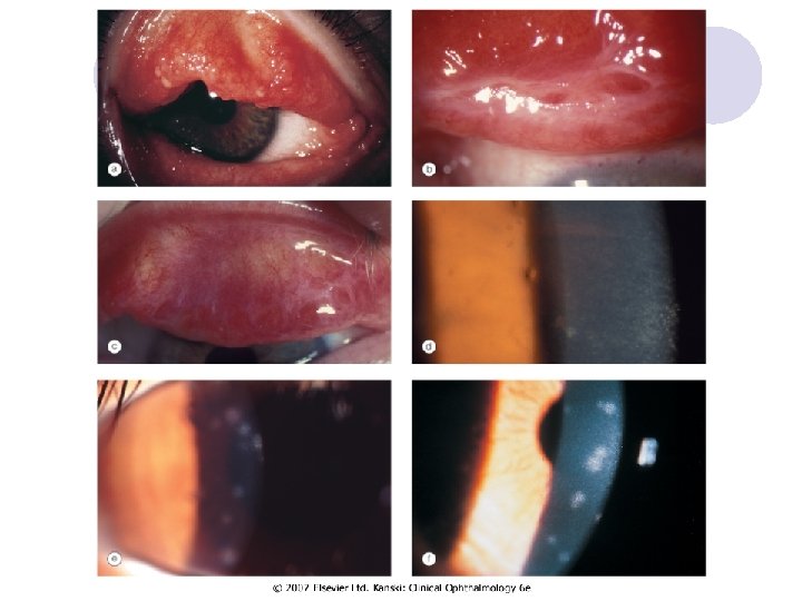
Eyelids - blepharitis l Blepharitis - anterior and posterior Chronic anterior blepharitis l Anterior blepharitis affects the area surrounding the bases of the eyelashes and may be staphylococcal or seborrhoeic l
Eyelids - blepharitis Burning, grittiness and mild photophobia with remissions and exacerbations l Symptoms are usually worse in the mornings l
Eyelids - anterior blepharitis Staphylococcal blepharitis l Hard scales and crusting mainly located around the bases of the lashes (collarettes). l Madarosis, trichiasis and poliosis in severe long-standing cases. l Seborrhoeic blepharitis l Hyperaemic and greasy anterior lid margins with sticking together of lashes l The scales are soft and located anywhere on the lid margin and lashes l
Chronic seborrhoeic blepharitis
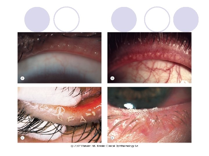
Eyelids – posterior blepharitis l Chronic posterior blepharitis l Caused by meibomian gland dysfunction
Eyelids – posterior blepharitis l Signs of meibomian gland dysfunction : Capping of meibomian gland orifices with oil globules Pouting, recession, or plugging of the meibomian gland orifices l Hyperaemia and telangiectasis of the posterior lid margin l Pressure on the lid margin results in expression of meibomian fluid that may be turbid or appear like toothpaste l The tear film is oily and foamy and froth may accumulate on the lid margins or inner canthi l l
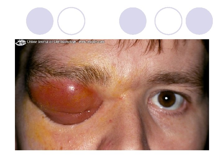
Orbit – preseptal cellulitis Infection of the subcutaneous tissues anterior to the orbital septum. l Causes ¡ Skin trauma - laceratio, insect bites (S. aureus or S. pyogenes) ¡ Spread of local infection - from an acute hordeolum or dacryocystitis. ¡ From remote infection of the upper respiratory tract or middle ear by haematogenous spread l Signs - Unilateral, tender and red periorbital oedema l
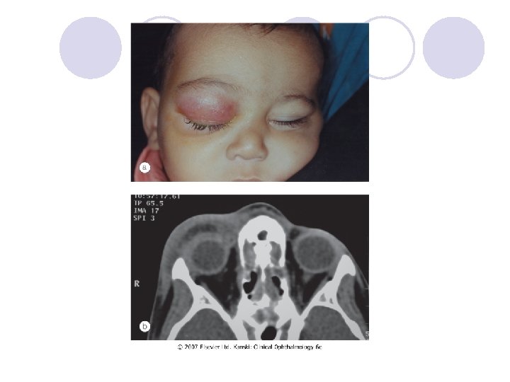
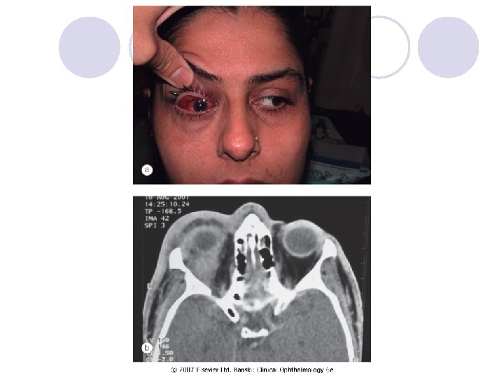
Orbit - Bacterial orbital cellulitis Life-threatening infection of the soft tissues behind the orbital septum, mainly in children l The most prevalent causative organisms are S. pneumoniae, S. aureus, S. pyogenes and H. influenzae. l Pathogenesis Sinus-related - ethmoidal, typically affects children and young adults. l Extension of preseptal cellulitis l Local spread from adjacent dacryocystitis, and mid-facial or dental infection l Haematogenous spread l l
Orbit - Bacterial orbital cellulitis l Presentation is with a rapid onset of severe malaise, fever, pain and visual impairment l Signs Unilateral, tender, warm and red periorbital oedema Proptosis, lid swelling Painful ophthalmoplegia Optic nerve dysfunction l l
Orbit - Bacterial orbital cellulitis l Complications Ocular complications - exposure keratopathy, raised intraocular pressure, occlusion of the central retinal artery or vein, endophthalmitis and optic neuropathy l Intracranial complications - meningitis, brain abscess and cavernous sinus thrombosis l Subperiosteal abscess - along the medial orbital wall l Orbital abscess in post-traumatic or postoperative cases. l
Dry Eye Disorders l There is inadequate tear volume or function resulting in an unstable tear film and ocular surface disease. Keratoconjunctivitis sicca (KCS) refers to any eye with some degree of dryness. l Xerophthalmia describes a dry eye associated with vitamin A deficiency. l Xerosis refers to extreme ocular dryness and keratinization that occurs in eyes with severe conjunctival cicatrization. l Sjögren syndrome is an autoimmune inflammatory disease which is usually associated with dry eyes. l
Dry Eye Disorders l Symptoms l feelings of dryness, grittiness and burning worsen during the day, transient blurring of vision, redness and crusting of the lids
Conjunctival injection is diffuse, beefy-red and more intense away from the limbus l Instillation of 10% phenylephrine drops will constrict the conjunctival and superficial episcleral vasculature l
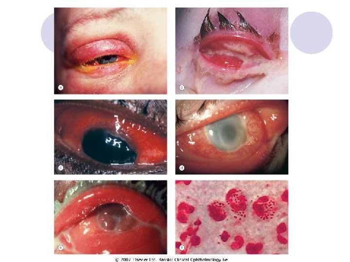
Conjunctivitis l Bacterial - H. influenzae, S. pneumoniae, S. aureus Papillary reaction over the tarsal plates l Mucopurulent discharge l l Gonococcal keratoconjunctivitis - pseudomembrane formation, Lymphadenopathy, Corneal ulceration
Conjunctivitis l Viral conjunctivitis Adenoviral keratoconjunctivitis - the most common external ocular viral infection l Sporadic or occur in epidemics in hospitals, schools and factories l Transmission of this highly contagious virus -respiratory or ocular secretions l Dissemination is by contaminated towels or equipment such as tonometer heads l
Conjunctivitis l Presentation Unilateral watering, redness, discomfort and photophobia l The contralateral eye is typically affected 1 -2 days later, but less severely l Eyelid oedema and tender pre-auricular lymphadenopathy. l Follicular conjunctivitis l
Conjunctivitis l l l Acute allergic rhinoconjunctivitis Seasonal allergic conjunctivitis (hay fever) - onset during the spring and summer The most frequent allergens are tree and grass pollens Perennial allergic conjunctivitis causes symptoms throughout the year with exacerbation in the autumn when exposure to house dust mites, animal dander and fungal allergens is greatest Presentation - redness, watering and itching, associated with sneezing and nasal discharge
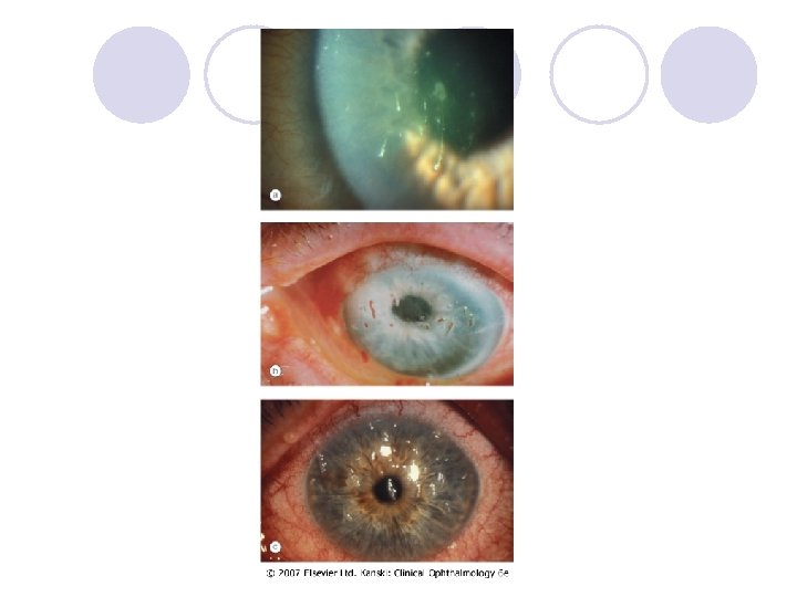
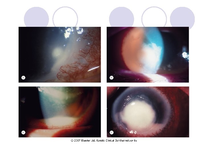
Cornea – infectious keratitis l l l l Keratitis – bacterial (P. aeruginosa , S. aureus, S. pyogenes) Risk factors - Contact lens wear, trauma Presenting symptoms - pain, photophobia, blurred vision and discharge Signs An epithelial defect, infiltrate around the margin, circumcorneal injection Stromal oedema and small hypopyon Progressive ulceration may lead to corneal perforation and endophthalmitis.
Cornea – infectious keratitis l Keratitis – fungal (stromal infiltrate with indistinct margins, surrounded by satellite lesions, hypopyon)
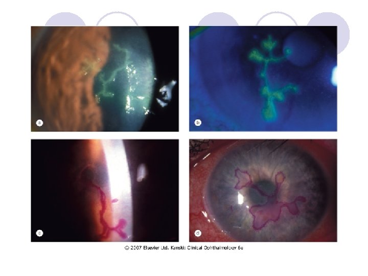
Cornea – infectious keratitis l Keratitis viral – herpes simplex virus l linear-branching (dendritic) ulcer, corneal sensation is reduced
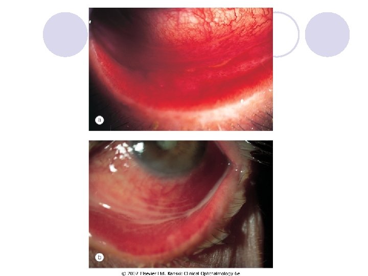
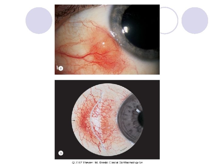
Episclera l Episcleritis – simple (sectoral or diffuse) , nodular – young, female Presentation - always sudden l The eye becoming red and uncomfortable within an hour of the start of an attack - hotness, pricking or generalized discomfort l Without systemic associations l
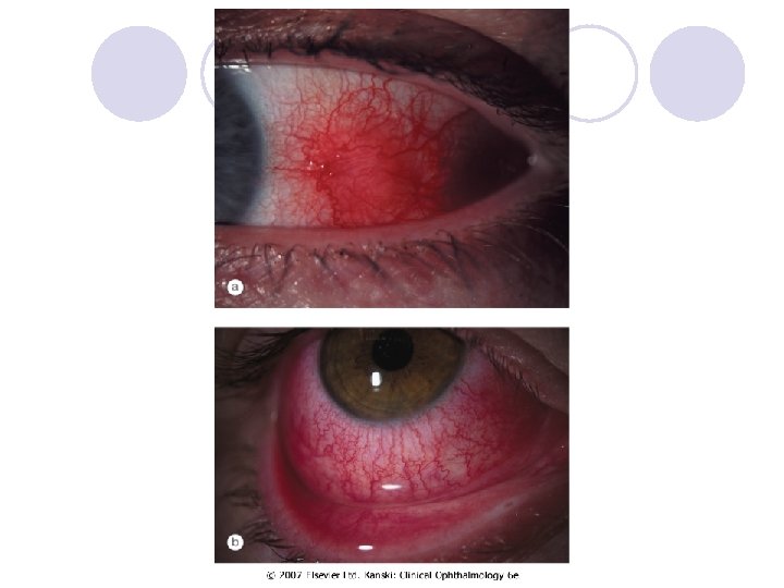
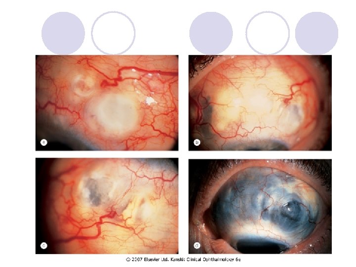
Sclera l Scleritis - oedema and cellular infiltration of the entire thickness of the sclera Anterior non-necrotizing scleritis – diffuze or nodular l Redness, pain which may spread to the face and temple l
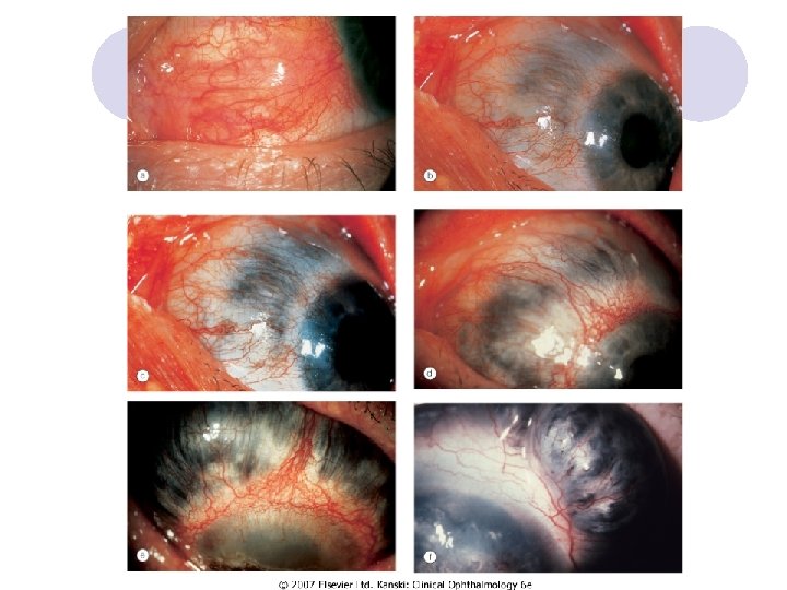
Sclera Necrotizing anterior scleritis with inflammation l pain - severe and persisten l Scleral thinning due to necrosis allows the blue choroid to show through the translucent hydrated scar tissue that has replaced normal sclera l
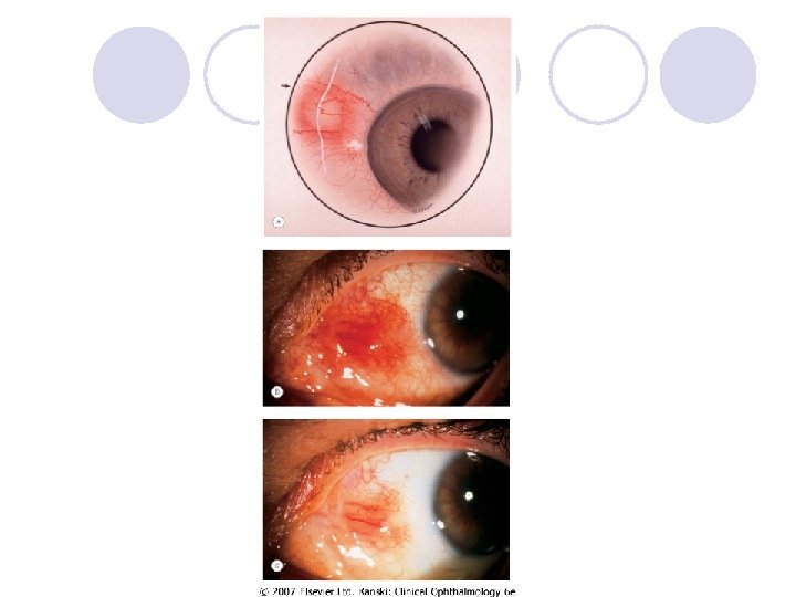
Sclera l Scleromalacia perforans Specific type of necrotizing scleritis without inflammation that typically affects elderly women with long-standing rheumatoid arthritis l Yellow scleral necrotic plaques near the limbus without l vascular congestion
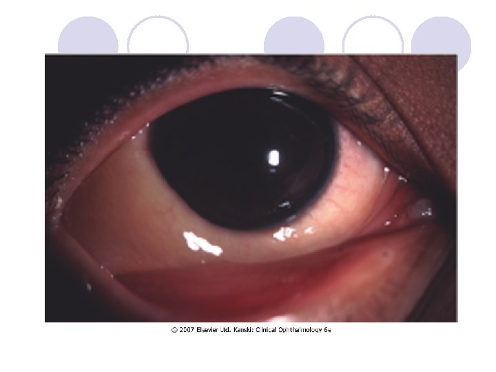
Glaucoma - Acute congestive angle closure
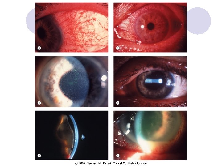
Uveitis Anterior uveitis may be subdivided into: l Iritis in which the inflammation primarily involves the iris. l Iridocyclitis in which both the iris and ciliary body are involved l Ciliary injection - peripheral hyperemia of the anterior l ciliary vessels which produces a deep red or rose color of the corneal stroma, and must be distinguished from hyperemia of the conjunctival vessels. May spread to the perilimbic corneal tissue. Called also ciliary flush.
Anterior uveitis l l l l Ciliary (circumcorneal) injection Miosis due to sphincter spasm Endothelial dusting by myriad of cells is present early and gives rise to a 'dirty' appearance Aqueous cells Aqueous flare reflects the presence of protein due to a breakdown of the blood-aqueous barrier Aqueous fibrinous exudate Hypopyon Posterior synechiae may develop quite quickly and must be broken down before they become permanent
Acute endophtalmitis Acute inflammation of all ocular structure l Endogennous or exogennous (surgery, trauma) l Signs - chemosis, corneal injection, relative afferent pupil defect, corneal haze, fibrinous exudate and hypopyon, vitritis with impaired view of the fundus l
Acute endophthalmitis
End !!!! l