
Red Cell Morphology Basic Introduction Reference Color Atlas

Red Cell Morphology Basic Introduction Reference: Color Atlas of Hematology by Eric Glassy, M. D.

RBC Classification - - - SIZE - - - • Normocyte • Microcyte • Macrocyte • Anisocytosis - - - PALLOR - - - • Normochromic • Hyperchromic • Polychromasia - - - SHAPE - - - • Acanthocytes • Burr Cells • Ovalocytes • Tear Drops • Stomatocytes • Spherocytes • Schistocytes • Target Cells • Sickle Cells - - INCLUSIONS - - - • NRBC’s • Howell-Jolly bodies • Basophilic Stippling • Pappenheimer bodies • Hemoglobin crystals

RBC Parameters Hct_ • MCV = X 10 RBC Microcytic < 80 f. L 80 – 100 f. L Macrocytic > 100 f. L Hgb_ • MCH = X 10 RBC 27 – 31 pg/cell Hgb_ • MCHC = X 100 Hct 32 – 36 g/d. L Hypochromic < 32 g/d. L Hyperchromic > 36 g/d. L

Normal RBC
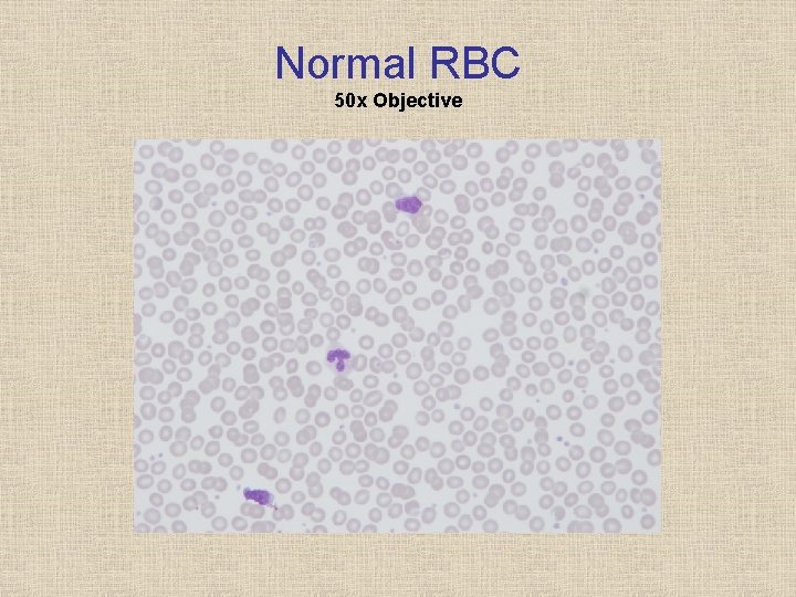
Normal RBC 50 x Objective
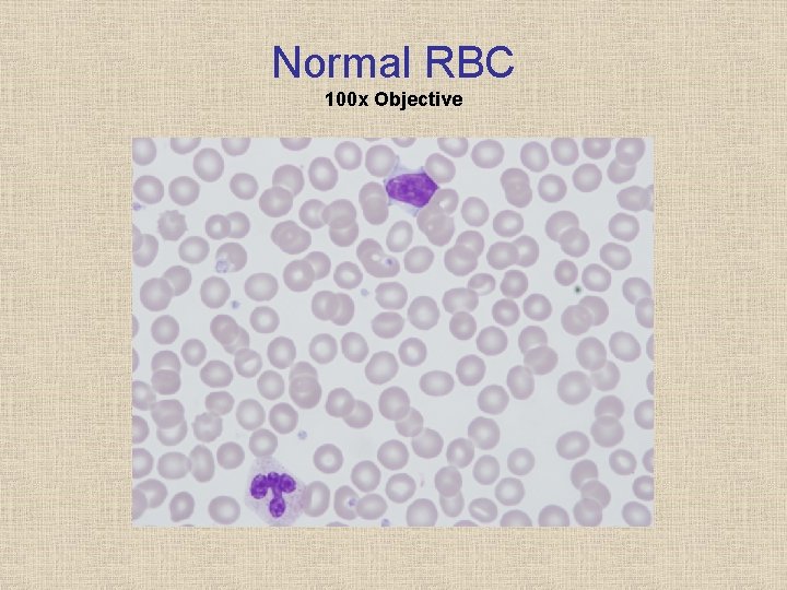
Normal RBC 100 x Objective
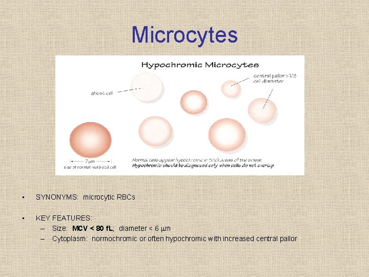
Microcytes • SYNONYMS: microcytic RBCs • KEY FEATURES: – Size: MCV < 80 f. L; diameter < 6 m – Cytoplasm: normochromic or often hypochromic with increased central pallor
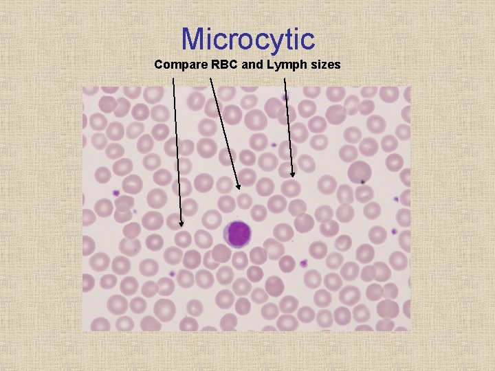
Microcytic Compare RBC and Lymph sizes

Macrocytes • SYNONYMS: macrocytic RBCs; macro ovalocyte (oval form) • KEY FEATURES: – Size: MCV > 100 f. L; diameter > 8 m – Shape: round or oval – Cytoplasm: normochromic, but may sometimes be hypochromic; no polychromasia

Macrocytes Compare RBC and Lymph sizes

Anisocytosis Size Variation • • RDW Guide (g/d. L) Slight = 16 20 Moderate = 20 26 Marked = > 26 Only use RDW as a guide

Hypochromia • SYNONYMS: hypochromic RBCs • KEY FEATURES: – Size: microcytic or macrocytic cells – Cytoplasm: central pallor > 1/3 of RBC diameter (increased central pallor); some cells contain so little hemoglobin that they are “ghost cells”
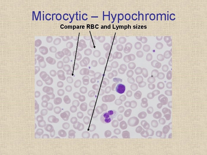
Microcytic – Hypochromic Compare RBC and Lymph sizes
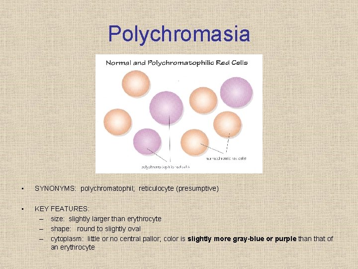
Polychromasia • SYNONYMS: polychromatophil; reticulocyte (presumptive) • KEY FEATURES: – size: slightly larger than erythrocyte – shape: round to slightly oval – cytoplasm: little or no central pallor; color is slightly more gray-blue or purple than that of an erythrocyte

Polychromic
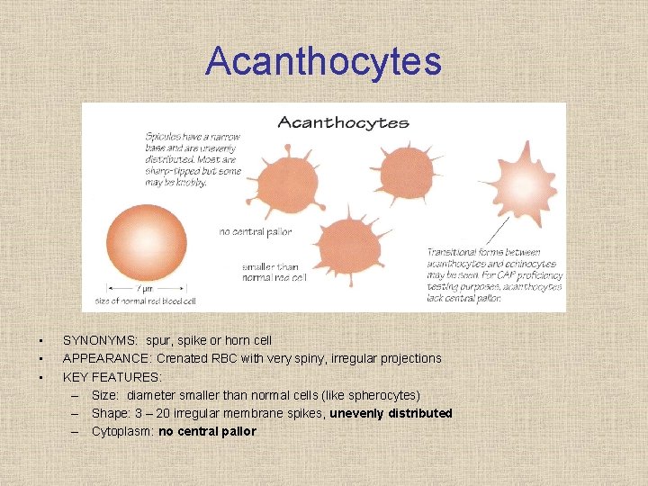
Acanthocytes • • • SYNONYMS: spur, spike or horn cell APPEARANCE: Crenated RBC with very spiny, irregular projections KEY FEATURES: – Size: diameter smaller than normal cells (like spherocytes) – Shape: 3 – 20 irregular membrane spikes, unevenly distributed – Cytoplasm: no central pallor
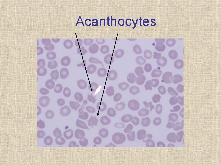
Acanthocytes
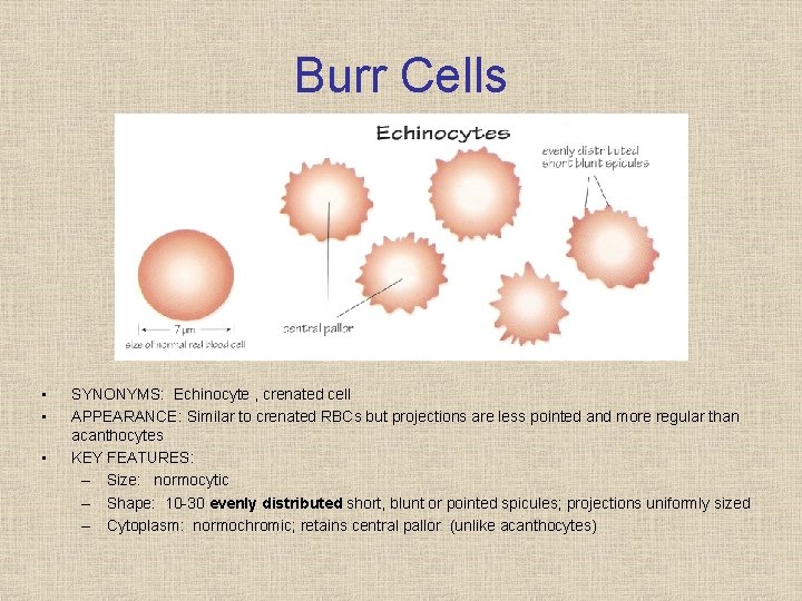
Burr Cells • • • SYNONYMS: Echinocyte , crenated cell APPEARANCE: Similar to crenated RBCs but projections are less pointed and more regular than acanthocytes KEY FEATURES: – Size: normocytic – Shape: 10 30 evenly distributed short, blunt or pointed spicules; projections uniformly sized – Cytoplasm: normochromic; retains central pallor (unlike acanthocytes)
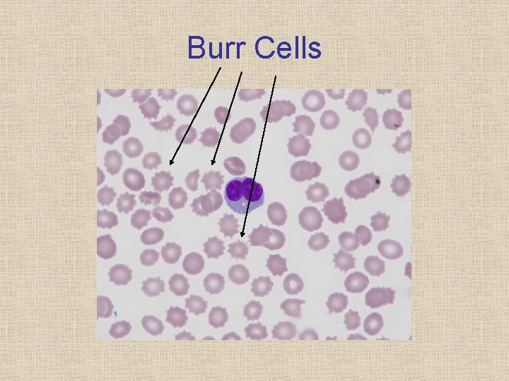
Burr Cells
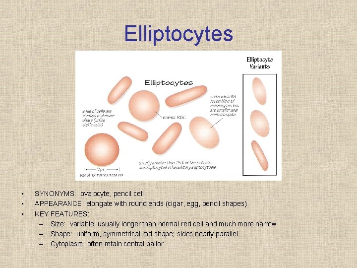
Elliptocytes • • • SYNONYMS: ovalocyte, pencil cell APPEARANCE: elongate with round ends (cigar, egg, pencil shapes) KEY FEATURES: – Size: variable; usually longer than normal red cell and much more narrow – Shape: uniform, symmetrical rod shape; sides nearly parallel – Cytoplasm: often retain central pallor
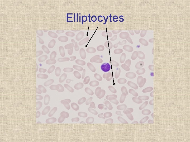
Elliptocytes
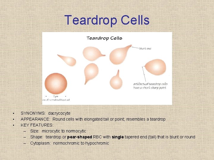
Teardrop Cells • • • SYNONYMS: dacrycocyte APPEARANCE: Round cells with elongated tail or point; resembles a teardrop KEY FEATURES: – Size: microcytic to normocytic – Shape: teardrop or pear-shaped RBC with single tapered end (tail) that is blunt or round – Cytoplasm: normochromic to hypochromic
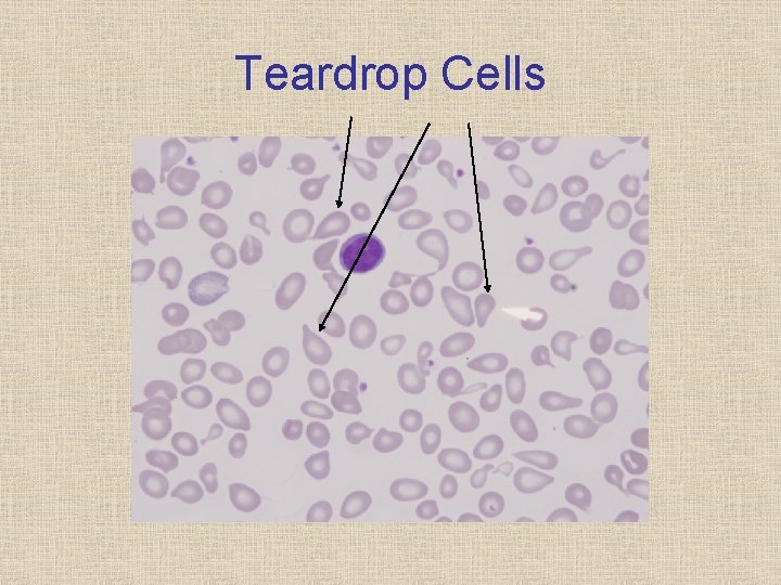
Teardrop Cells
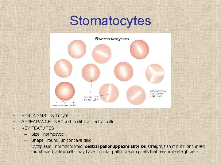
Stomatocytes • • • SYNONYMS: hydrocyte APPEARANCE: RBC with a slit like central pallor KEY FEATURES: – Size: normocytic – Shape: round; uniconcave disc – Cytoplasm: normochromic; central pallor appears slit-like, straight, fish mouth, or curved rod shaped; a few cells may have tri polar pallor creating cells that resemble sleigh bells
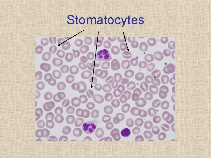
Stomatocytes
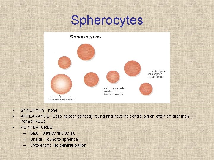
Spherocytes • • • SYNONYMS: none APPEARANCE: Cells appear perfectly round and have no central pallor; often smaller than normal RBCs KEY FEATURES: – Size: slightly microcytic – Shape: round to spherical – Cytoplasm: no central pallor

Spherocytes 50 x Objective
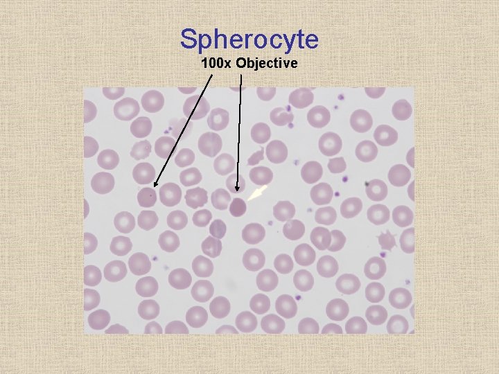
Spherocyte 100 x Objective
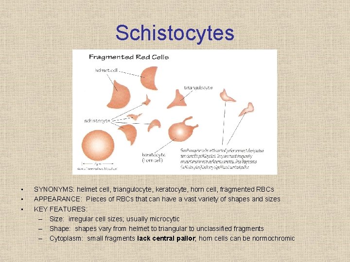
Schistocytes • • • SYNONYMS: helmet cell, triangulocyte, keratocyte, horn cell, fragmented RBCs APPEARANCE: Pieces of RBCs that can have a vast variety of shapes and sizes KEY FEATURES: – Size: irregular cell sizes; usually microcytic – Shape: shapes vary from helmet to triangular to unclassified fragments – Cytoplasm: small fragments lack central pallor; horn cells can be normochromic
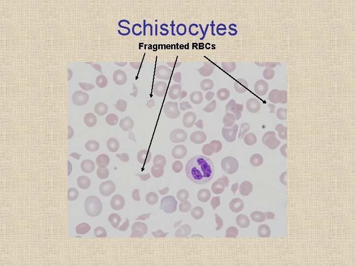
Schistocytes Fragmented RBCs
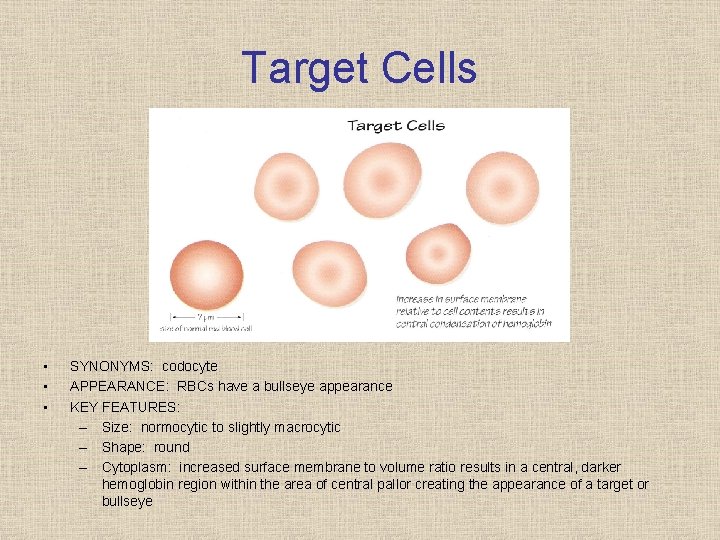
Target Cells • • • SYNONYMS: codocyte APPEARANCE: RBCs have a bullseye appearance KEY FEATURES: – Size: normocytic to slightly macrocytic – Shape: round – Cytoplasm: increased surface membrane to volume ratio results in a central, darker hemoglobin region within the area of central pallor creating the appearance of a target or bullseye
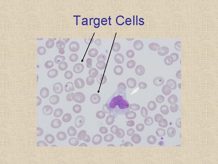
Target Cells
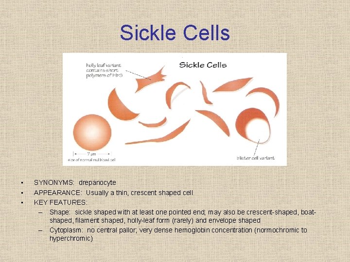
Sickle Cells • • • SYNONYMS: drepanocyte APPEARANCE: Usually a thin, crescent shaped cell KEY FEATURES: – Shape: sickle shaped with at least one pointed end; may also be crescent shaped, boat shaped, filament shaped, holly leaf form (rarely) and envelope shaped – Cytoplasm: no central pallor; very dense hemoglobin concentration (normochromic to hyperchromic)
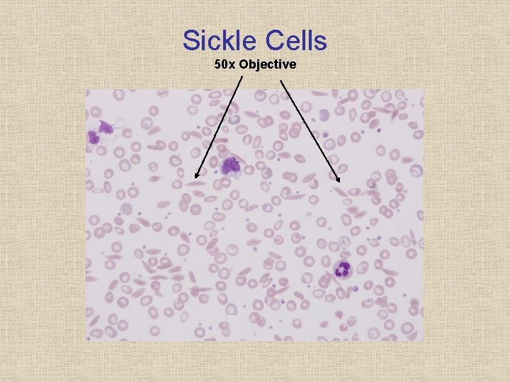
Sickle Cells 50 x Objective
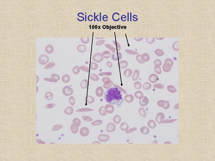
Sickle Cells 100 x Objective
RBC Inclusions • • • NRBCs Howell Jolly Bodies Basophilic Stippling Pappenheimer Bodies Hemoglobin Crystals

Nucleated Red Blood Cells • • • SYNONYMS: NRBCs APPEARANCE: RBC with a small, pyknotic nucleus with dense chromatin KEY FEATURES: – Size: 8 10 m size; slightly larger than a mature RBC – Cytoplasm: varies depending on stage of maturation; usually more blue gray
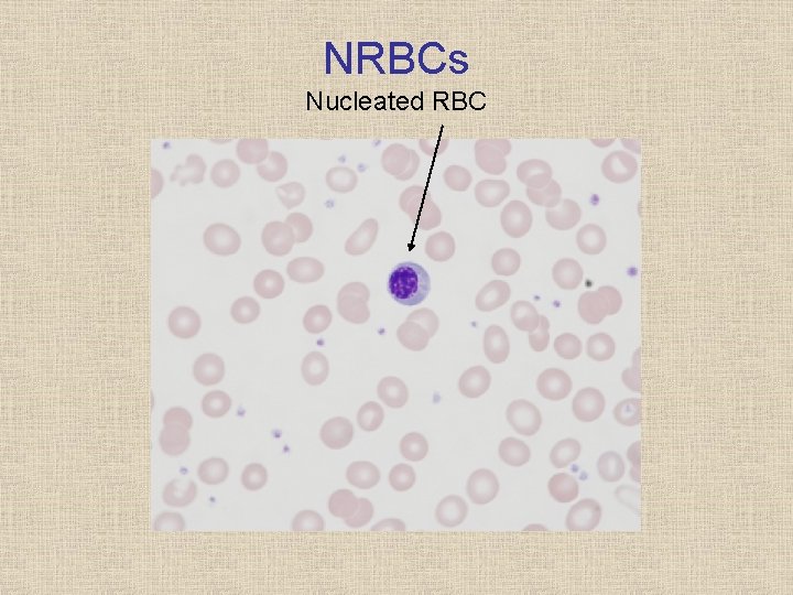
NRBCs Nucleated RBC
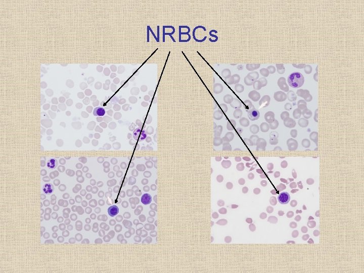
NRBCs
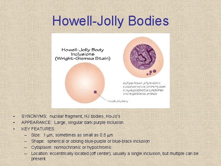
Howell Jolly Bodies • • • SYNONYMS: nuclear fragment, HJ bodies, Ho Jo’s APPEARANCE: Large, singular dark purple inclusion KEY FEATURES: – Size: 1 m; sometimes as small as 0. 5 m – Shape: spherical or oblong blue purple or blue black inclusion – Cytoplasm: normochromic or hypochromic – Location: eccentrically located (off center); usually a single inclusion, but multiple can be present

Howell Jolly Bodies Howell-Jolly Bodies Pappenheimer Bodies (Iron)
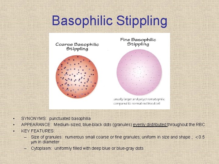
Basophilic Stippling • • • SYNONYMS: punctuated basophilia APPEARANCE: Medium sized, blue black dots (granules) evenly distributed throughout the RBC KEY FEATURES: – Size of granules: numerous small coarse or fine granules; uniform in size and shape ; 0. 5 m in diameter – Cytoplasm: uniformly filled with deep blue or blue gray dots
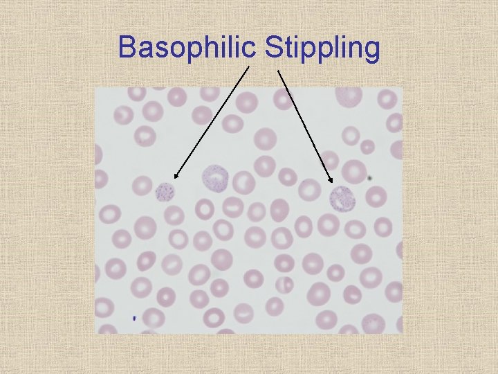
Basophilic Stippling
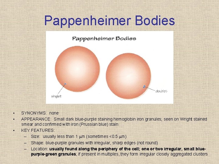
Pappenheimer Bodies • • • SYNONYMS: none APPEARANCE: Small dark blue purple staining hemoglobin iron granules; seen on Wright stained smear and confirmed with iron (Prussian blue) stain KEY FEATURES: – Size: usually less than 1 m (sometimes <0. 5 m) – Shape: blue purple granules with irregular, sharp edges (not round) – Location: usually found along the periphery of the cell; one or two irregular, small bluepurple-green granules; if present in multiples, they form irregular closely aggregated clusters
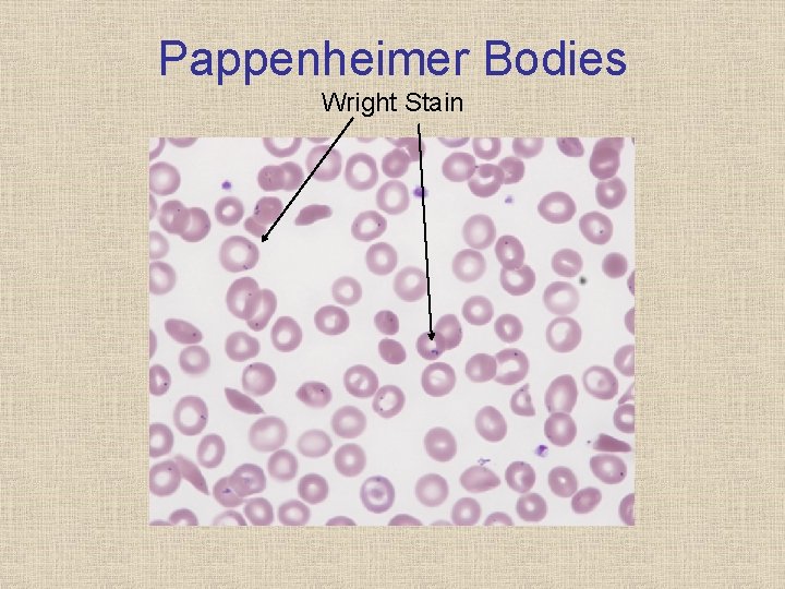
Pappenheimer Bodies Wright Stain
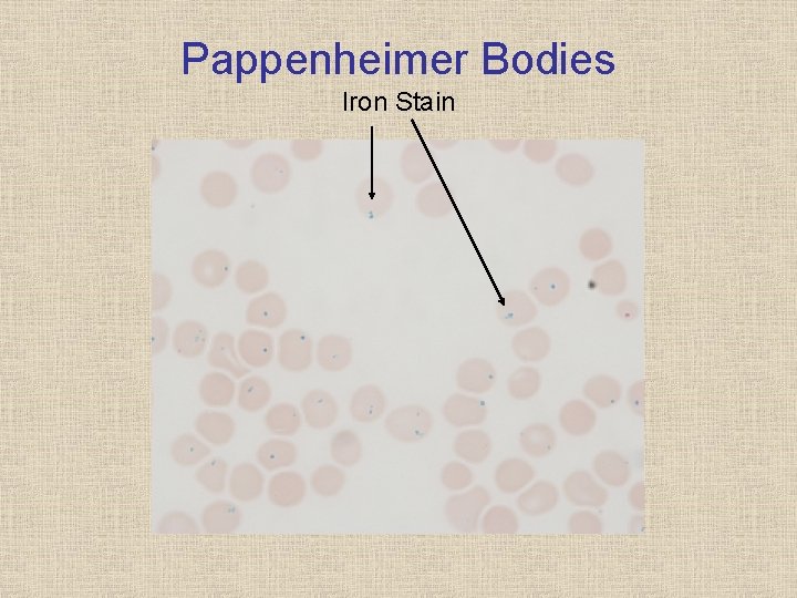
Pappenheimer Bodies Iron Stain
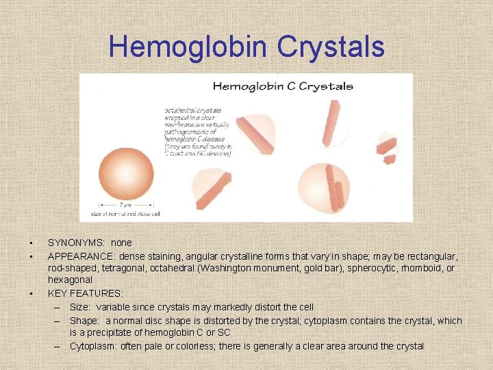
Hemoglobin Crystals • • • SYNONYMS: none APPEARANCE: dense staining, angular crystalline forms that vary in shape; may be rectangular, rod shaped, tetragonal, octahedral (Washington monument, gold bar), spherocytic, rhomboid, or hexagonal KEY FEATURES: – Size: variable since crystals may markedly distort the cell – Shape: a normal disc shape is distorted by the crystal; cytoplasm contains the crystal, which is a precipitate of hemoglobin C or SC – Cytoplasm: often pale or colorless; there is generally a clear area around the crystal
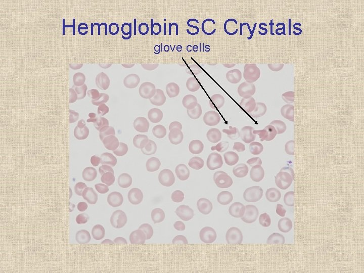
Hemoglobin SC Crystals glove cells
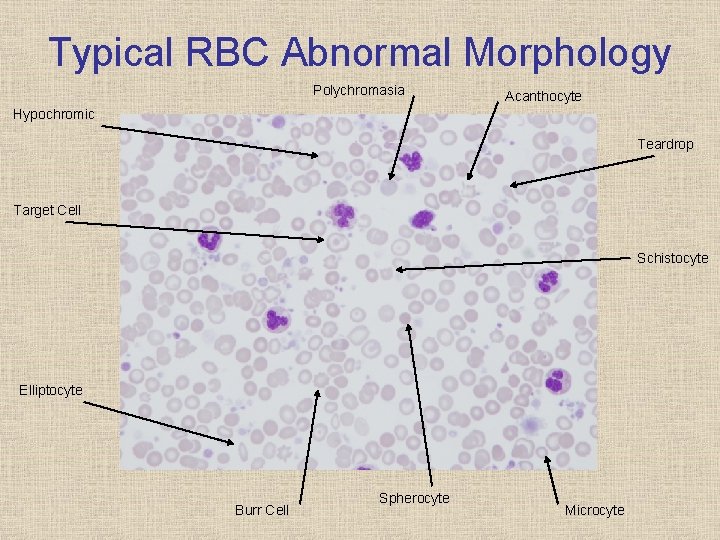
Typical RBC Abnormal Morphology Polychromasia Acanthocyte Hypochromic Teardrop Target Cell Schistocyte Elliptocyte Burr Cell Spherocyte Microcyte

Any Questions?
- Slides: 50