Red Blood Cells AnemiaErythrocytosi s Transfusion Phlebotomy blood
 수혈의학 (Transfusion)")
적혈구의 구조와 생리 혈액학 Red Blood Cells (Anemia/Erythrocytosi s) 수혈의학 (Transfusion)

Phlebotomy: blood specimen collection Source from Amazon. com

Tube Guide Order of Draw Name Laboratory use Number of inversions Collection volume Plain 7 ml 면역부(Cold, Cryo, Drug Test) 혈액은행(X-matching, ) None 7 ml CTAD 2. 7 ml 혈액부(PT, APTT) 면역부 (Protein C, Protein S, W. F(Ag, Activity)) 3~4 2. 7 ml 5 6 ml, 9. 5 ml 8 3 ml SST 6 ml (Serum Separation Tube) 면역부 생화학부 SST 9. 5 ml EDTA 3 ml Needle 21 G Luer Adapter Blood Collection Set 23 G (Butterfly Needle) Blood collection Technique 혈액부(CBC) 생화학부(Hb. A 1 C) 면역부(PLT associated Ab, CD 4, CD 8, CSA) 혈액은행(Direct Coombs’ test) 비고
")
TABLE 3 -6 Order of Draw: Evacuated Tube and Syringe 1 Blood-culture tubes (yellow) 2 Coagulation sodium citrate tube (blue stopper) 3 Serum tubes with or without clot activator or gel separator 4 Heparin tubes with or without gel (green stopper) 5 Ethylenediaminetetraacetic acid (EDTA) tubes (lavender stopper) 6 Glycolytic inhibitor tubes (gray stopper) Henry’s Clinical Diagnosis and Management

올바른 채혈 순서 1. Blood culture 2. Citrate tube 3. Plain tube or SST tube 4. Heparin tube 5. EDTA tube 6. Antiglycolytic tube

Peripheral blood & Bone marrow aspirates

Normal hematopoiesis

Normal hematopoiesis

Normal hematopoiesis

Diagnostic hematology WBC / platelet RBC
 수혈의학 (Transfusion)")
적혈구의 구조와 생리 혈액학 Red Blood Cells (Anemia/Erythrocytosi s) 수혈의학 (Transfusion)

Erythropoiesis Normoblast BM Reticulocyte RBC PB

Erythropoiesis Normoblast BM RBC and its precursors Reticulocyte RBC PB

Structure of Red Blood Cells • Biconcave disk shape – Diameter 7. 5 -8 um – Morphological changes for oxygen transport

Morphological changes for oxygen transport

• Lipid Bilayer – Membrane skeleton • RBCs shape & reversible deformability – Diffuse, passive, & active transport – RBC membrane defect • Congenital spherocytosis

Normoblast BM Reticulocyte RBC PB
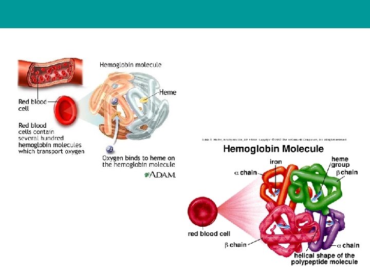

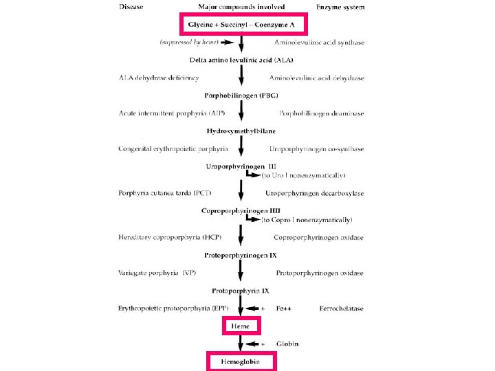
Heme = a porphyrin ring + an iron atom Hemoglobin = one heme + one polypeptide chain
, liver, etc – Succinyl")
Heme and Hemoglobin • Heme – Synthesis : bone marrow(85%), liver, etc – Succinyl co. A + glycine [mitochondrion] – ALA synthetase • Hemoglobin – One heme group + one polypeptide chain – 1 g of Hb binds 1. 34 m. L of oxygen – 600 g (normal adult red blood cells) of Hb • 800 m. L of oxygen
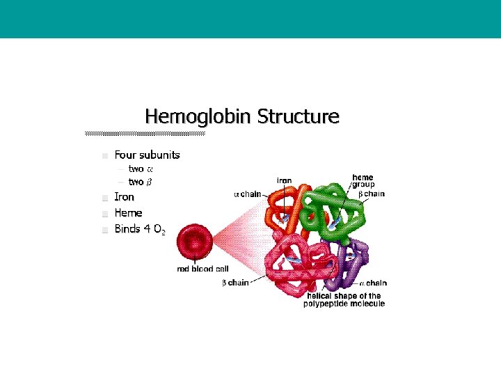
 = two pairs of polypeptide chain + each")
• Normal Human hemoglobin(Hb A) = two pairs of polypeptide chain + each chain carries a heme portion

• Three normal hemoglobins – Hb A α 2β 2 – Hb A 2 α 2δ 2 – Hb F α 2γ 2 • Globin chain genes – – – Gene α β γ δ Number 2/2 1/1 1/1 Chromosome 16 11 11 11

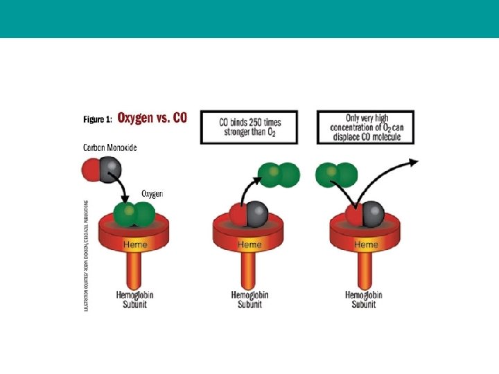
• Main function of hemoglobin “ Oxygen delivery” 관여하는 factor 1. Hemoglobin molecule • Amino acid substitution 2. Blood flow, rate of diffusion of oxygen, oxygen affinity • Oxygen Dissociation Curve – Relationship btw the amount of oxyhemoglobin(% saturation) and partial pressure of oxygen(oxygen tension)

• Oxygen Dissociation Curve • p. H, 2, 3 -DPG, temp. • P 50 • Oxygen affinity of Hb • Partial pressure of oxygen at the Hb is half-oxygenated • Inversely related to O 2 affinity • 37’C, PCO 2 40 mm. Hg, p. H 7. 4 • p 50 27 mm. Hg • Ferrous hemoglobin(2+) • Ferric hemoglobin(3+) • loss of oxygen delivery
![Iron Metabolism • Iron – – • Heme iron [ Hb, myoglobin, heme proteins]](http://slidetodoc.com/presentation_image/4ff5c8733133ad12b2ffc9fbb01c1890/image-29.jpg "Iron Metabolism • Iron – – • Heme iron [ Hb, myoglobin, heme proteins]")
Iron Metabolism • Iron – – • Heme iron [ Hb, myoglobin, heme proteins] Non-heme iron [ferric state] Iron distributed in six compartments – – – Hemoglobin(1 ml packed RBC contains 1 mg iron) Storage iron (ferritin, hemosiderin) Myoglobin Labile iron pool Tissue iron Transport iron
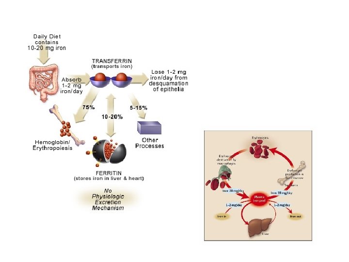

• Absorption of iron – Balance • Daily losses (M: 1 mg, 2 mg F) – Mucosal receptor on duodenum/upper jejunum – Normal state ; ingested iron 5 ~ 20% absorbed – Mechanism unclear – Absorbed iron • Sequestrated within the mucosal epi. Cell • Enter the circulation
 – Synthesis in the liver")
• Iron transport…Transferrin – Plasma iron carrier protein(transport) – Synthesis in the liver – Inversely regulated by body’s iron stores – Normal state • 1/3 of binding sites of transferrin are saturated with iron – Transferrin-iron complex to erythrocyte • Majority of iron – heme systhesis • Small amount of residual non-heme iron [ferritin]
 – 20 ~ 60% of normoblasts – Number")
– Small ferritin granule—sideroblast(1 ~ 5) – 20 ~ 60% of normoblasts – Number of sideroblasts • Availability of iron • The incorporation of iron into heme Normoblast Sideroblas t Ringed sideroblast

• Storage of iron – – In the RBCs… splenic macrophages Hb catabolized into iron, protophophyrin, globin Iron is stored in RES system (spleen, BM, liver) Two forms- ferritin(major), hemosiderin – s-Ferritin reflects body iron storages • Ferritin in RES system leaks into circulation • Decrease s-ferritin– valid indication of decreased iron stores • Not useful s-ferritin – Acute phase reactant, prolonged elevated – Underlying inflammatory disease, neoplasia, liver disease

• Soluble transferrin receptor • Bone marrow …Prussian blue stain – Storage iron – Sideroblast
- Slides: 35