Reconstructive Flap 101 Basic Principles Imaging and Beyond

Reconstructive Flap 101: Basic Principles, Imaging, and Beyond e. Ed. E#: e. Ed. E-88 E Supsupin 1, I Alava 2, D Freet 3, E Bonfante 1, S Garza 1, Y Weinstock 2 Institutions: 1 University of Texas Houston Department of Diagnostic & Interventional Imaging, Houston, TX, 2 University of Texas Houston Department of Otorhinolaryngology, Head & Neck Surgery, Houston, TX, 3 University of Texas Houston Department of Plastic Surgery, Houston, TX

Author disclosures None

Reconstructive flaps in head &neck surgery ü Basics of flaps ü Case illustrations (including imaging): q Flaps for restitution of function and cosmesis q Flap for cosmesis

Define “Flap” A “Flap” can be defined as a piece of tissue that is moved from one part of the body to another part of the body with a blood supply intact.

Flaps v A flap is referred to as a “Pedicled” flap if its blood supply is left intact at its origin when it is moved to the new site. v A flap is referred to as a “Free” flap if the blood supply of the donor tissue to be moved is severed from its origin and then re-connected to a new artery and vein at the recipient site. v A flap can be made of any type of tissue: bone, cartilage, fascia, subcutaneous tissue, skin, or any combination of these.

Flaps v Flaps are required for reconstruction of large composite defects resulting from trauma or cancer extirpation. v A flap is referred to as a “Free” flap if the blood supply of the donor tissue to be moved is severed from its origin and then re-connected to a new artery and vein at the recipient site. v A flap can be made of any type of tissue: bone, cartilage, fascia, subcutaneous tissue, skin, or any combination of these.

Flaps v Flaps can be used for cosmetic purposes, functional purposes, or a combination of both depending on the needs of the patient.

Flaps v Flaps can be used for cosmetic purposes, functional purposes, or a combination of both depending on the needs of the patient.

What are commonly used flaps for head and neck reconstruction? Ø Pectoralis major Ø Latissimus dorsi Ø Rectus Abdominis Ø Radial Forearm Ø Free Fibula Ø Free Iliac Crest

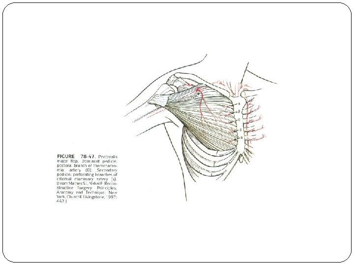
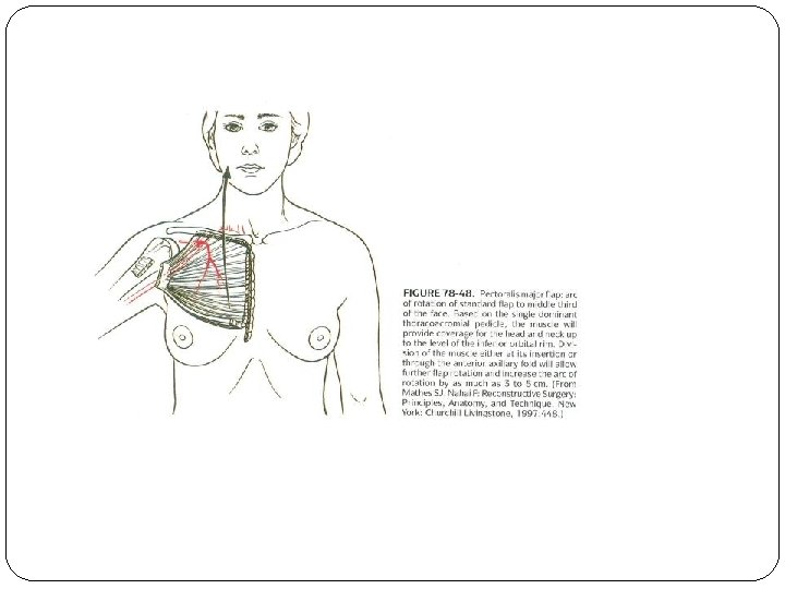
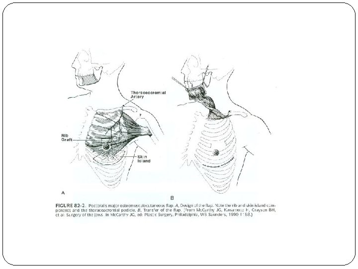
Pectoralis major Ø Is considered a “work horse” in head and neck reconstruction Ø Can be used as a muscle-only flap or can include skin Ø Most commonly transferred as a pedicled flap based on the pectoral branch of the thoracoacromial arterial trunk Ø Can be used for intra-oral lining, head and neck skin resurfacing or tubed for creation of a
. (2006). Plastic Surgery Volum The Head and Neck,")
From: Mathes, S. J. (Ed. ). (2006). Plastic Surgery Volum The Head and Neck, Part 2. Philadelphia, PA: Saunders Elsevier.

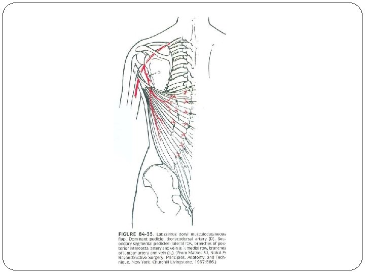
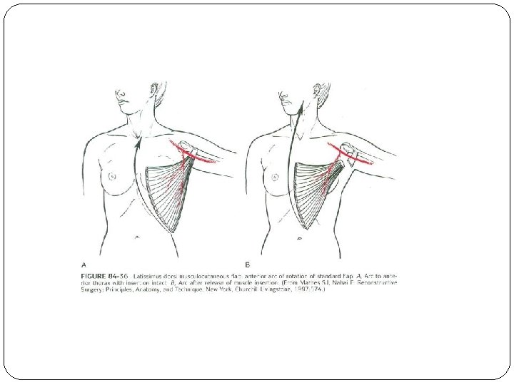
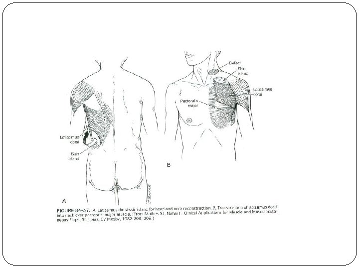
Latissimus Dorsi Ø Can be used as a pedicled flap or free flap Ø Large bulky muscle or muscle and skin flap based on the thoracodorsal branch of the subscapular artery Ø In head and neck reconstruction, this flap is used to fill large defects. Ø Can provide a very large amount of skin if needed Ø Not commonly used secondary to its bulky nature
. (2006). Plastic Surgery Volume 3: The Head and")
From: Mathes, S. J. (Ed. ). (2006). Plastic Surgery Volume 3: The Head and Neck, Part 2. Philadelphia, PA: Saunders Elsevier.

Rectus abdominis Ø Always used as a free flap Ø Muscle or muscle and skin flap based on the deep inferior epigastric artery Ø Used to fill large defects such as that of a total glossectomy Ø Disadvantage is abdominal wall weakness.
. (2006). Plastic Surgery Volume 3: The Head and")
From: Mathes, S. J. (Ed. ). (2006). Plastic Surgery Volume 3: The Head and Neck, Part 2. Philadelphia, PA: Saunders Elsevier.

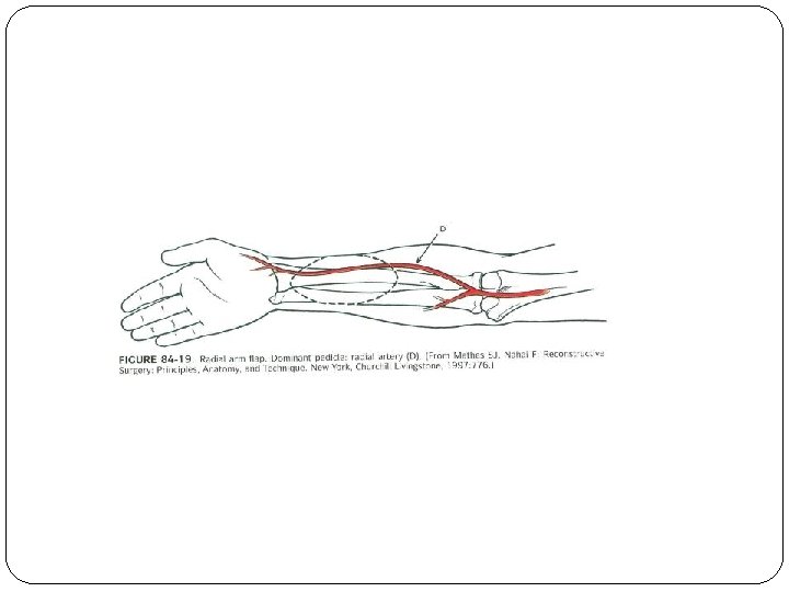
Radial Forearm Ø This is a free flap based on the radial artery. Ø Thin fasciocutaneous (fascia and skin) flap that can be used for intra-oral lining, used to resurface a tongue defect or can be tubed to create a neopharynx Ø Donor site can be problematic (poor healing).
. (2006). Plastic Surgery Volume 3: The Head and")
From: Mathes, S. J. (Ed. ). (2006). Plastic Surgery Volume 3: The Head and Neck, Part 2. Philadelphia, PA: Saunders Elsevier.
. (2006). Plastic Surgery Volume 3: The Head and")
From: Mathes, S. J. (Ed. ). (2006). Plastic Surgery Volume 3: The Head and Neck, Part 2. Philadelphia, PA: Saunders Elsevier.
. (2006). Plastic Surgery Volume 3: The Head and")
From: Mathes, S. J. (Ed. ). (2006). Plastic Surgery Volume 3: The Head and Neck, Part 2. Philadelphia, PA: Saunders Elsevier.

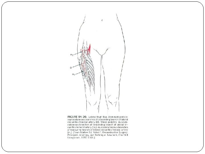
Anteriolateral thigh Ø Free flap that is based on the descending branch of the lateral circumflex femoral artery. Ø Fasciocutaneous flap that can be made very large for large complex defects. Ø Can be used for intra-oral lining, can be tubed for neo-pharynx, can be used for head and neck resurfacing, or can be a combination of any of these Ø Minimal donor site morbidity
. (2006). Plastic Surgery Volume 3: The Head and")
From: Mathes, S. J. (Ed. ). (2006). Plastic Surgery Volume 3: The Head and Neck, Part 2. Philadelphia, PA: Saunders Elsevier.
. (2006). Plastic Surgery Volume 3: The Head and")
From: Mathes, S. J. (Ed. ). (2006). Plastic Surgery Volume 3: The Head and Neck, Part 2. Philadelphia, PA: Saunders Elsevier.
. (2006). Plastic Surgery Volume 3: The Head and")
From: Mathes, S. J. (Ed. ). (2006). Plastic Surgery Volume 3: The Head and Neck, Part 2. Philadelphia, PA: Saunders Elsevier.

Free Fibula Ø Free flap based on the peroneal artery that uses a portion of the fibula and sometimes the overlying skin of the leg for complex reconstruction of the mandible and intra-oral lining Ø An excellent source of a large amount of bone. Ø Minimal donor site morbidity
. (2006). Plastic Surgery Volume 3: The Head and")
From: Mathes, S. J. (Ed. ). (2006). Plastic Surgery Volume 3: The Head and Neck, Part 2. Philadelphia, PA: Saunders Elsevier.
. (2006). Plastic Surgery Volume 3: The Head and")
From: Mathes, S. J. (Ed. ). (2006). Plastic Surgery Volume 3: The Head and Neck, Part 2. Philadelphia, PA: Saunders Elsevier.
. (2006). Plastic Surgery Volume 3: The Head")
 From: Mathes, S. J. (Ed. ). (2006). Plastic Surgery Volume 3: The Head and Neck, Part 2. Philadelphia, PA: Saunders Elsevier.
. (2006). Plastic Surgery Volume 3: The Head and")
From: Mathes, S. J. (Ed. ). (2006). Plastic Surgery Volume 3: The Head and Neck, Part 2. Philadelphia, PA: Saunders Elsevier.

Free Iliac Crest Ø Free flap that is based on the deep circumflex iliac artery. Ø Can be bone only or can be bone and skin Ø Used mostly for mandibular reconstruction. Skin can be used for intra-oral lining. Ø Donor site can be very painful.

Image: AO Foundation

Flap for subtotal glossectomy Necrotic, malignant infiltrating mass in the left tongue/floor of mouth. Resection of the mass and free flap reconstruction was planned.

Flap for subtotal glossectomy Tongue mass is resected. Neck dissection prior to extirpation of mass. Apron flap is lifted up (yellow arcs).
 with")
Flap for subtotal glossectomy Flap is harvested from the anterolateral thigh Flap (bracket) with its vascular (arrows) pedicle is shown.

Flap for subtotal glossectomy Harvested flap from thigh Free flap sawn into tongue stump and floor of mouth Immediate postoperative result

Flap for total glossectomy Images taken Intraoperatively and 1 month after surgery This case illustrates the use of free flap mainly for restoration of function.

Flap for layngopharyngectomy This patient presented with advanced transglottic laryngeal squamous cell cancer that ruptured into the skin (brackets & circle). The mass has obliterated the upper airway requiring tracheostomy (red arrow). Total laryngopharyngectomy with Free flap reconstruction was planned.

Flap for layngopharyngectomy Large flap harvested from anterolateral thigh for free flap reconstruction and creation of neopharynx

Flap for layngopharyngectomy Defect after neck dissection and extirpation of mass

Flap for layngopharyngectomy Creation of tube/neopharynx from free flap harvested from thigh

Flap for layngopharyngectomy Creation of tube/neopharynx from free flap harvested from thigh Uvula – yellow arrow

Flap for layngopharyngectomy This case illustrates the use of free flap for creation of neopharynx and closure of a large soft tissue defect. Flap is employed both for restitution of function and cosmesis.

Pectoralis flap A diabetic woman with poor glycemic control who developed multicompartmental abscesses in the suprahyoid neck and necrotizing fasciitis (CT). Full thickness loss of tissue in the face and neck after debridement with exposed mandible (arrow). Flap reconstruction is required to close the defect.

Pectoralis flap is used to fill the soft tissue defect.

Pectoralis flap - postop Free flap used to fill the soft tissue defect in the left face and neck, and create the lining of the oral cavity. There is persistent enhancement in the masticator space on MR (circle) a year after surgery, with no clinical signs of active infection.

Pectoralis myocutaneous flap - postop M F Postoperative CT showing the pectoralis myocutaneous flap (arrows). M – muscle FFat In this case the flap serve both purposes of restoration of function and cosmesis

Total glossectomy and mandibular resection with free flap reconstruction This patient presented with oral tongue mass, later proven to be squamous cell carcinoma. The plan was resection of the tongue mass including the mandible, with free flap reconstruction.

Total glossectomy and mandibular resection with free flap reconstruction Courtesy of James Melville DDS Resection of tongue and mandible

Total glossectomy and mandibular resection with free flap reconstruction Courtesy of James Melville DDS Specimen removed

Total glossectomy and mandibular resection with free flap reconstruction Courtesy of James Melville DDS After resection of tongue and mandible Harvesting flap from thigh

Total glossectomy and mandibular resection with free flap reconstruction Courtesy of James Melville DDS Mandibular reconstruction with Titanium chain

Total glossectomy and mandibular resection with free flap reconstruction Courtesy of James Melville DDS Flap implantation

Total glossectomy and mandibular resection with free flap reconstruction Courtesy of James Melville DDS Immediate postoperative result – this case illustrates use of flap for both restoration of function and cosmesis

Example of free flap reconstruction for cosmesis Preoperative CT with cosmetic defect on the right (bracket) after prior parotidectomy 10 years earlier for adenoid cystic carcinoma. Free flap reconstruction was decided Flap harvested from after a negative workup for the thigh disease recurrence. Neck dissection in preparation for Immediate postsurgical free flap reconstruction result SCM – sternocleidomastoid muscle CCA – common carotid artery bifurcation

Example of free flap reconstruction for cosmesis Before After

Summary �Reconstructive flaps in the head and neck are used for restitution of function or cosmesis, or both, depending on the needs of the patient. �Case illustrations are provided.

Bibliography of illustrations Slides 11 -14; 16 -19; 21; 23 -26; 28 -31; 33 -36; 38 Mathes, S. J. (Ed. ). (2006). Plastic Surgery Volume 3: The Head and Neck, Part 2. Philadelphia, PA: Saunders Elsevier. Slide 36: “Reconstruction: Harvesting. ” AOfoundation. org. Web. 23 Mar. 2015.
- Slides: 64