RECOMBINATION Contents Introduction Recombination Types of recombination Homologous

RECOMBINATION

Contents • Introduction • Recombination • Types of recombination • Homologous recombination • Holliday model • Enzymes involved in homologous recombinatio • Sites specific recombination

Introduction The previous seminar on repair and mutaion of DNA dealt mainly with small changes in DNA sequences resulting from errors in replication or damage to DNA. The DNA sequence of a chromosome can change in large segments as well, by the processes of recombination and transposition

Accurate DNA replication and repair of DNA damage are essential to maintaining genetic information and ensuring its accurate transmission from parent to offspring. Recombination plays an important role in this process by allowing genes to be reassorted into different combinations. Genetic recombination results in the exchange of genes between paired homologous chromosomes during meiosis From the standpoint of evolution, however, it is also important to generate genetic diversity. However, increasing genetic diversity is not the only role

Recombination is also an important mechanism for repairing damaged DNA. Recombination is involved in rearrangements of specific DNA sequences that alter the expression and function of some genes during development and differentiation. Thus, recombination plays important roles in the lives of individual cells and organisms, as well as contributing to the genetic diversity of the species.
 from two parental DNA molecules or")
Recombination is the production of new DNA molecule(s) from two parental DNA molecules or different segments of the same DNA molecule. Transposition is a highly specialized form of recombination in which a segment of DNA moves from one location to another, either on the same chromosome or a different chromosome.

TYPES OF RECOMBINATION General or homologous recombination: occurs between DNA molecules of very similar sequence. Illegitimate or nonhomologous recombination : occurs in regions where no large-scale sequence similarity is apparent. Site-specific recombination occurs between particular short sequences (about 12 to 24 bp) present on otherwise dissimilar parental molecules. Replicative recombination, which generates a new copy of a segment of DNA.

Figure 1. Types of natural recombination.

General Recombination General recombination is an integral part of the complex process of meiosis in sexually reproducing organisms. It results in a crossing over between pairs of genes along a chromosome, which are revealed in appropriate matings. The chiasmata that link homologous chromosomes during meiosis are the likely sites of the crossovers that result in recombination. General recombination also occurs in nonsexual organisms when two copies of a chromosome or chromosomal segment are present. Also, the retrieval system for post-replicative repair involves general recombination. The mechanism of recombination has been intensively studied in bacteria and fungi, and some of the enzymes involved have been well characterized.
 Crossing over often produces an exchange of genetic material.")
FIGURE 2 Crossing over. (a) Crossing over often produces an exchange of genetic material. (b) The homologous chromosomes of a grasshopper are shown during prophase I of meiosis. Many points of joining (chiasmata) are evident between the two homologous pairs of chromatids. These chiasmata are the physical manifestation of prior homologous recombination (crossing over) events.

Figure 3 Models of recombination In copy choice, recombination occurs during the synthesis of daughter DNA molecules. DNA replication starts with one parental DNA template and then switches to a second parental molecule, resulting in the synthesis of recombinant daughter DNAs containing sequences homologous to both parents. In breakage and rejoining, recombination occurs as a result of breakage and crosswise rejoining of parental DNA molecules.

Figure 4 Experimental demonstration of recombination by breakage and rejoining

Figure 4 Homologous recombination by complementary base pairing Parental DNAs are broken at staggered sites, and overlapping single-stranded regions are exchanged via base pairing with homologous sequences. The result is a heteroduplex region, in which the two DNA strands are derived from different parental molecules.

Figure 5. The Holliday model for homologous recombination Single-strand nicks are introduced at the same position on both parental molecules. The nicked strands then exchange by complementary base pairing, and ligation produces a crossed-strand intermediate called a Holliday junction.

Holliday Model One of the first plausible models to account for the preceding observations was formulated by Robin Holliday. The key features of the Holliday model are the formation of heteroduplex DNA; The creation of a cross bridge; its migration along the two heteroduplex strands, termed branch migration; the occurrence of mismatch repair; and the subsequent resolution, or splicing, of the intermediate structure to yield different types of recombinant molecules
 Nationality British")
Robin Holliday Born 6 November 1932 Died 9 April 2014 (aged 81) Nationality British Occupation Molecular biologist Known for Holliday junction

Figure 7. A prototype mechanism for genetic recombination

Enzymatic cleavage and the creation of heteroduplex DNA This partially heteroduplex double helix is a crucial intermediate in recombination, and has been termed the Holliday structure. Or Holliday Junction.

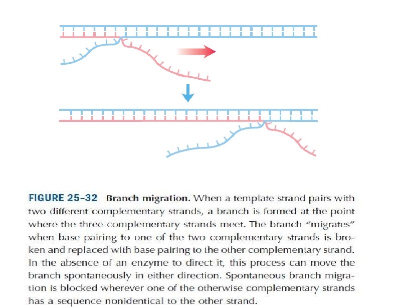
Branch migration The Holliday structure creates a cross bridge, or branch, that can move, or migrate, along the heteroduplex. This phenomenon of branch migration is a distinctive property of the Holliday structure.

Figure 8 Branch migration, the movement of the crossover point between DNA complexes. (After T. Broker, Journal of Molecular Biology 81, 1973, 1; from J. D. Watson et al. , Molecular Biology of the Gene, 4 th ed. Copyright © 1987 by Benjamin Cummings. )

Resolution of the Holliday structure The Holliday structure can be resolved by cutting and ligating either the two originally exchanged strands or the originally unexchanged strands. The former generates a pair of duplexes that are parental, except for a stretch in the middle containing one strand from each parent. If the two parents had different alleles in this stretch, as indicated here, then the DNA will be heteroduplex. The latter resolution step generates two duplexes that are recombinant, with a stretch of heteroduplex DNA.
 The Holliday structure shown in an extended form. (b) The rotation")
Figure 9 (a) The Holliday structure shown in an extended form. (b) The rotation of the structure shown in part a can yield the form depicted in part c. Resolution of the structure shown in part c can proceed in two ways, depending on the points of enzymatic cleavage, yielding the structures shown in part d. The dotted lines show which segments will rejoin to form recombinant strands for each particular cleavage scheme. The strands are shown linearly in part e and can be repaired to the forms shown in part f. (From H. Potter and D. Dressler, Cold Spring Harbor Symposium on Quantitative Biology 43, 1970, 970. Cold Spring Harbor Laboratory, Cold Spring Harbor, NY. )
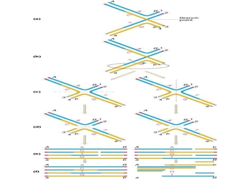

Figure 10. Isomerization and resolution of Holliday junctions are resolved by cutting and rejoining of the crossed strands. If the. Holliday junction formed by the initial strand exchange is resolved, the resulting progeny are heteroduplexes but are not recombinant for genetic markers outside of the heteroduplex region. Two rotations of the crossed-strand molecule, however, produce an isomer in which the unbroken parental strands, rather than the initially nicked strands, are crossed. Cutting and rejoining of the crossed strands of this isomer yield progeny that are recombinant heteroduplexes.
 A duplex is cut")
Meselson-Radding model Figure 11. The Meselson. Radding heteroduplex model. (a) A duplex is cut on one chain. (b)DNA polymerase displaces one chain. (c) The resulting single chain displaces its counterpart in the homolog. (d) This displaced chain is enzymatically digested. (e) Ligation completes the formation of a Holliday junction, which is genetically asymmetric in that only one of the two duplexes has a region of potentially heteroduplex DNA. If the junction migrates, heteroduplex DNA can arise on both duplexes. (f) Resolution of the junction occurs as in the Holliday model. (From F. W. Stahl, “The Holliday Junction on Its Thirtieth Anniversary, ” Genetics 138, 1994, 241– 246. )

Application of the Holliday model to genetic crosses Figure 12. When a mismatch is generated within a heteroduplex region, mismatch repair converts the mismatch into either the wild-type or the mutant sequence.

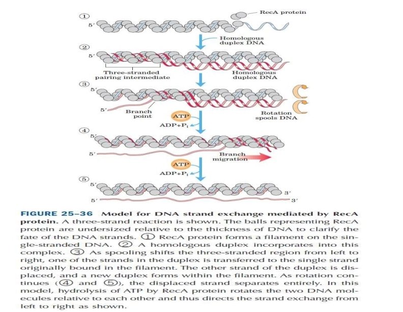
Enzymes Involved in Homologous Recombination Rec. A : which promotes the exchange of strands between homologous DNAs that causes heteroduplexes to form. • The action of Rec. A can be considered in three stages: I. First, the Rec. A protein binds to single-stranded DNA, , II. forming a complex between the two DNAs. III. The Rec. A protein then catalyzes strand exchange, with the single strand originally coated with Rec. A displacing its homologous strand to form a heteroduplex. Thus, the Rec. A protein is capable of catalyzing, by itself, the strand exchange reactions that are central to the formation of Holliday junctions.

Figure 12. Function of the Rec. A protein Rec. A initially binds to single-stranded DNA to form a protein-DNA filament. The Rec. A protein that coats the single-stranded DNA then binds to a second, double-stranded DNA molecule to form a non-base-paired complex. Complementary base pairing and strand exchange follow, forming a heteroduplex region.

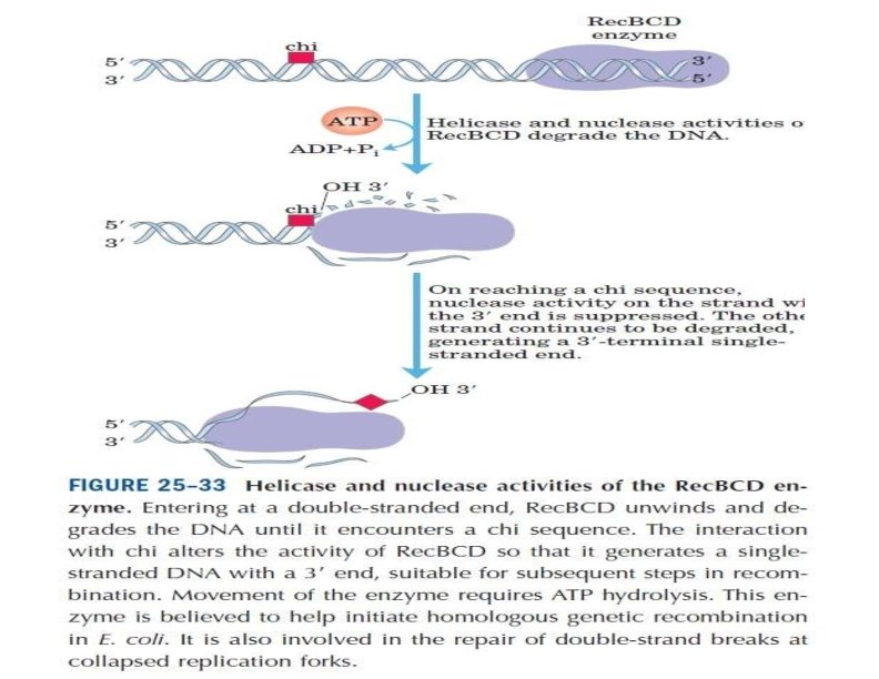
Rec. BCD enzyme Most recombination events in E. coli also require the Rec. BCD enzyme, which is a complex of three proteins (Rec. B, C, and D). The properties of Rec. BCD are consistent with the hypothesis that it initiates recombination by providing the single-stranded DNA to which Rec. A binds. Rec. BCD accomplishes this task by unwinding and nicking double-stranded DNA. The Rec. BCD complex binds to the end of a DNA molecule and then acts as a helicase, transiently unwinding the DNA as it travels along the molecule. When it encounters a specific nucleotide sequence (GCTGGTGG, called a chi site), Rec. BCD acts as a nuclease to introduce a single-strand nick. It then continues to unwind the double helix, forming a displaced single strand to which Rec. A can bind to initiate strand exchange.

Figure 13. Initiation of recombination by Rec. BCD The E. coli Rec. BCD complex binds to the end of a DNA molecule and unwinds the DNA as it travels along the molecule. When it encounters a specific sequence (called a chi site), it nicks the DNA strand. Continued unwinding then forms a displaced single strand to which Rec. A can bind

Ruv A, B, C Once a Holliday junction is formed, three other E. coli proteins (Ruv. A, B, and C) become involved in : Ruv. A and Ruv. B act as a complex to drive the migration of the site at which the strands cross in the Holliday junction, thereby varying the extent of the heteroduplex region and the position at which the crossed strands will be cut and rejoined. Ruv. C then resolves Holliday junctions by cleaving the crossed DNA strands. Rejoining of the cleaved strands by ligation completes the process, yielding two recombinant molecules.

Figure 13. Branch migration catalyzed by Ruv. A and Ruv. B. From Eggleston, A. K. and West, S. C. (1996) Trends in Genetics 12: 20 -25.

Figure 14 Branch migration and resolution of Holliday junctions Two E. coli proteins (Ruv. A and Ruv. B) together catalyze the movement of the crossedstrand site in Holliday junctions (branch migration). Ruv. C resolves the Holliday junctions by cleaving the crossed strands, which are then joined by ligase.

Figure 8. 20. Resolution requires cleavage by Ruv. C dimers. Adapted from Eggleston, A. K. and West, S. C. (1996) Trends in Genetics 12: 20 -25.

Site-Specific Recombination Results in Precise DNA Rearrangements is a very different type of process: recombination is limited to specific sequences. Recombination reactions of this type occur in virtually every cell, filling specialized roles that vary greatly from one species to another. Examples include : I. regulation of the expression of certain genes and II. promotion of programmed DNA rearrangements in embryonic development or in the replication cycles of someviral and plasmid DNAs. Each site-specific recombination system consists of an enzyme called a recombinaseand a short (20 to 200 bp), unique DNA sequence where the recombinase acts (the recombination site).

FIGURE 25– 38 A site-specific recombination reaction. Tyr recombinase

Fig. 15 A surface contour model of a four-subunit integraseclass recombinase called the Cre recombinase, bound to a Holliday intermediate (shown with light blue and dark blue helix strands). Theprotein has been rendered transparent so that the bound DNA is visible (derived from PDB ID 3 CRX).

Effects of site-specific recombination

References Cooper - Recombination in DNA Sequences – “The Cell” – 2 nd Edition. Nelson L. David(University of Wisconsin–Madison), Cox M. Michael (University of Wisconsin–Madison)- DNA Recombination(page no. 983 -991), “Principles of Biochemistry”- 4 th Edition

- Slides: 43