RECENT ADVANCES IN RAPID MICROBIAL IDENTIFICATION AND CHARACTERIZATION

RECENT ADVANCES IN RAPID MICROBIAL IDENTIFICATION AND CHARACTERIZATION TECHNIQUES Mackenzie Slifierz Hiu Ching Lai James Feiner

PUBLIC HEALTH EMERGENCIES Hurricane Katrina, 2005 At least five level 3 biosafety labs within hurricane zone.

PUBLIC HEALTH EMERGENCIES Hurricane Ike, 2008 Galveston Nation Labs, a level 4 biosafety lab working with potential bioterrorism pathogens including hantavirus, anthrax, and ebola virus.
 PCR-based Methods")
PRESENTATION OVERVIEW 1. Traditional Detection Methods 2. Advanced Rapid Detection Methods: i) PCR-based Methods ii) Nanotechnology iii) Immunoassays iv) Other Rapid Microbial Methods 3. General advantages and limitations 4. Conclusion

TRADITIONAL DETECTION METHODS Culturing bacteria: colony morphology, colour, and size. Staining: Gram stain, spore stain, flagella, cell morphology. Biochemical analysis: carbohydrate utilization and fermentation. Inhibition: Bile-salts, antibiotic resistance, dye tolerance.

Traditional Detection Methods
")
RAPID DETECTION TECHNIQUES What is a rapid detection technique? A rapid microbial method (RMM) is any technique which can identify or characterize a microorganism in hours or minutes as opposed to days or weeks. Some techniques that can be used for rapid analysis: PCR-based Methods Nanotechnology Immunoassays Spectroscopy Chromatography

Rapid Identification and Characterization Techniques PCR-based Methods

PCR: POLYMERASE CHAIN REACTION Amplification of microbial DNA. Detection only requires very little DNA. Sample is directly from food/clinical sources. No Culturing. Advantages: Low cost (PCR machine: $500) Quick assay (3. 5 hrs)
 estimates the quantity of microorganisms in the sample:")
Real-Time PCR (RT-PCR) estimates the quantity of microorganisms in the sample:

Designing Primers can be designed from broad-range/highly conserved bacterial 16 S ribosomal genes. This allows detection of most bacteria but also specieslevel identification. Primers can target genes that encode proteins that differentiate known bacteria. Further detection from the amplified DNA.
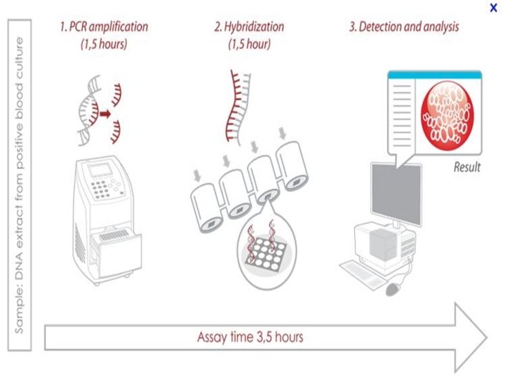

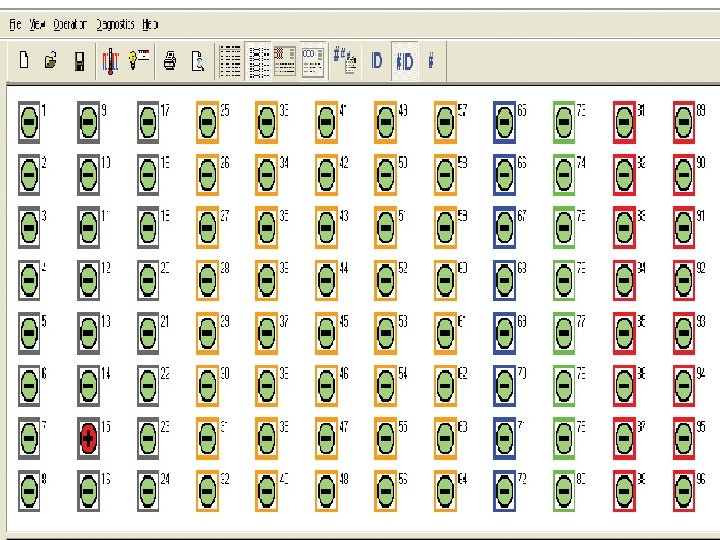
DIRECT METHOD Design specific fluorescently labeled oligonucleotide probes to differentiate between microorganisms in the sample: Specific Species (eg E. coli) Gram Differentiation Spore formation Combine with microarray: allows for many PCR reactions in a single rapid procedure. Quick Efficient Automated

HRMA: HIGH-RESOLUTION MELTING ANALYSIS Design three conserved primers for three hypervariable regions (V 1, V 3, and V 6) within the 16 S r. RNA gene. Double stranded DNA is “melted” and then binds to a fluorescent dye.
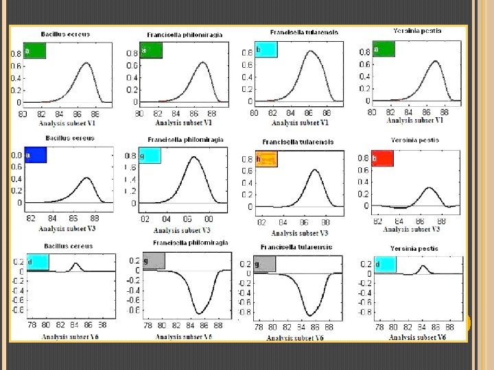

Compare combinations of V 1, V 3, V 6:

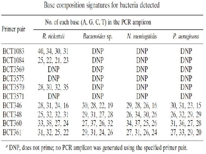
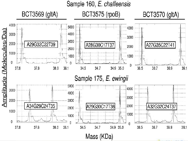
EIMS: ELECTROSPRAY IONIZATION MASS SPECTROMETRY Primers designed for conserved bacterial 16 S ribosomal genes. DNA is amplified by PCR. Ionize the amplified DNA for mass spectrometry analysis. Detect the mass of amplified DNA and compare to a database.

DGGE: DENATURING GRADIENT GEL ELECTROPHORESIS Use specific primers to target a region of the bacterial genome and amplify with PCR. Amplified DNA is run through a denaturing gel. Different bacterial DNA has different DGGE patterns due to nucleotide content and the secondary structures formed during partial denaturing. Can determine genus of unknown microorganisms by comparing known DGGE profile. (eg. Nitrosomonas which oxidizes NH 3)

Lactobacillus Bacteroides

Rapid Identification and Characterization Techniques Nanotechnology

NANOTECHNOLOGY Nanotechnology is the creation and manipulation of matter at the nanoscale. Nano-constructs capable of self-assembly and specific activity: imitation of natural molecular structures. Extreme sensitivity reduces needed sample size.

NANOROCKETS Self-propelled microtubes: capture-and-release bacteria. 8 um long; 0. 5 um diameter. 300 body-lengths/second. Microengine: platinum-catalyzed oxidation of H 2 O 2 fuel. Lectin receptor selectively bind bacterial polysaccharides. Lectin-bacterial bond broken by low-p. H gylcine solution. Mass production: membrane template electrodepostion. Nanorockets are inexpensive and reusable.

NANOROCKETS Rapid capture and isolation of pathogenic bacteria in complex media. Duel action: magnetic attachment and release of therapeutic molecules. H 2 O 2 fuel makes bacteria non-cultural but they remain viable and thus testable. Biochips: nanorockets scour samples for pathogens in microfluidic channels.

SURFACE-ENHANCED-RAMAN-SCATTERING Raman spectroscopy: Viruses bound to Ag substrate. Exposed to an infrared laser. Laser scattered by viral particles. Spectrograph records signal. Virus spectral barcodes made. Silver nanorod substrate: Enhances spectra cross section by orders of magnitude. Oblique angle deposition: cheap and simple procedure. Deposits nanorods in random arrays at an uniform angle.
: Each share conserved spectra due to")
SURFACE-ENHANCED-RAMAN-SCATTERING Three strains of the influenza virus (flu): Each share conserved spectra due to close relation. Highlighted: non-conserved spectra correlate to variable nucleic acids and surface proteins. Fast method for detection and classification of viruses, bacteria or toxins. 1300 1200 1100 1000 900 800 700 600 Raman Shift (cm-1) 500 Monitoring of pollutants.

Biomarker Proteins CANTILEVERS Cantilevers are horizontal beams anchored on one end: Antibody Bent Cantilever Mass bends the cantilever downwards. A counterbalancing force can restore the original position, resulting in a certain resonance frequency. The resonance frequency is determined electronically. Cantilevers can be manufactured at the nanoscale: Made in dimensions as small as: 5 um x 2 um x 30 nm. Antibodies attached to cantilevers can detect the binding of single viral or bacterial particles. Ideal for airborne virus detection or microfluidic biochips.
: Sample Piezoelectric scanner A")
CANTILEVERS Photodiode Laser Cantilevers and Atomic Tip Force Microscopy (AFM): Sample Piezoelectric scanner A nano-pin attached to a cantilever traces cell surfaces. Pin-cantilever movement is detected by laser deflection. Produces detailed high resolution topological images of cells and profiles of molecular bond strength. Rapid microbial detection applications: Imaging completed in minutes: location of cell-surface markers. Capable of analyses in aqueous solutions and in vivo.

CANTILEVERS Omp. F: a porin protein of E. coli’s outer membrane: Forms channels composed of β – strands (circled). Left: X-ray crystallography image. Right: AFM image made with constant force microscopy. Scale bar: 50 Å.

CANTILEVERS AFM image of a Saccharomyces cerevisiae yeast cell trapped in a microporous membrane, scale: 1 um.
 400 300 200 100 0 200 300 400")
CANTILEVERS 600 500 Force (p. N) 400 300 200 100 0 200 300 400 Extension (nm) 500 Force spectrometry on a typical bacterial cell: Left: a cantilever attached to a membrane polysaccharide encounters resistance as it pulls away from the molecule. Right: corresponding force-extension curve shows the growing molecular force offered by the polysaccharide as it is stretched.

Rapid Identification and Characterization Techniques IMMUNOASSAYS

IMMUNOASSAYS: RAPID QUANTIFICATION OF FOODBORNE PATHOGENS Multiplex immunoflourescent technique: Antibody-Antigen interaction provides specificity and the conjugated flourescent quantum dots allow for rapid detection of multiple pathogens.

QUANTUM DOTS

Multiplex Immunoflourescent Assay

RESULTS

IMMUNOFLOURESCENT ASSAY Advantages Disadvantages Can quantify the number of microorganisms Relatively quick (< 2 hrs) Simultaneous detection of multiple microorganisms Does not require culturing Sensitive and specific Expensive Current methods require technical expertise Requires lab equipment, but there is a potential for portable equipment

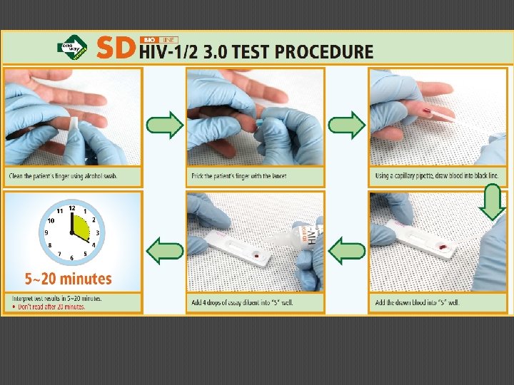
IMMUNOASSAYS: DIPSTICK ASSAY FOR HIV-1 AND HIV-2 SD Bioline HIV-1/2 3. 0 Test One step, rapid, immunochromatographic test that can distinguish between HIV-1 and HIV-2, and can detect all isoforms of each! Sample: human serum/plasma (10 microliter sample) or whole blood (20 microliter sample). Evaluated by WHO: 99. 3% specificity, 100% sensitivity Time to run test: 5 -20 minutes Price per test: $0. 85 – 1. 10 USD

DIRECT SANDWICH ELISA

RESULTS

DIPSTICK IMMUNOASSAYS Advantages Relatively cheap Rapid results Requires very little sample Very specific and sensitive Stable over broad temperature range Easy to transport Does not require technical expertise to use Disadvantages Qualitative only Invasive procedure Disposal Accidental infections

IMMUNOASSAYS: USING CHEWING GUM TO DETECT MALARIA Researchers at UCLA have received funding from the Bill and Melinda Gates Foundation to develop an innovative way to detect malaria using chewing gum (MALi. VA). The technique: chewing gum containing antibodies detects malaria-specific antigens in the saliva. The gum is then blotted onto a paper to give a visual result. This method will be ideal for undeveloped countries where electrical lab equipment and technical expertise is not readily available.

Rapid Identification and Characterization Techniques Other Rapid Microbial Methods

Computational Analysis and Databases The use of computational analysis provides highthroughput results in a short amount of time. Data can be shared in online databases. MALDI – TOF Mass Spectrometry Matrix-Assisted Laser Desorption Ionisation Time-of. Flight Species-specific mass spectra of peptides, protein, or other organic molecules by mass spectrometry.

MALDI – TOF Mass Spectrometry Very common in clinical biology laboratory: Efficient (multiple samples per run) Cost-effective ($1. 80 USD/sample) Rapid (<10 mins)

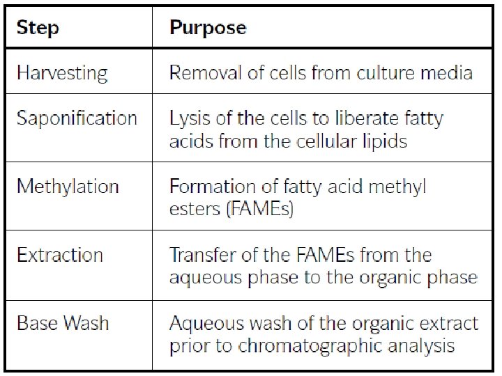
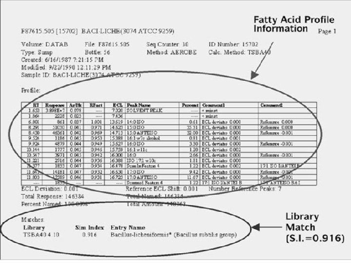
FAME: FATTY ACID METHYL ESTER Cellular fatty acid analysis by Gas Chromatography. MIDI Sherlock Microbial Identification System. Comparing fatty acid components to database.

Rapid Identification and Characterization Techniques Conclusion

Overall Advantages and Disadvantages of Rapid Microbial Methods: Advantages Results are rapid! Ease-of-use testing. Tests are made to be very sensitive and specific. The tests can be portable. Reduces workload on medical laboratories. Can test for microorganisms that cannot be cultured. Allows for earlier detection and faster treatment. Disadvantages Expensive investment in new lab equipment. May require additional technical expertise. Potential for abuse: may be used for bioterrorism. Censorship of scientific data can hinder new developments in this field.

THE FUTURE! Phasing out of culture-based microscopy: Can’t compete with superior techniques and lower cost methods. Unable to rectify its own flaws. Biology Museum Bacterial Cultures Take-home diagnosis and treatment kits: Prepackaged reagents and protocols: available at your local superstore! Rapidly identify disease-causing pathogens and recommend or provide the appropriate treatment. Cheap and mass produced, affordable to the developing nations: improvement in global health.

QUESTIONS?
- Slides: 55