
Reactive Thiols in Redox Signaling SFRBM Worshop Boston
Reactive Thiols in Redox Signaling SFRBM Worshop Boston University Medical Center November 15, BU-NHLBI Cardiovascular 2006 Proteomics Center Denver, CO Richard A. Cohen, MD Director, Vascular Biology Unit Boston University School of Medicine racohen@bu. edu

Outline • Introduction: Reactive thiol proteome • MS screening for reactive thiols with ICAT • S-glutathiolation labeling and detection methods • S-glutathiolation in signaling
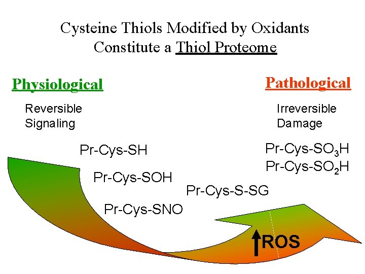
Cysteine Thiols Modified by Oxidants Constitute a Thiol Proteome Pathological Physiological Reversible Signaling Irreversible Damage Pr-Cys-SH Pr-Cys-SO 3 H Pr-Cys-SO 2 H Pr-Cys-S-SG Pr-Cys-SNO ROS
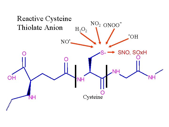
Reactive Cysteine Thiolate Anion H 2 O 2 NO 2 ONOO- . OH . NO SH S- O O SNO, SOx. H O NH OH NH NH O Cysteine NH
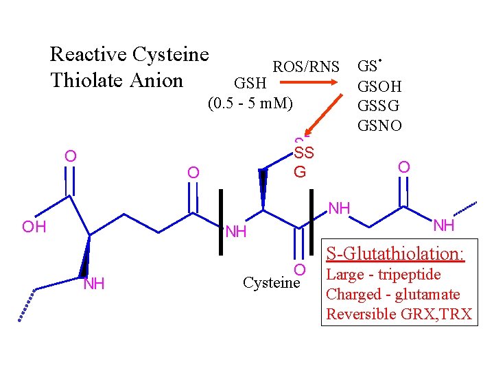
Reactive Cysteine Thiolate Anion ROS/RNS GSH (0. 5 - 5 m. M) - O S SH SS SG O O NH OH NH NH O Cysteine . GS GSOH GSSG GSNO NH S-Glutathiolation: Large - tripeptide Charged - glutamate Reversible GRX, TRX
MS screening for reactive thiols with ICAT
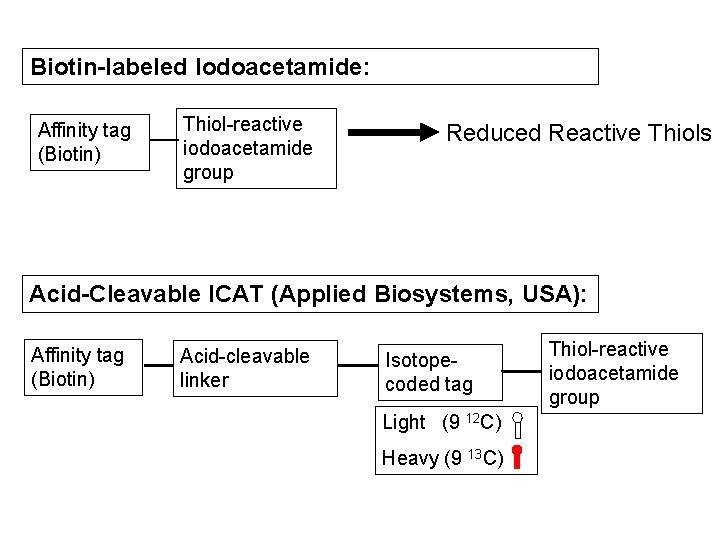
Biotin-labeled Iodoacetamide: Affinity tag (Biotin) Thiol-reactive iodoacetamide group Reduced Reactive Thiols Acid-Cleavable ICAT (Applied Biosystems, USA): Affinity tag (Biotin) Acid-cleavable linker Isotopecoded tag Light (9 12 C) Heavy (9 13 C) Thiol-reactive iodoacetamide group
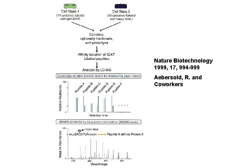
Nature Biotechnology 1999, 17, 994 -999 Aebersold, R. and Coworkers
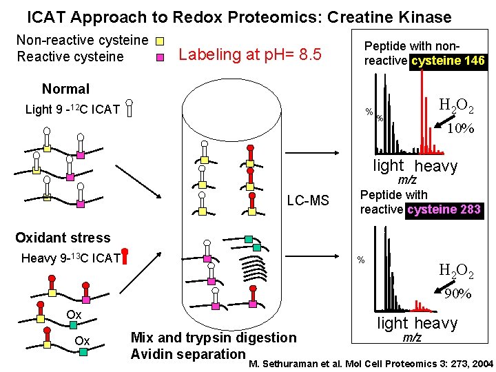
ICAT Approach to Redox Proteomics: Creatine Kinase Non-reactive cysteine Reactive cysteine Labeling at p. H= 8. 5 Peptide with nonreactive cysteine 146 Normal Light 9 -12 C ICAT % H 2 O 2 % 10% light heavy LC-MS m/z Peptide with reactive cysteine 283 Oxidant stress Heavy 9 -13 C ICAT % H 2 O 2 90% Ox Ox Mix and trypsin digestion Avidin separation light heavy m/z M. Sethuraman et al. Mol Cell Proteomics 3: 273, 2004
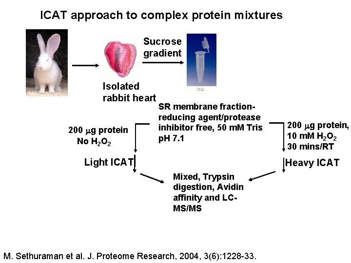
ICAT approach to complex protein mixtures Sucrose gradient Isolated rabbit heart 200 g protein No H 2 O 2 SR membrane fractionreducing agent/protease inhibitor free, 50 m. M Tris p. H 7. 1 Light ICAT 200 g protein, 10 m. M H 2 O 2 30 mins/RT Heavy ICAT Mixed, Trypsin digestion, Avidin affinity and LCMS/MS M. Sethuraman et al. J. Proteome Research, 2004, 3(6): 1228 -33.
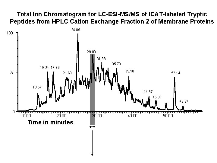
Total Ion Chromatogram for LC-ESI-MS/MS of ICAT-labeled Tryptic Peptides from HPLC Cation Exchange Fraction 2 of Membrane Proteins 24. 89 100 29. 00 31. 38 35. 70 16. 34 17. 86 % 39. 18 21. 60 52. 14 13. 57 44. 87 46. 81 54. 47 0 10. 00 20. 00 Time in minutes 30. 00 40. 00 50. 00
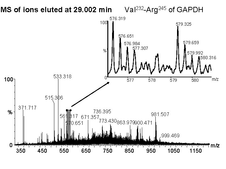
MS of ions eluted at 29. 002 min 100 Val 232 -Arg 245 of GAPDH 576. 319 579. 325 576. 651 % 533. 318 100 0 579. 659 576. 984 577. 307 579. 992 580. 316 578 579 580 m/z 515. 306 % 371. 717 736. 395 981. 507 561. 317 671. 357 773. 430863. 979900. 471 570. 651 999. 469 0 350 450 550 650 750 850 950 1050 1150 m/z
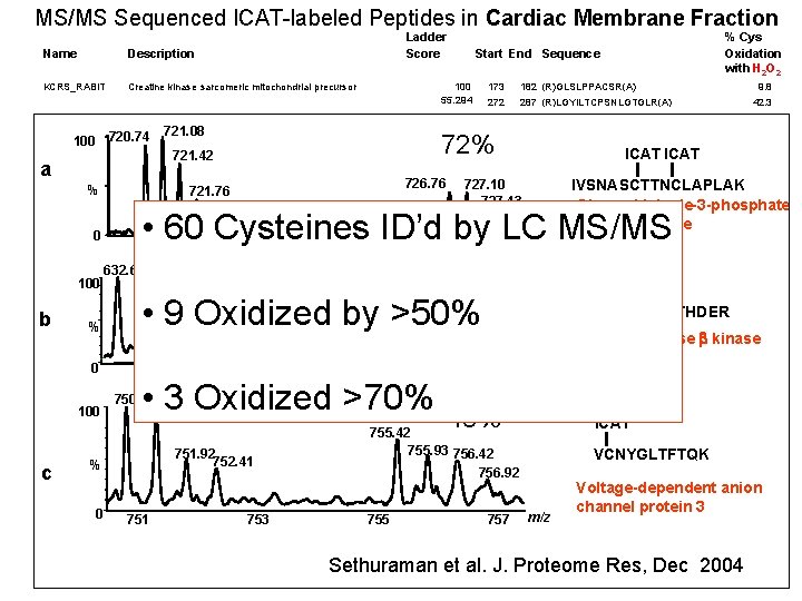
MS/MS Sequenced ICAT-labeled Peptides in Cardiac Membrane Fraction Ladder Score Name Description KCRS_RABIT Creatine kinase sarcomeric mitochondrial precursor 100 a Fructose biphosphate aldolase % 42. 3 52. 941 287 (R)LGYILTCPSNLGTGLR(A) 9. 8 69. 863 232 245 (R)VPTPNVSVVDLTCR(L) 26. 9 64. 835 143 159 (K)IVSNASCTTNCLAPLAK(V) 72. 2 49. 485 69 40. 5 ICAT 86 (R)VNPCIGGVILFHETLYQK(A) 12. 3 dehydrogenase • 60 Cysteines ID’d by LC MS/MS Sarcoplasmic endoplasmic reticulum Ca 2+ ATPase 0 723 721 725 Polymeric immunoglobulin receptor precursor 632. 67 633. 00 KPBB_RABIT b 182 (R)GLSLPPACSR(A) 272 201 207 (R)CQYVTEK(V) 726. 7683. 871727. 10 IVSNASCTTNCLAPLAK 24. 3 56. 364 331 341 (R)ALANSLACQGK(Y) 24. 1 727. 43 Glyceraldehyde-3 -phosphate 50. 633 437 451 (K)VGEATETALTCLVEK(M) 32. 5 727. 94 721. 76 ATA 2_RABIT 100 173 72% 721. 42 ALFA_RABIT PIGR_RABIT 100 55. 294 721. 08 phosphate dehydrogenase Glyceraldehyde 720. 74 G 3 P_RABIT % Cys Oxidation with H 2 O 2 Start End Sequence % K 6 PF_RABIT 633. 34 633. 65 Phosphofructose kinase muscle type Voltage 633 dependent anion selective channel protein 635 1 POR 2_RABIT 750. 91 Voltage-dependent anion-selective channel protein 2 751. 42 100 372 397 (K)VDGDTCSLNEFTITGSTYAPIGEVHK(D) 15. 1 79. 07 727 468 476 m/z(R)ANACNSVIK(Q) 28. 6 25. 62 353 374 (R)SPPVLKGFPGGSVTIRCPYNPK(R) 16. 279 587 595 (K)ARCPVPRRR(Q) 36. 082 595 612 (R)RQWYPLSRKLRTSCPEPR(L) 62% 635. 68 29. 508 276 287 636. 01 32. 836 832 844 636. 35 59. 13 636. 68 108 128 • 9 Oxidized by >50% Phosphorylase B kinase beta regulatory chain 0 POR 1_RABIT POR 3_RABIT 44. 138 • 3 Oxidized >70% Voltage-dependent anion-selective channel protein 3 120 30. 1 66. 019 46 64 (K)SCSGVEFSTSGSSNTDTGK(V) 96. 364 64 74 (K)VCNYGLTFTQK(W) 48. 6 77 (K)LDKPSVVNWMCYRK(T) 43. 9 48% 28. 767 64 755. 42 57. 143 172 755. 93 756. 42 75. 676 Vitamin K dependent protein S precurson fragment 32. 727 DHSA_HUMAN NUFM_RAT ODO 1_HUMAN 753 Succinate dehydrogenase mitochondrial pre 47. 273 755 kinase<5 66. 99 Hemoglobin beta 1 chain Malate dehydrogenase 62. 3 (R)GITNLCVIGGDGSLTGADTFR(S) Phosphorylase 45. 6 c. HBB_RABIT% 751 (K)NIIYYKCNTHDER(E) 224637235 (K)YQIDPDACFSAK(V) m/z Plasma retinol binding protein MDHM_RAT 0 <5 NIIYYKCNTHDER 630 Phosphatidylcholine-sterol acyltransferase [Precursor] PRTS_RABIT 13. 7 (R)QTLCSLLPRESR(S) 81. 967 RETB_RABIT FA 10_RABIT ICAT 59. 223 LCAT_RABIT 751. 92 Coagulation factor 752. 41 50. 746 334 756. 92 88 442 92 757 648 (K)CNENYTTDFIFNLYSEEGK(G) 138 (R)EHINLGCDVDFDIAGPSIR(G) ICAT 181 (R)QRQEELCLSR(Q) VCNYGLTFTQK 344 (R)RNVAPACLPQK(D) 95 (K)LSELHCDK(L) 48 54. 38 28. 1 39 49. 6 Voltage-dependent anion 89. 6 channel protein 3 104 (K)GCDVVVIPAGVPR(K) 26 452 (K)QKKHCLVTVEK(G) m/z 64. 384 233 246 (R)GVIALCIEDGSIHR(I) 26. 7 56. 164 233 246 (R)GVIALCIEDGSIHR(I) 26. 7 64. 706 7 22 (K)TTGLVGLAVCDTPHER(L) Sethuraman 46. 575 et al. 278 J. Proteome Res, Dec 2004 2 -oxoglutarate dehydrogenase E 1 component, mitochondrial 291 (R)FGLEGCEVLIPALK(T) NADH-ubiquinone oxidoreductase 24 99. 6 31. 5 8. 5
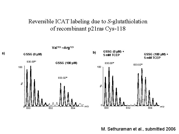
Reversible ICAT labeling due to S-glutathiolation of recombinant p 21 ras Cys-118 Val 103 –Arg 123 a) GSSG (0 μM) + 5 m. M TCEP b) GSSG (0 μM) 830. 003+ GSSG (100 μM) + 5 m. M TCEP 830. 003+ 833. 023+ 100 % % 833. 023+ 0 830 832 834 m/z M. Sethuraman et al. , submitted 2006
• S-glutathiolation labeling and detection methods • S-glutathiolation in signaling
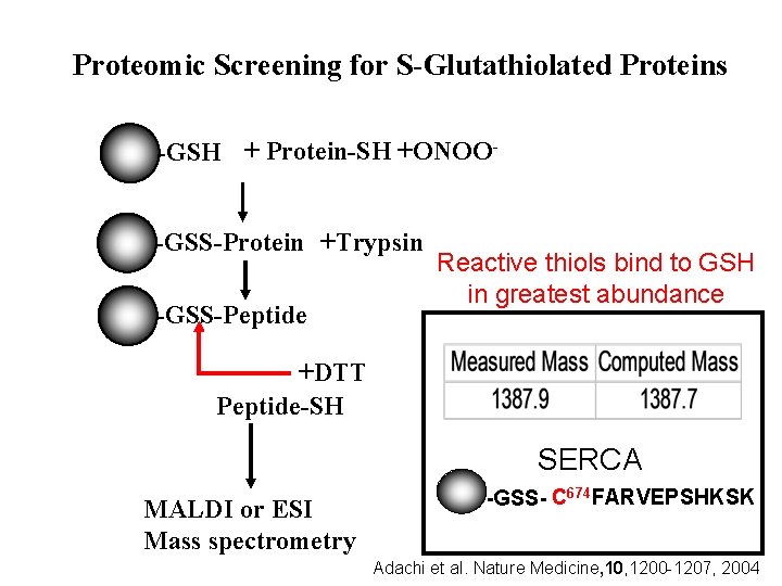
Proteomic Screening for S-Glutathiolated Proteins -GSH + Protein-SH +ONOO-GSS-Protein +Trypsin -GSS-Peptide Reactive thiols bind to GSH in greatest abundance +DTT Peptide-SH SERCA MALDI or ESI Mass spectrometry -GSS- C 674 FARVEPSHKSK Adachi et al. Nature Medicine, 10, 1200 -1207, 2004
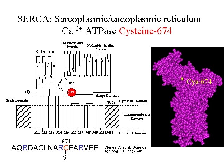
SERCA: Sarcoplasmic/endoplasmic reticulum Ca 2+ ATPase Cysteine-674 Phosphorylation Domain B - Domain P (1) Nucleotide - binding Domain Cys-674 D 351 294 Y 674 C Hinge Domain Stalk Domain (997) Cytosolic Domain Transmembrane Domain M 1 M 2 M 3 M 4 M 5 M 6 M 7 M 8 M 9 M 10 M 11 Luminal Domain 674 AQRDACLNARCFARVEP S- Olesen C, et al. Science 306: 2251 -5, 2004
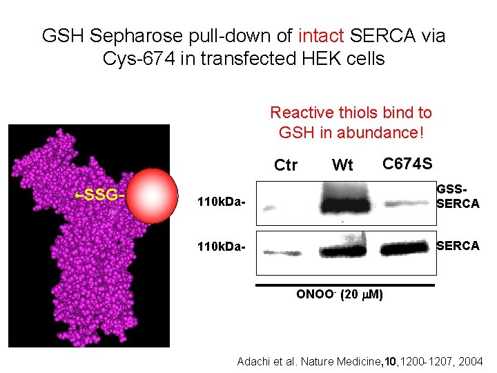
GSH Sepharose pull-down of intact SERCA via Cys-674 in transfected HEK cells Reactive thiols bind to GSH in abundance! Ctr -SSG- Wt C 674 S 110 k. Da- GSSSERCA 110 k. Da- SERCA ONOO- (20 M) Adachi et al. Nature Medicine, 10, 1200 -1207, 2004
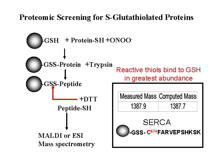
Proteomic Screening for S-Glutathiolated Proteins -GSH + Protein-SH +ONOO-GSS-Protein +Trypsin -GSS-Peptide Reactive thiols bind to GSH in greatest abundance +DTT Peptide-SH SERCA MALDI or ESI Mass spectrometry -GSS- C 674 FARVEPSHKSK
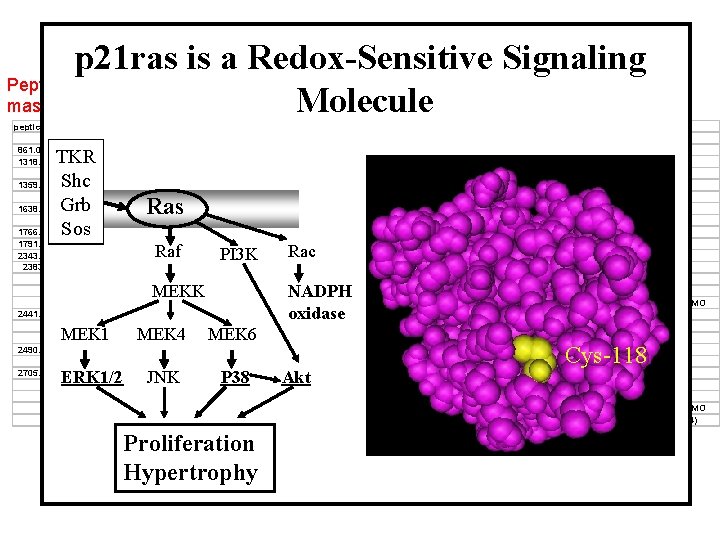
Peptide masses ONOO- -induced Glutathiolation of Proteins in p 21 ras is a. Rabbit Redox-Sensitive Aortic Homogenate. Signaling Molecule peptides Protein MW 861. 0504 1318. 672 titin Na-K-ATPase Alpha subunit 1359. 659 PP 2 A subunit. A, PR 65 regulatory beta L-Ca-channel 1638. 793 MLCK Na+ Channel alpha 1766. 626 ROCK-1 1791. 699 RYR 3 2343. 926 PP 2 A subunit. A, PR 65 regulatory beta 2383. 89 Ig. G Ras n. NOS ROCK-1 2441. 977 Ig. G Ras n. NOS 2490. 252 Myosin Na-K-ATPase Alpha subunit 2705. 113 Ras L-Ca-channel n. NOS RYR 3 TKR Shc Grb Sos Raf MEKK MEK 1 ERK 1/2 MEK 4 JNK 110. 05 114. 3 60. 97 149. 73 125. 7 225. 73 158. 33 550. 32 60. 97 11. 71 20. 91 160. 85 158. 33 11. 71 20. 91 160. 85 PI 3 K MEK 6 114. 3 20. 91 149. 73 160. 85 550. 32 P 38 Proliferation Hypertrophy Measured Mass Computed Mass Error Da Da ppm 1317. 664 1358. 651 1637. 785 1765. 618 1790. 691 2342. 918 2382. 882 2440. 969 2489. 244 2704. 105 Rac NADPH oxidase Akt 1317. 707 1317. 732 1358. 64 1358. 724 1637. 771 1637. 801 1765. 833 1790. 926 2343. 222 2383. 137 2383. 166 2383. 104 2383. 154 2441. 142 2441. 171 2441. 109 2489. 179 2489. 195 2704. 297 2704. 352 2704. 335 2704. 358 2704. 16 Start -33 -52 9 -53 8 -10 -122 -131 -130 -107 -119 -93 -114 -71 -83 -57 26 20 -71 -92 -85 -94 -21 552 525 334 1186 1049 1159 740 1956 494 37 168 920 447 37 168 920 109 673 102 18 829 3376 2086 End 563 535 345 1196 1063 1173 754 1970 514 59 188 939 465 59 188 939 129 694 126 39 850 3399 2106 Sequence ATKTPVSDLRCK ILAAQGCK CVSSPHFQVAER RASFHLECLKR DLEVVEGSAARFDCK RFPCCQVSIESGKGK QCSMLDVDLKQSQQK CPTTLKELISQTMIR RKSELPQDPHTKKLEAHCR GLEWIACIRGGGSGSTNYASWAK QHKLRKLNPPDESGRGCMSCK MREGDELCGQEEAFRTWAKK DEMEQKCRTSNIKLDKIMK GLEWIACIRGGGSGSTNYASWAK QHKLRKLNPPDESGRGCMSCK MREGDELCGQEEAFRTWAKK YAAWMIYTYSGLFCVTVNPYK ACVINGMQLKDMDPSELVEALR VKDSDDVPMLNKCDVGNKCDLAAR NCELDKNQRQCVEYALKARPLR CALMEMRHPNSLQEERKSYKVR KMLPIGLNMCTPGDQELISLKSR SQIAFPKMVASCCRFLCYFCR Cys-118 (Mallis RJ, Thomas JA, Biochem J 2001) IAA, MO IAA IAA IAA, MO IAA(4)
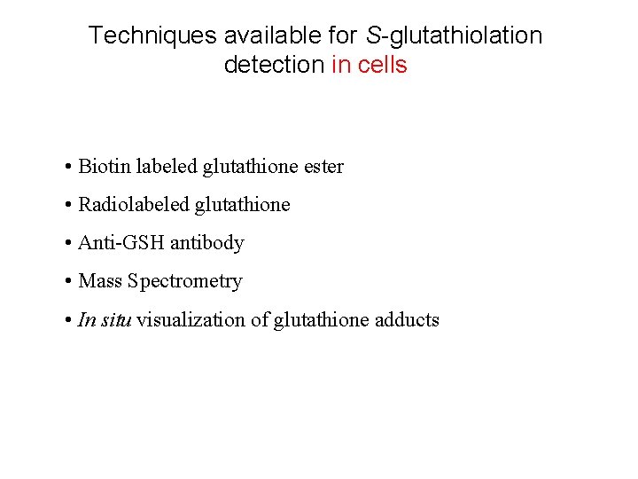
Techniques available for S-glutathiolation detection in cells • Biotin labeled glutathione ester • Radiolabeled glutathione • Anti-GSH antibody • Mass Spectrometry • In situ visualization of glutathione adducts
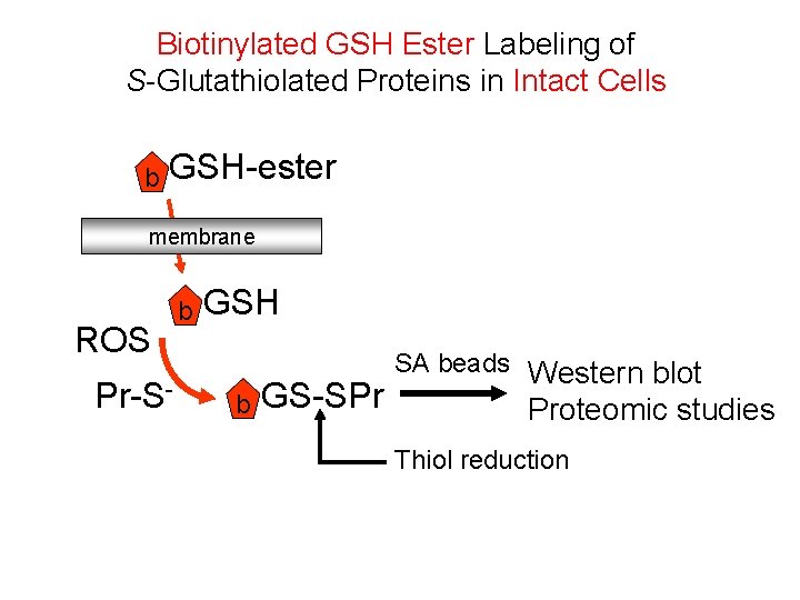
Biotinylated GSH Ester Labeling of S-Glutathiolated Proteins in Intact Cells b GSH-ester membrane ROS Pr-S- b GSH b GS-SPr SA beads Western blot Proteomic studies Thiol reduction
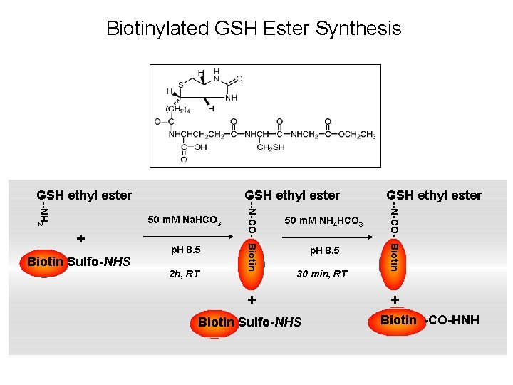
Biotinylated GSH Ester Synthesis 50 m. M Na. HCO 3 + Biotin Sulfo-NHS p. H 8. 5 2 h, RT 50 m. M NH 4 HCO 3 p. H 8. 5 30 min, RT + Biotin Sulfo-NHS GSH ethyl ester --N-CO- Biotin --NH 2 GSH ethyl ester + Biotin -CO-HNH
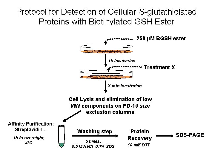
Protocol for Detection of Cellular S-glutathiolated Proteins with Biotinylated GSH Ester 250 µM BGSH ester 1 h incubation Treatment X X min incubation Cell Lysis and elimination of low MW components on PD-10 size exclusion columns Affinity Purification: Streptavidin… 1 h to overnight, 4°C Washing step 5 times: 0. 5 M Na. Cl 0. 1% SDS Protein Recovery 10 m. M DTT SDS-PAGE
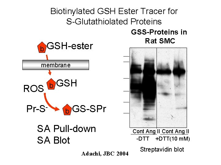
Biotinylated GSH Ester Tracer for S-Glutathiolated Proteins b GSH-ester GSS-Proteins in Rat SMC membrane ROS Pr-S- b GSH b GS-SPr SA Pull-down SA Blot Adachi, JBC 2004 Cont Ang II -DTT +DTT(10 m. M) Streptavidin blot
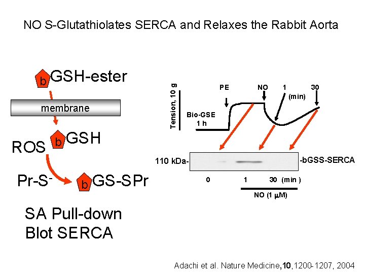
b GSH-ester membrane ROS Pr-S- b Tension, 10 g NO S-Glutathiolates SERCA and Relaxes the Rabbit Aorta PE NO 1 30 (min) Bio-GSE 1 h GSH -b. GSS-SERCA 110 k. Da- b GS-SPr 0 1 30 (min ) NO (1 M) SA Pull-down Blot SERCA Adachi et al. Nature Medicine, 10, 1200 -1207, 2004
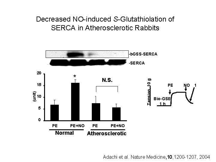
Decreased NO-induced S-Glutathiolation of SERCA in Atherosclerotic Rabbits -b. GSS-SERCA 20 * N. S. (unit) 15 10 5 Tension, 10 g -SERCA PE NO 1 Bio-GSE 1 h 0 PE PE+NO Normal PE PE+NO Atherosclerotic Adachi et al. Nature Medicine, 10, 1200 -1207, 2004
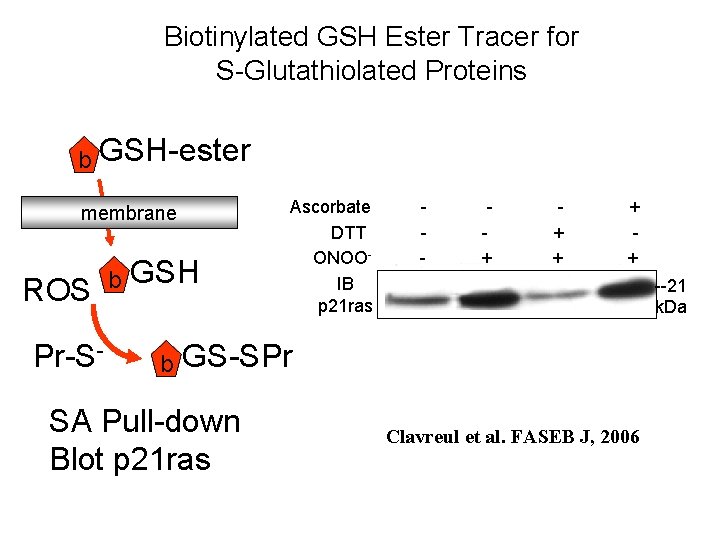
Biotinylated GSH Ester Tracer for S-Glutathiolated Proteins b GSH-ester membrane ROS Pr-S- b GSH b Ascorbate DTT ONOOIB p 21 ras - + + + --21 k. Da GS-SPr SA Pull-down Blot p 21 ras Clavreul et al. FASEB J, 2006
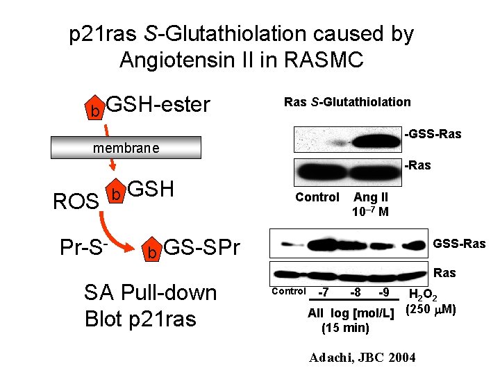
p 21 ras S-Glutathiolation caused by Angiotensin II in RASMC b GSH-ester Ras S-Glutathiolation -GSS-Ras membrane -Ras ROS Pr-S- b GSH b Control Ang II 10– 7 M GS-SPr SA Pull-down Blot p 21 ras GSS-Ras Control -7 -8 -9 H 2 O 2 AII log [mol/L] (250 M) (15 min) Adachi, JBC 2004
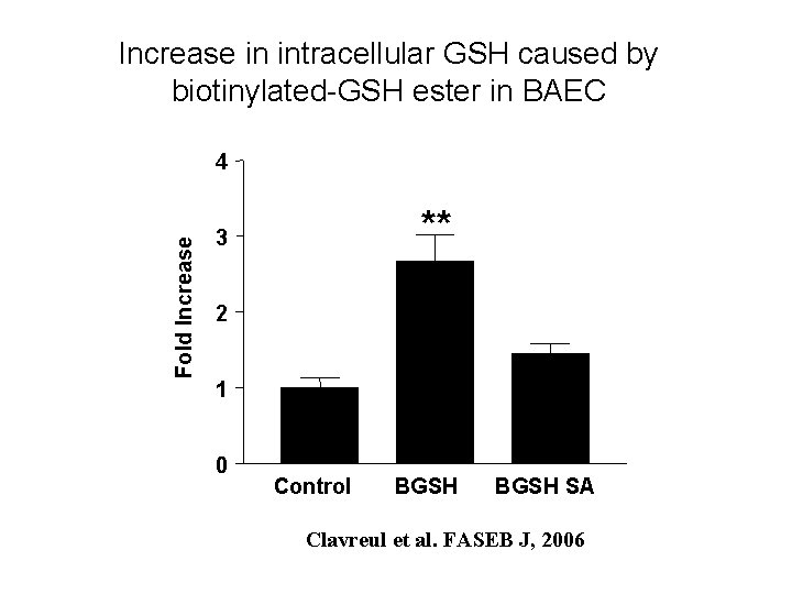
Increase in intracellular GSH caused by biotinylated-GSH ester in BAEC Fold Increase 4 ** 3 2 1 0 Control BGSH SA Clavreul et al. FASEB J, 2006
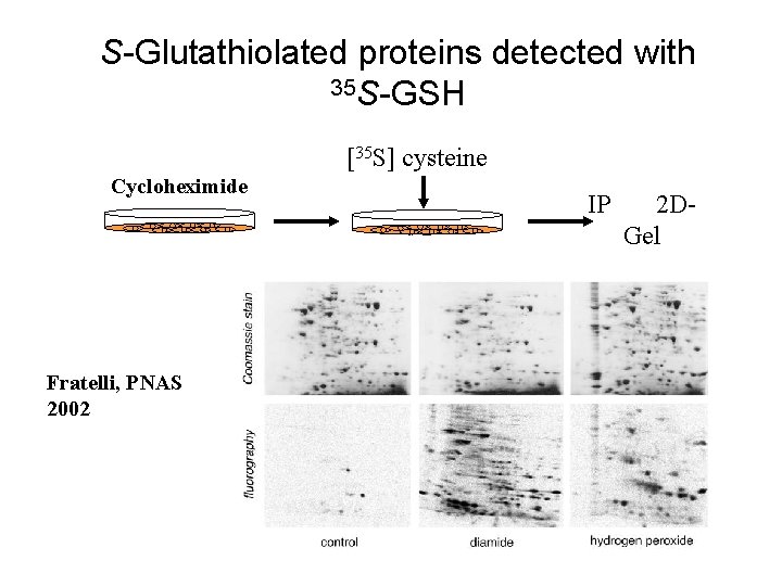
S-Glutathiolated proteins detected with 35 S-GSH [35 S] cysteine Cycloheximide Fratelli, PNAS 2002 IP 2 DGel
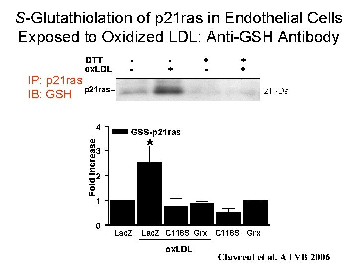
S-Glutathiolation of p 21 ras in Endothelial Cells Exposed to Oxidized LDL: Anti-GSH Antibody DTT ox. LDL IP: p 21 ras-IB: GSH + + --21 k. Da GSS-p 21 ras * Fold Increase 4 - 3 2 1 0 Lac. Z C 118 S Grx ox. LDL Clavreul et al. ATVB 2006
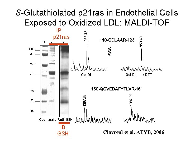
1 2 3 118 -CDLAAR-123 953. 43 IP p 21 ras 953. 32 S-Glutathiolated p 21 ras in Endothelial Cells Exposed to Oxidized LDL: MALDI-TOF SSG 100 75 50 37 Ox. LDL + DTT 1397. 63 20 1397. 69 150 -QGVEDAFYTLVR-161 25 15 - Coomassie Anti -GSH IB GSH Clavreul et al. ATVB, 2006
Acknowledgements: Vascular Biology Unit: Nicolas Clavreul Ardelle Jia Ying Takeshi Adachi Reiko Matsui Maha Sethuraman Bingbing Jiang Mengwei Zang Xiaoyong Tong Markus Bachschmid Stefan Schildknecht Tyler Heibeck Hossein Bayat Rebecca Zee Robert Weisbrod Shanqin Xu Karlene Maitland Chaomei Shi Xuiyun Hou Cardiovascular Proteomics Center Mark Mc. Comb Claire Dauly Peter O’Connor Cheng Zhao Cathy Costello University of Kansas Christian Schoeneich Viktor Sharov
- Slides: 34