
Radiology Pathology Correlation Patient Presentation CASEY MORRISON MS
- Slides: 15
Radiology + Pathology Correlation Patient Presentation CASEY MORRISON, MS 4 SEPTEMBER 7 TH, 2017
H. E. A 69 y. o. female who presented to the ED with a 2 -3 month history of general malaise, 15 lb weight loss + complaints of R shoulder/RUQ pain Came in after her PCP called to discuss abnormal labs results from her recent visit: Alk Phos-169, AST-94, ALT-23. She was unable to schedule outpatient RUQ U/S until more than a week later PMHx: HTN, HLD, GERD PSHx: Sacrocolpopexy, Hysterectomy with bilateral salpingo-oopherectomy, Lumpectomy of R breast in 2008 for DCIS Fam. Hx: Mother- COPD; Father- CAD, thyroid cancer; Sister: Breast cancer
Imaging Considering her history of breast cancer, RUQ pain, labs derangements and recent wt. loss + malaise a CT abdomen was ordered to rule out abdominal pathologies including primary and metastatic lesions to the abdomen CT is a very sensitive test for many abdominal diseases including GU stones, appendicitis, diverticulitis, abdominal aortic aneurysms, bowel obstructions. Thus it is often used in the work up of abdominal pain and to determine stage of cancer, follow progression, and evaluate for metastases
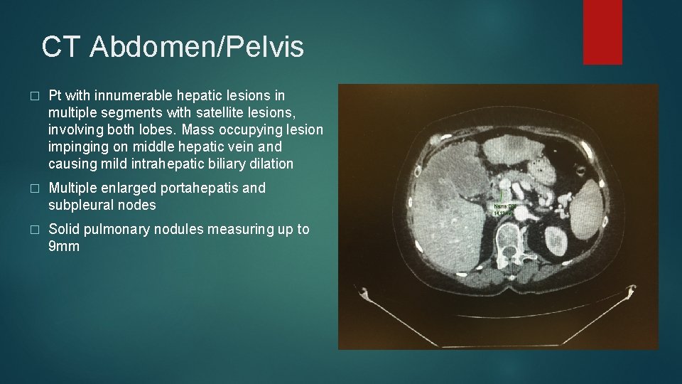
CT Abdomen/Pelvis � Pt with innumerable hepatic lesions in multiple segments with satellite lesions, involving both lobes. Mass occupying lesion impinging on middle hepatic vein and causing mild intrahepatic biliary dilation � Multiple enlarged portahepatis and subpleural nodes � Solid pulmonary nodules measuring up to 9 mm
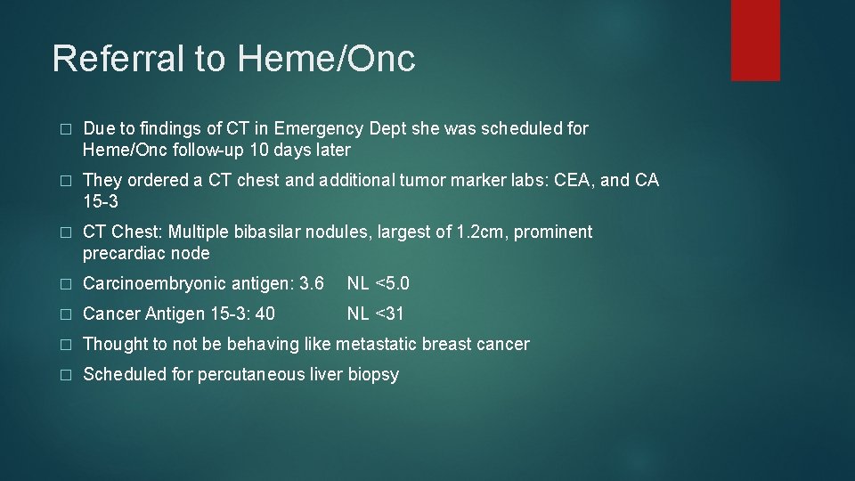
Referral to Heme/Onc � Due to findings of CT in Emergency Dept she was scheduled for Heme/Onc follow-up 10 days later � They ordered a CT chest and additional tumor marker labs: CEA, and CA 15 -3 � CT Chest: Multiple bibasilar nodules, largest of 1. 2 cm, prominent precardiac node � Carcinoembryonic antigen: 3. 6 NL <5. 0 � Cancer Antigen 15 -3: 40 NL <31 � Thought to not be behaving like metastatic breast cancer � Scheduled for percutaneous liver biopsy
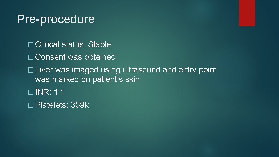
Pre-procedure � Clincal status: Stable � Consent was obtained � Liver was imaged using ultrasound and entry point was marked on patient’s skin � INR: 1. 1 � Platelets: 359 k
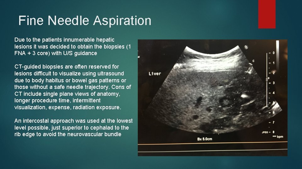
Fine Needle Aspiration Due to the patients innumerable hepatic lesions it was decided to obtain the biopsies (1 FNA + 3 core) with U/S guidance CT-guided biopsies are often reserved for lesions difficult to visualize using ultrasound due to body habitus or bowel gas patterns or those without a safe needle trajectory. Cons of CT include single plane views of anatomy, longer procedure time, intermittent visualization, expense, radiation exposure. An intercostal approach was used at the lowest level possible, just superior to cephalad to the rib edge to avoid the neurovascular bundle
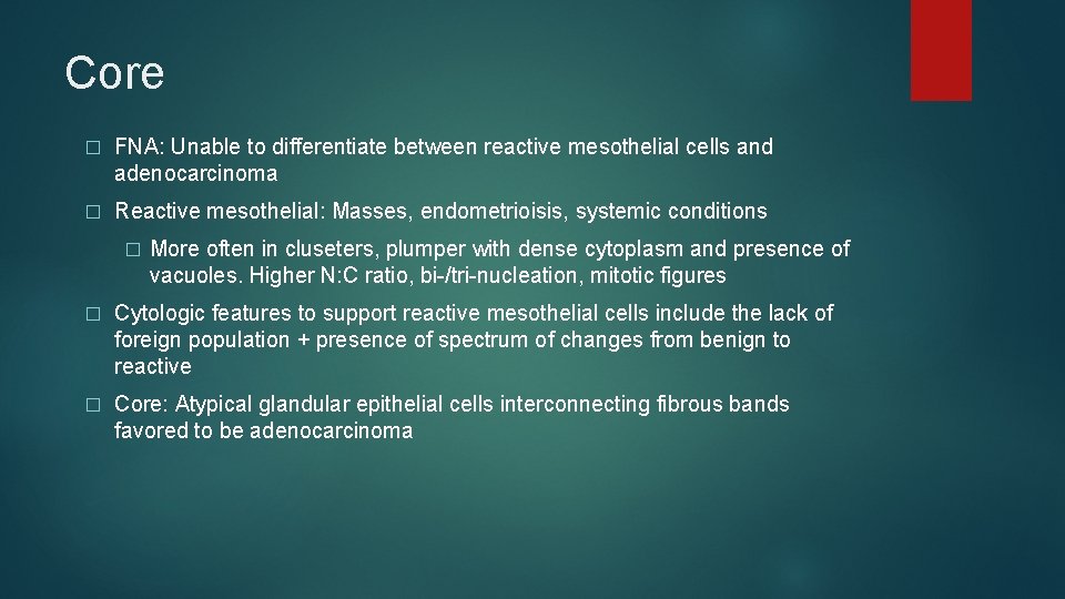
Core � FNA: Unable to differentiate between reactive mesothelial cells and adenocarcinoma � Reactive mesothelial: Masses, endometrioisis, systemic conditions � More often in cluseters, plumper with dense cytoplasm and presence of vacuoles. Higher N: C ratio, bi-/tri-nucleation, mitotic figures � Cytologic features to support reactive mesothelial cells include the lack of foreign population + presence of spectrum of changes from benign to reactive � Core: Atypical glandular epithelial cells interconnecting fibrous bands favored to be adenocarcinoma
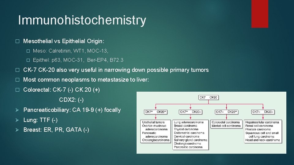
Immunohistochemistry � Mesothelial vs Epithelial Origin: � Meso: Calretinin, WT 1, MOC-13, � Epithel: p 63, MOC-31, Ber-EP 4, B 72. 3 � CK-7 CK-20 also very useful in narrowing down possible primary tumors � Most common neoplasms to metastasize to liver: � Colorectal: CK-7 (-) CK 20 (+) CDX 2: (-) Ø Pancreaticobiliary: CA 19 -9 (+) focally Ø Lung: TTF (-) Ø Breast: ER, PR, GATA (-)
Cholangiocarcinoma � Malignant neoplasm arising from the epithelial cells of the biliary tract with cytologic features of cholangioctyes � 2 nd most common primary liver malignancy behind Hepatocellular carcinoma, representing 3% of all GI malignancies in the US. � Typically presents with painless in jaundice in 7 th decade of Life. � Risk factors include some overlapping with HCC: NAFLD, HBV, HCV but also include primary sclerosis cholangitis, liver flukes endemic to Asia, chronic choledocholithiasis/cholangitis, and Caroli’s disease
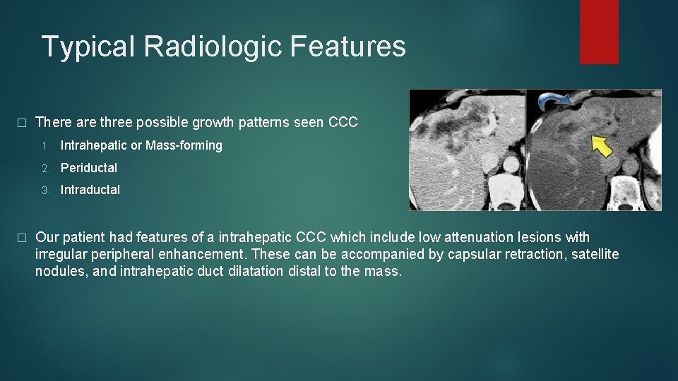
Typical Radiologic Features � � There are three possible growth patterns seen CCC 1. Intrahepatic or Mass-forming 2. Periductal 3. Intraductal Our patient had features of a intrahepatic CCC which include low attenuation lesions with irregular peripheral enhancement. These can be accompanied by capsular retraction, satellite nodules, and intrahepatic duct dilatation distal to the mass.
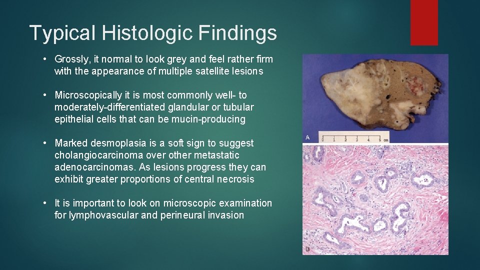
Typical Histologic Findings • Grossly, it normal to look grey and feel rather firm with the appearance of multiple satellite lesions • Microscopically it is most commonly well- to moderately-differentiated glandular or tubular epithelial cells that can be mucin-producing • Marked desmoplasia is a soft sign to suggest cholangiocarcinoma over other metastatic adenocarcinomas. As lesions progress they can exhibit greater proportions of central necrosis • It is important to look on microscopic examination for lymphovascular and perineural invasion
Staging � There are multiple staging systems that exist for intrahepatic cholangiocarcinoma including the Union for International Cancer Control/American Joint Cancer Committee Okabayashi. � Systems use factors including tumor size, Number of lesions, bilateral vs unilaterality, and vascular invasion to calculate stage
Treatment &Prognosis � If possible the gold standard of treatment is resection. Ability to resect is the number one determiner of prognosis. � Even with resection prognosis and 5 -yr survival is poor � 10 -44% 5 -yr survival � Combined systemic chemo therapy is another potential option � Some studies have shown radiation + transplant has achieved a 5 -yr survival of 65% Our patient is not a surgical candidate considering bilobular involvement and her distant metastases but she is considering single agent chemotherapy with Gemcitabine as well as referral to Palliative for her related-pain, nausea, and anorexia.
References � Carr, Brian I. . "Tumors of the Liver and Biliary Tree. " Harrison's Principles of Internal Medicine, 19 e Eds. Dennis Kasper, et al. New York, NY: Mc. Graw. Hill, 2014, http: //accessmedicine. mhmedical. com/content. aspx? bookid=1130§ionid=69857977. � Mc. Gahan, John P. , et al. "Role of FNA and core biopsy of primary and metastatic liver disease. " International journal of hepatology 2013 (2013). � Shaw, Colette, and Susan Shamimi‐Noori. "Ultrasound and CT‐directed liver biopsy. " Clinical Liver Disease 4. 5 (2014): 124 -127. � Vinay Kumar MBBS, MD, FRCPath, Abul K. Abbas MBBS, and Jon C. Aster MD, Ph. D. Robbins Basic Pathology, Chapter 16, 637 -677