Radiology of common brain diseases 366 RAD 1

Radiology of common brain diseases 366 RAD 1

Scope Radiology of common brain diseases Lecture 1 Lecture 2 n Intracranial bleeding n Intracranial tumors n Brain ischemia n Brain infections n Brain edema n Brain inflammation 2

Objectives Radiology of common brain diseases At the end of the lecture the student is expected to: 1. identify cerebral infarction on images and know the clinical deficits associated with each type of infarction. 2. understand the imaging findings of the different types of intracranial bleeding. 3. recognize the cerebral edema signs on neuroimaging and differentiate between its types. 3

4

5

Intracranial Bleeding Extradural n Subarachnoid n Intraventricular n Intraparenchymal n 6

7
")
EDH n n n n Blood collection between inner table and dura. Biconvex (lentiform) Occur at site of impact 95% unilateral, supratentorial Does not cross sutures Can cross falx and tentorium Skull fracture in 90% Air seen in 20% 8

EDH Arterial 90%, Venous 10% n Nontraumatic-rare n Lucid interval-50% n C/F: headache, nausia, vomiting, convulsions, herniation. n 9

SDH n n n Blood collection between dura and arachnoid. Crescent shape Supratentorial Cross sutures, but not dural attachments May extend along falx and tentorium 10

SDH Trauma is the most common cause n Acute: 6 hr-3 d n Subacute: 3 d-3 w n Chronic: >3 w n 11

SDH 12

SDH 13

14

15

Blood in the suprasellar cistern Blood in the interhemispheric fissure Blood in the ambient cistern Blood in the sylvian fissures 16
 n Nontraumatic n C/F:")
SAH Blood between pia and arachnoid n Traumatic (most common) n Nontraumatic n C/F: headache, vomiting, blurred vision, neck rigidity n Complications: hydrocephalus (acute/delayed), vasospasm, rebleeding. n 17

18

19

Parenchymal bleed Causes: HTN, trauma, AVM, aneurysm, permaturity, tumors, infarction, coagulopaty. n 20

Trauma 21

22

Trauma 23

24

Courtesy: Osborn AG 25

Courtesy: Smithuis R 26

27

28

29

30

31

32

33

34

35

36

37

T 2 WI FLAIR DWI 38

FLAIR DWI 39

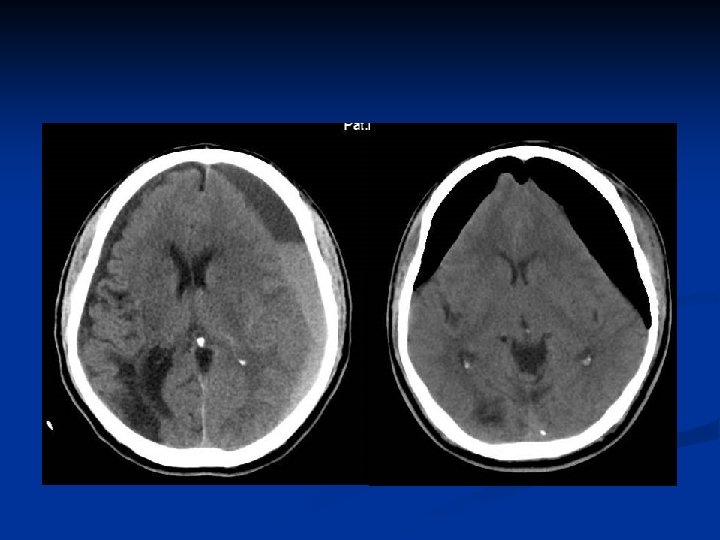
Brain edema n Vasogenic: n n Trauma/infection/inflammation/tumors Cytotoxic: n Ischemia/trauma • Both could be generalized or localized • Both may co-exist 40

Brain edema n Imaging findings: Hypodensity on CT n Low signal on T 1, high signal on T 2 & FLAIR n Loss of GM/WM interface n Compressed ventricles n Effaced sulci & cisterns n Dense cerebellum n Brain herniation n Vascular compression-ischemia n 41

Brain edema 3 w later 42

Brain edema 43

Brain edema 44

Brain edema 45

Brain edema Vasogenic Cytotoxic Location White matter Gray&White matter DWI Non-restricted Restricted Shape Finger-like Diffuse 46

The cause of this hematoma is: A. B. C. D. Courtesy of Chong V. Anticoagulation Hypertension Ruptured aneurysm Trauma 47

This CT shows: A. Epidural B. Subdural C. Subarachnoid D. Intraparenchymal 48

? ? Normal 49

n n n SDH IVH SAH Pneumocephal us Edema 50

This CT shows: A. Subdural hematoma B. Subarachnoid hemorrhage C. MCA infarction D. All of the above 51

This CT shows: A. B. C. D. Epidural hematoma Subdural hematoma MCA infarction Normal brain 53

Compressed lateral ventricle Obliterated CSF space Normal sulci Subdural hematoma 54

Intracranial bleeding Brain infarctions Brain edema 55
- Slides: 55