Radiology Michael Lavoie Middlesex Community College Veterinary Assistant


 should")


-dog")


")


- Slides: 57
Radiology Michael Lavoie Middlesex Community College Veterinary Assistant Program 6/6/12
Radiograph Safety n n n Guidelines for determining who may be exposed in a working situation include: No person under 18 years old shall operate x-ray equipment or be exposed to X-rays in the work place. Pregnant employees should be advised against exposure after being informed of the associated hazards. Radiology assignments should be rotated among the staff, and practice owners should maintain a radiation exposure log. Signage should be posted prohibiting the presence of unauthorized persons (employees, contractors, or owners) in the X-ray area while radiographs are being taken.
Radiograph Safety n n n The mandatory use of personal protective equipment (PPE) should be discussed. Monitoring and testing exposure is an important part of this program. Exposed staff must know when to wear and test their personal dosimetry devices.
n n X-ray exposure can be from the primary beam or from scatter radiation. Your program should clearly explain that operators and assistants must not allow any part of their own body -- even with the proper shielding of their PPE -to be exposed to the primary beam. Scatter or secondary radiation will often travel upwards toward the head or torso of the operator or other personnel involved in a procedure. Looking away during exposure may help to protect delicate eye tissue.
These safe practices require protective actions while using x-rays: n n n Require all exposed employees to wear their own radiation exposure badges or rings. Failure to follow this requirement should lead to termination. All operators and assistants must either be behind a protective barrier, such as a wall or lead shield, or they must wear PPE. No part of a technician’s or operator’s body is allowed in the primary beam during an exposure, even when wearing protection. An operator or technician should never be on the Xray table.
n n An operator must not hold a tube house during its operation, even on portable units. When film must be held, cassette holders can be made to fit a practice’s specifications. The use of animal-restraining devices can help to minimize exposure to staff. Sedation is recommended for fractious animals. The restraint of an animal during radiographic exposure cannot be listed as a primary duty in an employee’s job description.
Help keep x-ray activities in your clinic safe by following these guidelines: n n n Indicate safe working zones. Place an area radiation badge one to two feet outside the designated safe zones. Send your monitoring lab an unexposed blank badge as a control. Maintain good housekeeping in the area. Monitor and inspect all the PPE at least monthly. Document these inspections and retain the records for at least one year. Immediately replace any PPE that is cracked, punctured, or worn
X-ray Machines n n Animal radiography often requires modifications to human radiology equipment. For vets to perform an x-ray on a small animal, the x-ray machine can be positioned over a table, while free-floating machines can be used for larger, standing animals. A free-floating x-ray machine can move up and down, so it can also x-ray the lower extremities. This can accommodate cows, horses and other large animals.
CT machines n n n A CT is another commonly used test in animal radiography. CTs provide hi-resolution cross section images of the inside of an animal. The image is much like an x-ray, but it is three dimensional and provides a clearer picture than a traditional x-ray.
CT Machines and MRI n n n Since an animal must lie on a table during the scan, the CT procedure poses a problem for large animals. A large animal CT table can accommodate animals up to 2, 000 pounds (about 907 kg). MRIs are also used in animal radiography, but they can be very expensive and are consequently not used as often. (unless there is a neurologist on staff)
General Key Points for Canine Radiology n n n Canine Thorax - VD, DV, and Lateral views include all lung fields on the radiograph Lateral view-most important to extend forelimbs to exclude musculature from the radiographic field. Sandbag around both forelimbs, sandbag over neck, and sandbag around both hindlimbs. (This works well if the animal is under anesthesia)
n Central x-ray beam penetrates the body at the level of the mid-cardiac silhouette. The light field is collimated to include the entire lung fields.
DV Restraint for Chest Radiographs n n DV view (most appropriate for cardiac concerns)-dog rests on sternum and hindquarters. Do not pull forelimbs forward; allow them to rest at the dog's sides. Hindquarters must be level The light field is collimated to include the entire lung fields.
VD Restraint for Chest Radiographs n n n VD view-dog is in dorsal recumbency. Sandbags around each of the four limbs and one bag over the neck. The light field is collimated to include the entire lung fields.
Lateral Restraint for Abdomen Radiographs n n Lateral view-dog is in right lateral recumbency. Hind legs are extended caudally to prevent them from overlying the bladder. Sandbag as for lateral thorax. Central beam penetrates approximately the last rib, but it is more accurate to include the cranial most or caudal most border on the film
VD Restraint for Abdomen Radiographs n n Dog is in dorsal recumbency. Sandbags as for VD thorax. Central beam penetrates approximately through the umbilicus, but it is more accurate to include the cranial most or caudal most border on the film The light field is collimated to include as much abdomen as possible with the cranial or caudal border of interest included.
Lateral Pelvic Radiographs in Dogs n n Lateral view-Dog lying on affected side (to prevent image magnification and distortion by placing side of interest closest to the film). Affected leg is neutral, unaffected leg is pulled back a moderate amount dorsally with the goal of keeping the pelvis lateral
Frog Leg View for Pelvic Radiographs in Dogs n n n VD frog-leg view Dog is in dorsal recumbency. Sandbags as for VD thorax. Central beam penetrates the pelvis through the pelvic inlet (at the level of the hip joints). The light field is collimated to include the entire pelvis with the proximal ½ of the femurs.
Straight Leg View for Pelvic Radiographs in Dogs n n VD straight leg view-Dog is in dorsal recumbency. Sandbag forelimbs as for VD thorax. Extend femurs until the hocks rest on the folded sandbag. Use a Velcro® strap or tape to rotate the femurs towards each other until they are parallel with each other (be careful not to over rotate dogs with marked muscle atrophy).
Straight Leg View for Pelvic Radiographs in Dogs n n n The hocks, stifles and hips should all be in line with the pelvis. Central x-ray beam penetrates the pelvis at the level of the hip joints, over the spine. The light field is collimated to include the entire pelvis and femurs, including the stifle joints.
General Key Points in Feline Radiology n n n Feline Whole Body-VD and lateral views include the entire cat from the thoracic inlet to the pelvis. If a special area of interest is noted in either area, then coned down views of either the thorax or abdomen should be obtained. Cats can (usually) fit on a whole large radiograph plate
Lateral Whole Body View in Cats n n n Lateral view-The cat is placed in right lateral recumbency. The forelimbs are extended cranially, the hindlimbs caudally The central x-ray beam is directed to the last rib and the light field is collimated to include thoracic inlet to the pelvis
VD Whole Body View in Cats n n VD view-The cat is placed in dorsal recumbency with the forelimbs extended slightly cranially, and the hindlimbs extended slightly caudally. The central beam penetrates the animal at the level of the last rib with the light field collimated to include thoracic inlet to the pelvis, and right and left skin edges.
Lateral Chest Radiographs in Cats n n n Cat is in right lateral recumbency. The forelimbs are extended cranially The hindlimbs are left neutral. Central beam penetrates at the cardiac silhouette. The light field is collimated to include the entire lung fields.
VD Chest Radiographs in Cats n n n Cat is in dorsal recumbency. Forelimbs are pulled cranially slightly; hindlimbs are restrained in a neutral position. The light field is collimated to include the entire lung fields.
DV Chest Radiographs in Cats n n Cat is in dorsal recumbency. Forelimbs are left along the body for support and hindquarters are level. Head is extended slightly to prevent too much weight on the sternum (causes rotation). The light field is collimated to include the entire lung fields.
Lateral Abdomen Radiographs in Cats n n Cat is in right lateral recumbency. The forelimbs are left neutral. The hindlimbs are extended caudally The light field is collimated to include the entire abdomen.
VD Abdomen Radiographs in Cats n n Cat is in dorsal recumbency. Forelimbs are pulled cranially slightly; hindlimbs are restrained in a neutral position. Central x-ray beam is directed towards umbilicus. The light field is collimated to include the entire abdomen.
Lateral View of the Pelvis in Cats n n n The cat is lying with the affected side down to prevent magnification and distortion of the area of interest. The affected leg is neutral; the unaffected leg is extended caudally The light field is collimated to include the entire pelvis and the proximal thirds of each femur.
VD View of the Pelvis in Cats n n The cat is in dorsal recumbency with the forelimbs pulled cranially slightly, hindlimbs are extended so the femurs are parallel. The central beam is directed towards the level of the hip joints and the light field is collimated to include the entire pelvis and the entire femurs.
Interesting Radiographs n n X-rays are passed through the patient to expose a piece of radiographic film on the other side of the patient. Thin areas of the patient allow a lot of x-rays through, which exposes the film and turns it black. Thicker body parts absorb more x-rays, so less hit the film, resulting in white areas. Thus white objects are dense like bone, and black objects are thin like air.
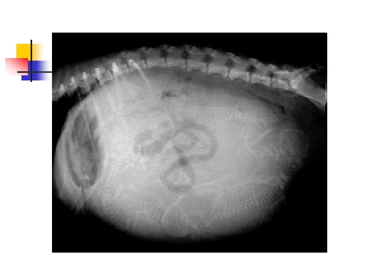
How Many Puppies? n n This dog's owner knew she was pregnant and was hoping for a big litter. Can you count how many puppies she is expecting? Here's a hint: the little puppy skeletons are visible. Try to count heads and backbones. It's harder than it looks because the puppies overlap each other!
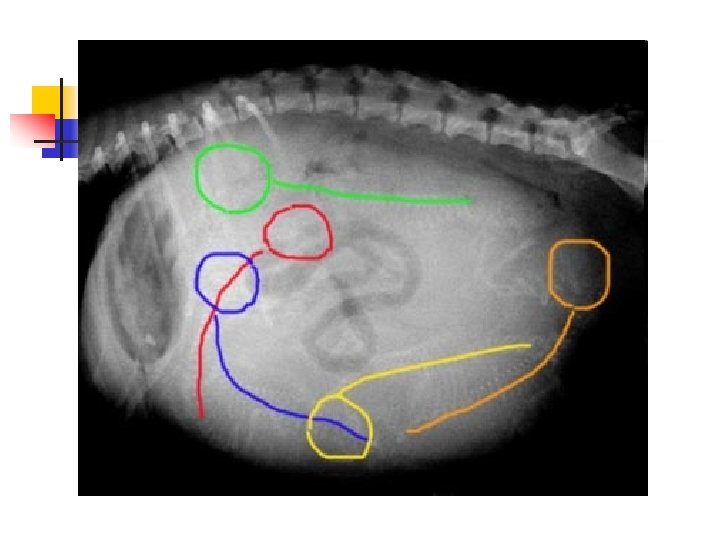
Here is the answer: n n There are five puppies. I have outlined the heads and backbones in different colors to point them out.
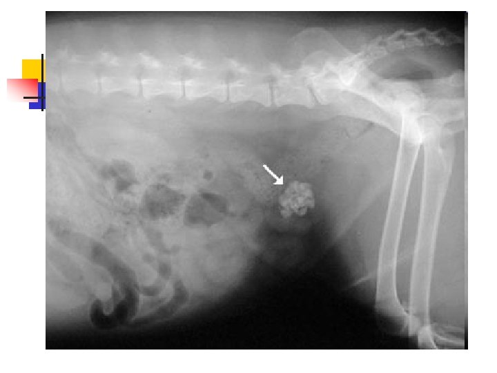
Bladder Stones n n n Bladder stones (also known as uroliths or cystic calculi) are hard, rock-like lumps of mineral that form in the urinary tract. Dogs and cats are different and usually form stones in the bladder. Because it is a bigger space, these stones tend to be much larger and usually cannot be passed, but have to be removed surgically or dissolved with special diets.
Bladder Stones n We suspect bladder stones when a dog has blood in the urine that doesn't respond to conventional antibiotic therapy, when we palpate a thickened bladder, when a dog strains to urinate, or when there are many crystals in a urine sample.
Bladder Stones n n Because most stones contain calcium, they are usually visible on a radiograph as white (bone density) objects in the area of the bladder. Sometimes there is a single large stone, and sometimes there are literally hundreds of tiny stones like sand or aquarium gravel.
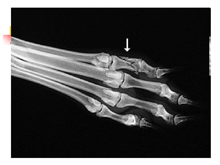
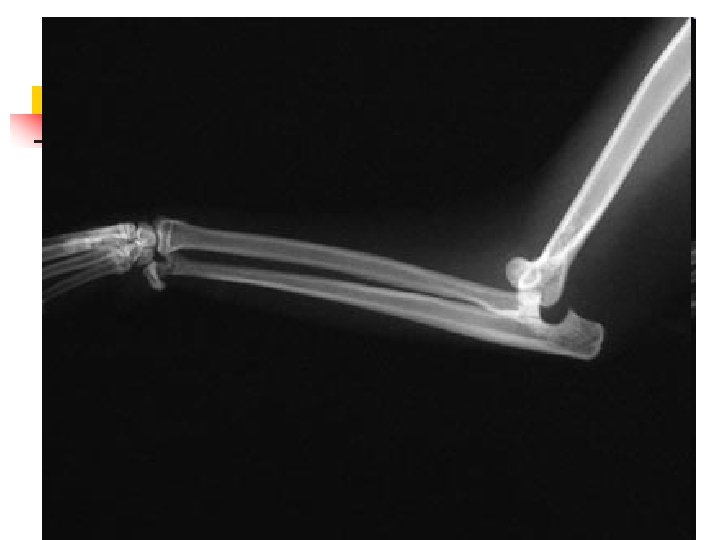
When Animals Limp n n n The good new is that 9 out of 10 limping animals have what we call soft tissue injuries a sprain, a pull, a bruise and only require exercise restriction to heal. However, 1 out of 10 animals with lameness has something more serious, and we can tell which ones by close observation of the patient walking and careful physical examination. In those cases, radiographs are used to make a diagnosis.
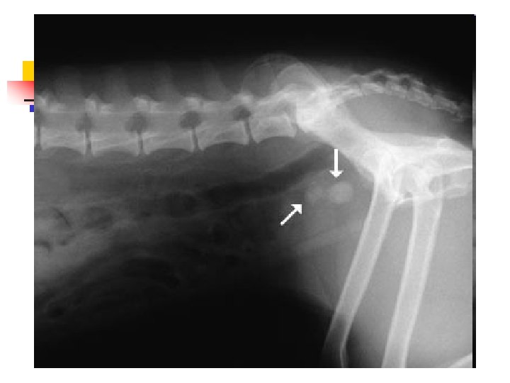
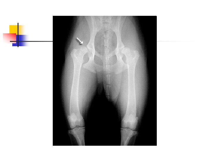
Hip Dysplasia n n Hip dysplasia is a looseness in the hip joint. The hip is a ball-and-socket joint and the head (ball) of the femur (thigh bone) normally should be deep within the hip socket. When hip dysplasia is present, the ball moves in and out of the socket with ease. Over time arthritis (degenerative joint disease, osteoarthritis) sets in as the body tries to stabilize the loose joint.
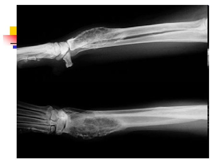
When Lameness isn’t Simple n n Sometimes when an animal limps the cause turns out to be something more serious than a simple injury. The doctors palpated a firm, painful lump in the leg this dog was favoring. Radiographs showed that the bone was expanded in that area, with a motheaten, hollowed-out center. These are classic signs of a tumor in the bone, known as an osteosarcoma.
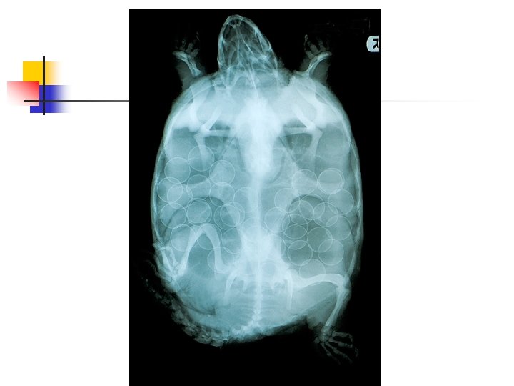
n n This snapping turtle was brought in after it had been hit by a car. Fortunately the injuries were minor and easily treated. She was diagnosed as pregnant, so she was released where she had been found as soon as possible so that she could lay her eggs. Can you count how many eggs?
Lab Equipment- Centrifuge n n n n Centrifuges are used to spin samples down at high rates of speed Blood and urine samples Used to separate or concentrate materials suspended in a liquid form Each time it is used it must be balanced Lid should be closed and secure, set for required spin time. No one should stand in front of centrifuge No one should stop motor or rotor manually.
Lab Equipment- refractometer n n Used to measure the weight of a liquid. Light weight and handheld Important to recalibrate on regular basis using distilled water If not properly calibrated will give inaccurate results.
Lab equipment- Blood Chemistry Analyzers n n Machines that run blood samples and measure routine blood chemistries Blood chemistries and CBCs may be run using whole blood, plasma, or serum Training is needed when using these machines Are easy to run, and give accurate numbers if used correctly, and quality controlled.
Serology Testing n n Used to provide a quick analysis for a common disease or virus A variety of reagants or chemicals are used to run each test, and should be kept with the test kit
Questions?