RADIOLOGY CASE HISTORIES Facial pain Trauma to orbit

RADIOLOGY

CASE HISTORIES: • Facial pain • Trauma to orbit • Allergic reaction • Bruit heard in neck • Neck mass inferiorly • Dysphagia • Hoarseness • Blurred vision • Epistaxsis

AP Caldwell View of Sinuses 2 4 3 1 5 6 1. Nasal Septum 2. Frontal Sinus 3. Maxillary Sinus 4. Ethmoid Sinus 5. Inferior Turbinate 6. Odontoid process

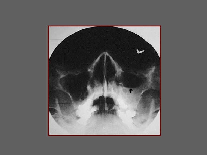
AP WATERS VIEW Sinuses 1. Frontal sinus 1 2. Ethmoid Sinus 10 3. Nasal Septum (bony) 4. Zygomatical-Frontal Suture 2 5. Maxillary Sinus 4 6. Zygoma 7. Zygomatic Arch 5 3 8. Mandible 9. Inferior orbital margin 6 9 7 8 10. Left orbit

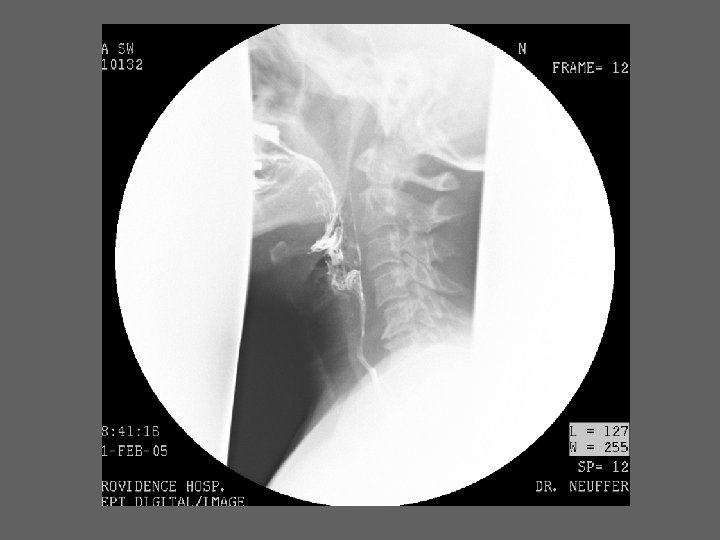
1. Frontal Sinus Lateral Sinus & Skull 2. Maxillary Sinus 3. Ethmoid Sinus 11 4. Spenoid Sinus 5. Sella Turcica 1 6. Occipital Bone 7. Mastoid Air Cells 3 8. Floor of posterior fossa 5 9. Anterior arch of C-1 7 10. Mandible 11. Coronal Suture 2 4 6 9 8 10

AP Townes SKULL 1 1 1. Parietal bone 2. Lambdoid suture 2 3 3. Foramen magnum 4. Petrous temporal bone 5. Mandible 6. Mastoid air cells 4 6 5

Base of the Skull 1. Lat. ptyergoid plate 2. Ethmoid Sinus 6 3. Odontoid Process 4. Sphenoid Sinus 2 5. Foramen ovale 6. Maxillary Sinus 7. Mastoid air cells 8. Ant arch of C-1 9. Margin of foramen magnum 1 10. Ext auditory canal 5 4 10 8 3 9 7

Airway 3 2 5 4 1 1. Calcified tracheal cartilage rings 2. Hyoid bone 3. Epiglottis 4. Thyroid cartilage 5. Cricoid cartilage

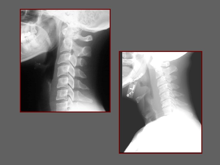
1 Lateral Neck 2 3 4 1. Stylo-hyoid ligament 2. Hyoid bone 3. Thyroid cartilage 4. Cricoid cartilage

3 Lateral Neck 1 2 4 4 1. Hard pallate 2. Soft pallate 3. Nasopharynx 4. Oropharynx

Arteriogram 2 3 1. Internal Carotid artery 2. Carotid Syphon 3. Maxillary artery 4. Occipital artery 5. External carotid artery 6. Common carotid artery 4 1 5 6

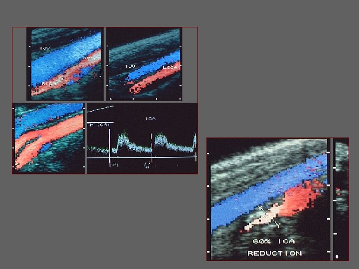
Color Doppler Ultrasound of Carotid Artery

SINUS-CT Axial view 1. Frontal Sinus 1

CT of Sinus Axial view 1 1. Ethmoid Sinus 2. Sphenoid Sinus 2 3 3. Carotid canal

1 CT of Sinus Axial view 4 2 5 3 1. Maxillary Sinus 2. Pterygoid plate 3. Nasopharynx 4. Nasal septum 5. Inferior turbinate

CT of Sinus 1 Axial View 2 4 4 3 3 1. Maxillary Sinus 2. Hard Palate 3. Mandible 4. Masseter muscle

2 Coronal view of Sinus 1 3 1. Fronto-nasal suture 2. Frontal sinus 3. Nasal bones

1 3 2 Coronal view of sinus 1. Ethmoid sinus 2. Inferior turbinate 3. Middle turbinate

1 Coronal view of sinus 2 1. Maxillary sinus 2. Nasal Septum

Coronal View of sinus 1 1. Sphenoid sinus 2. Hard Palatte 2
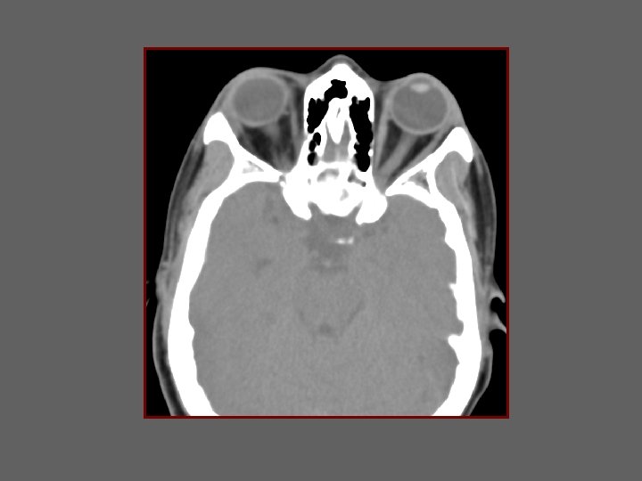

Axial scan of orbit 2 1 3 4 5 1. Retrorbital fat 2. Medial rectus 3. Lens 4. Lateral rectus 5. Optic nerve

SCAN LEVEL MAXILLARY SINUSES ZYGOMA LT ZYGOMA SPHENOID SINUS

SCAN LEVEL MANDIBULAR CONDYLE MAXILLA LT EXTERNAL AUDITORY MEATUS NASOPHARNYX MASTOIDS

SCAN LEVEL MASSETER MUSCLE MANDIBLE PTERYGOID MUSCLES PAROTID GLAND MASSETER MUSCLE LT

SCAN LEVEL SUBMANDIBULAR GLAND EPIGLOTTIS STERNOCLEIOMASTOID MUSCLE SUBCUTANEOUS FAT LT

SCAN LEVEL LT HYOID BONE JUGULAR VEIN COMMON CAROTID ARTERIES

SCAN LEVEL LT STERNOCLEIDOMASTOID MUSCLE THYROID CARTILAGE VOCAL CORD

SCAN LEVEL LT THYROID CARTILAGE COMMON CAROTID ARTERY JUGULAR VEIN CRICOID CARTILAGE

LT SCAN LEVEL THYROID GLAND CLAVICLE FAT TRACHEA ESOPHAGUS FAT

CASE HISTORIES: • Facial pain • Trauma to orbit • Allergic reaction • Bruit heard in neck • Neck mass inferiorly • Dysphagia • Hoarseness • Blurred vision • Epistaxsis

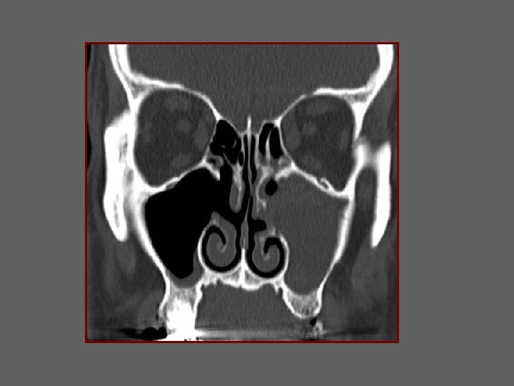
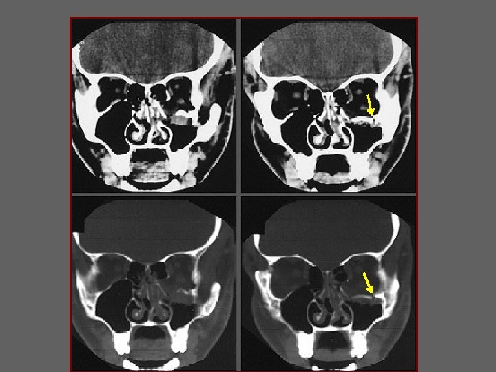
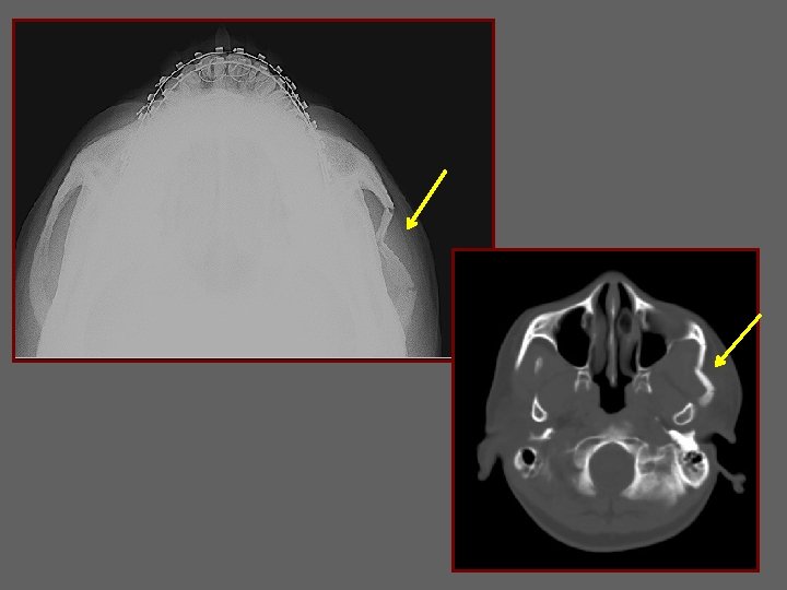
FACIAL PAIN AND NASAL CONGESTION
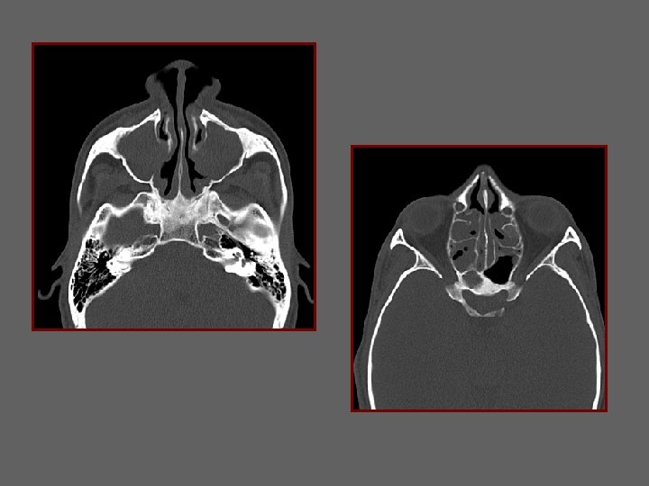
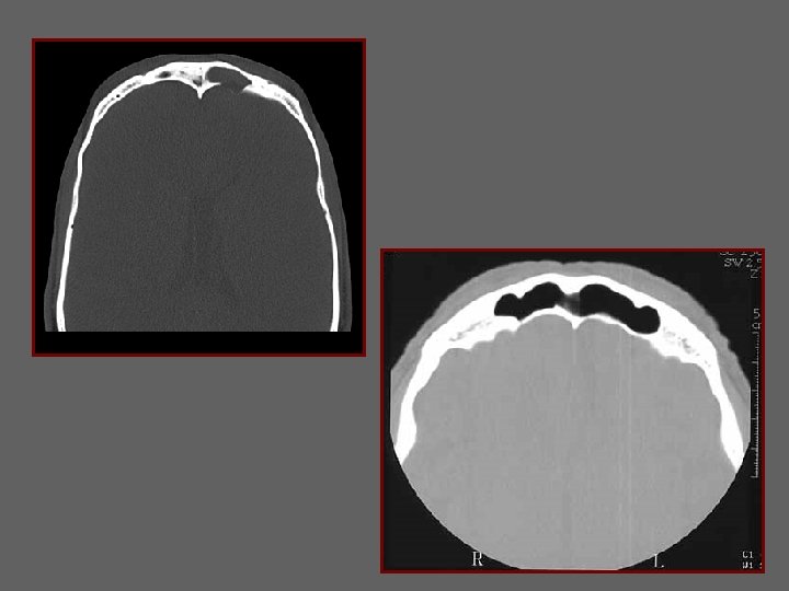

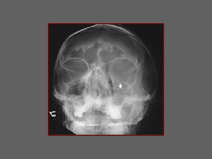
TRAUMA TO ORBIT

PT WITH ALLERGIC REACTION

ASYMTOMATIC BRUIT ON PHYSICAL EXAM

Arteriogram 2 3 1. Internal Carotid artery 2. Carotid Syphon 3. Maxillary artery 4. Occipital artery 5. External carotid artery 6. Common carotid artery 4 1 5 6

LATERAL CAROTID ARTERIOGRAM

Subtraction film

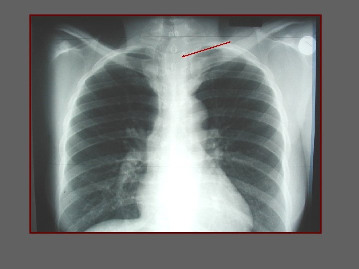
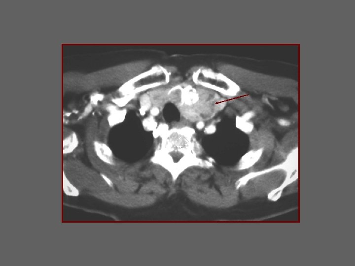
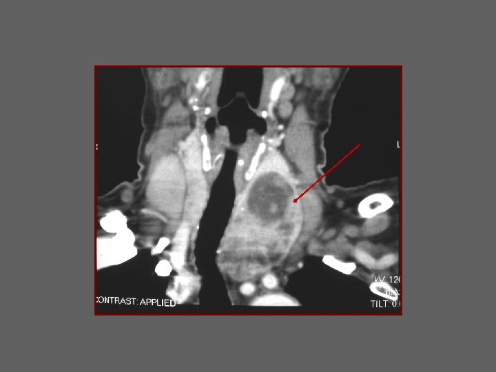
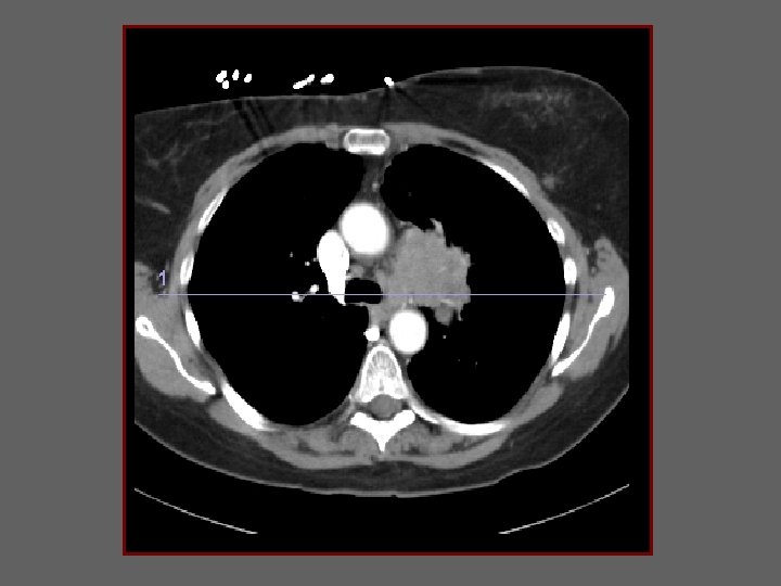
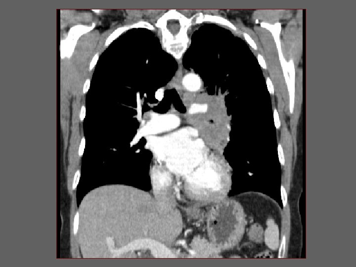
PALPABLE NECK MASS INFERIORLY

Thyroid Scan Nuclear Medicine

Thyroid Scan Nuclear Medicine

Thyroid

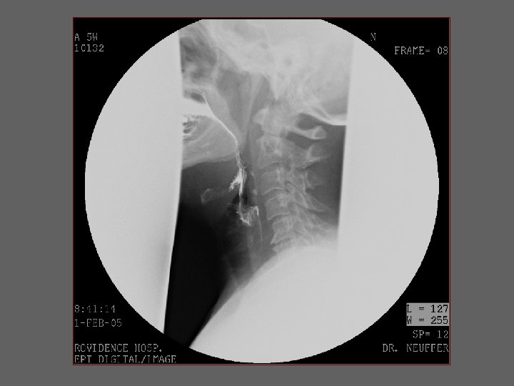
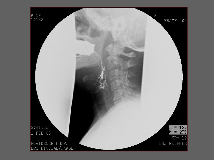
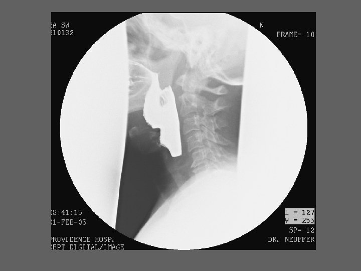
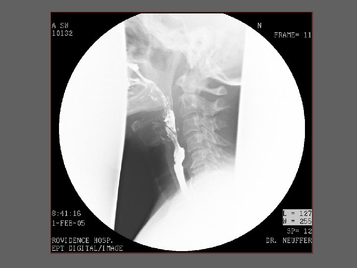
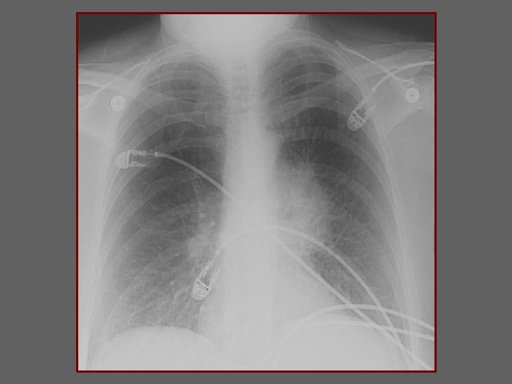
DYSPHAGIA

ASPIRATION NORMAL

HOARSENESS

VISUAL FIELD DEFECT BLURRED VISION

Normal skull Normal Sella

Normal Sella Mass

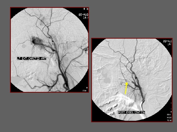
EPISTAXIS
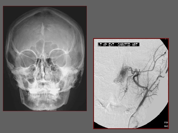

Vascular Tumor Normal
- Slides: 73