
Radiology 4 a case presentation Tim Herberg 61617

Radiology 4 a case presentation Tim Herberg 6/16/17
Clinical vignette • A. F. 70 y/o F presents w/ tachycardia 3 wks after R total knee replacement • Undergoes CTPA

Clinical vignette • 18 x 20 mm spiculated nodule in apical segment of RUL, 13 x 14 mm nodule in mid RUL
SPNs • Common, especially >50 • Ddx: granulomas, lung CA, solitary mets, abscess, hamartoma, AVM • Benign findings: no growth 2 yrs, calcification pattern (difuse, central, laminated, popcorn), small size • Patholic findings: recent growth, eccentric or stippled calcification pattern • Can be fooled by fluid in fissures, pleural plaques, skin lesions (nipple shadow, mole, lipoma) • mngt guidelines-Fleischner society

Clinical vignette • A. F. 70 y/o F w/ tachycardia 23 days after R total knee replacement • Undergoes CTPA-18 x 20 mm spiculated nodule in apical segment of RUL, 13 x 14 nodule in mid RUL • Undergoes PET on 6/1 and CT guided transthoracic lung biopsy on 6/8 via IR
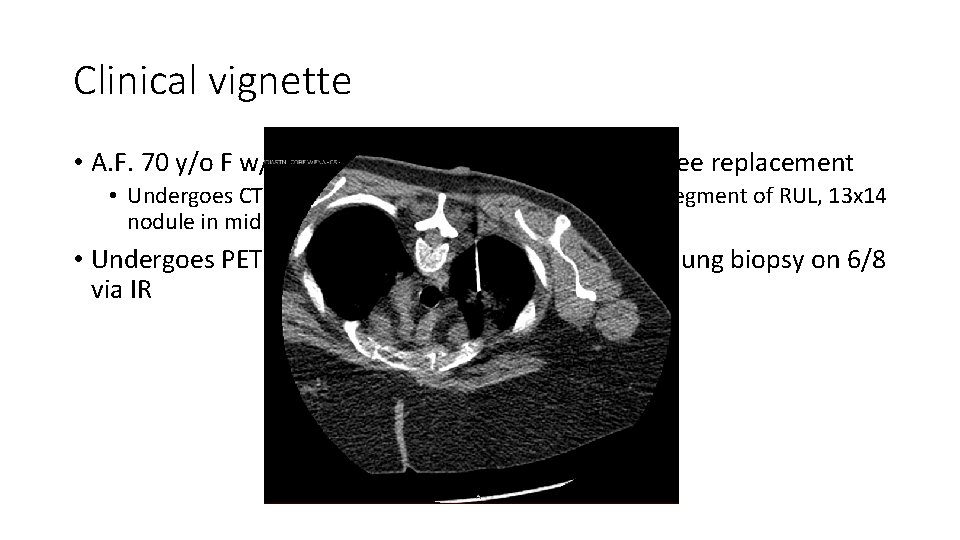
Clinical vignette • A. F. 70 y/o F w/ tachycardia 23 days after R total knee replacement • Undergoes CTPA-18 x 20 mm spiculated nodule in apical segment of RUL, 13 x 14 nodule in mid RUL • Undergoes PET on 6/1 and CT guided transthoracic lung biopsy on 6/8 via IR
Clinical vignette • A. F. 70 y/o F w/ tachycardia 23 days after R total knee replacement • Undergoes CTPA-18 x 20 mm spiculated nodule in apical segment of RUL, 13 x 14 nodule in mid RUL • Undergoes PET on 6/1 and CT guided transthoracic lung biopsy on 6/8 via IR • 1 day s/p lung biopsy developed right sided chest pain, tachycardia, and diaphoresis
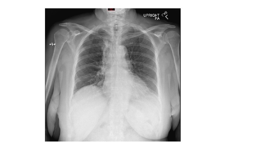
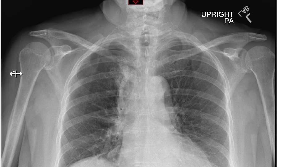

Diagnosis-PTX
Hospital course • Had pigtail chest tube placed with improvement of PTX and symptoms. Discharged the following day. Pathology revealed adenocarcinoma. Will f/u with heme/onc.
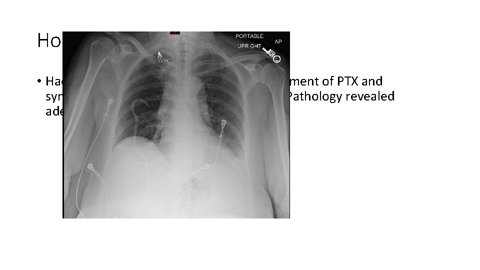
Hospital course • Had pigtail chest tube placed with improvement of PTX and symptoms. Discharged the following day. Pathology revealed adenocarcinoma. Will f/u with heme/onc.
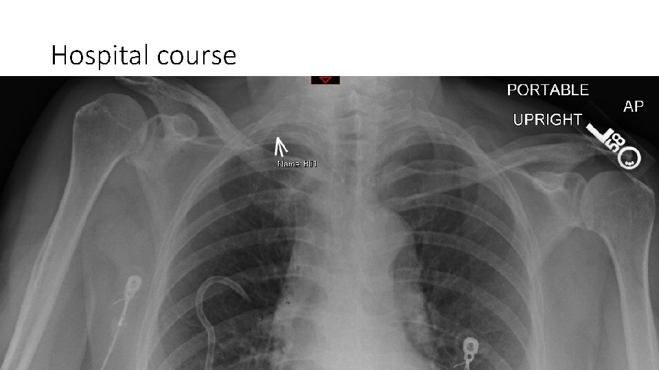
Hospital course • Had pigtail chest tube placed with improvement of PTX and symptoms. Discharged the following day. Pathology revealed adenocarcinoma. Will f/u with heme/onc.
Radiographic features, differential diagnosis for findings, etiologies, mngt • Transition point with peripheral lucency/lack of vascular lung markings • Upright vs supine • Ddx: • Etiologies: iatrogenic, idiopathic, asthma, COPD, trauma, pneumonia, neoplasm, marfans syndrome, smoking cocaine • Mngt:
References • UVA Radiology Tutorial: Introduction to chest radiology. https: //www. med-ed. virginia. edu/courses/rad/cxr/Index. html
- Slides: 15