Radiological syndroms Alveolar syndrome Bronchial syndrome Interstitial syndrome

Radiological syndroms Alveolar syndrome Bronchial syndrome Interstitial syndrome Vascular syndrome Mediastinal Syndrome

Alveolar syndrome

Pulmonary architecture : Morphological unit is the lobule 15 -25 mm

Acinus & lobule Lobule : morphological unit , smallest anatomical unit, ventilated by the centro lobular bronchiole accompanied by the centro lobular. Perilobular interstitial artery tissue dimension: 1 à 2, 5 cm composed of 3 to 5 acinus Acinus : functional unit 14 divisions between the trachea and the terminal bronchiole 1 et 1‘ bronchiole & centro lobular artery 2. bronchiole & terminal artery 3. respiratory bronchiole 4. Alveolar duct 5. Alveolar bag 6. Alveolus 7. Perilobular vein + lymphatic vessels Acinus

Features of alveolar syndrome • Blurred, heterogeneous opacity • Not well limited (except if there is contact with one fissure, then the consolidation is “systematised“) • With sometimes aeric bronchogram • No retraction in acute phase (but retraction is possible if chronic evolution)

Alveolar consolidation Blurred, heterogeneous Not well limited But sharp boundary if contact with a fissure Small fissure Right Upper Lobe Pneumonia

Systematised opacity with aeric bronchogram
")
Acute* alveolar syndrome - Main etiologies *The more frequent Located Diffuse Infections (bacterial, viral) Tuberculosis > 90 % of cases Localized pulmonary oedema Lung infarctus and embolism Lung traumatism Post radiotherapy (acute phase ) Mycosis Loeffler syndrome Alveolar sarcoidosis Broncho alveolar cancer Infections (bacterial, viral, opportunistic) Tuberculosis Pulmonary oedema > 90 % of cases Traumatism (contusion, fat embolism) Angeitis Pulmonary haemorrhage Loeffler syndrome, eosinophilic lung Hypersensibility pneumonia (allergic alveolitis)

Chronic* alveolar syndrome - Main etiologies * Much less common than acute should be referred to a pulmonologist Located Chronic pneumonia with eosinophilia Bronchiolo-alveolar cancer Lymphoma and haemopathy Diffuse bronchiolo-alveolar damage Diffuse Lymphoma and haemopathy Sarcoidosis Pulmonary alveolar proteinosis Lipidic pneumonia desquamative interstitial pneumonia

Young man with no medical history Fever, chills, sudden onset with right chest pain and purulent sputum Right upper lobe pneumonia (Streptoccocus pneumoniae) • Heterogeneous • Sharp boundary in contact with a fissure • Aeric bronchogramme • No retraction in acute phase
")
Right lower lobe pneumonia (S. pneumoniae)

Bacterial pneumonia are often caused by ear-nose-throat and dental infections and by Flu

Right upper lobe pneumonia F. 50 y Day 6 of a Flu t° 40°, cough and sore spot right chest very limited area of crackles in right scapular region

Rapid clinical improvement with Amoxicillin Slower radiological improvement Evolution day 10

Bifocal infection : RUL external segment and left lower lobe
 + in")
Cough, asthenia, loss of weight since 4 weeks AFB (acid fast bacilli) + in sputum Bilateral tuberculosis pneumonia

Retractile evolution with treatment

RUL Excavated pneumonia HIV - Pneumonia + adenopathies HIV+ RLL pneumonia + adenopathies HIV+ TB pneumonia is frequent in countries with high incidence of TB in HIV + patients, but also in HIV In case of AIDS : mediastinal adenopathies are often associated and TB pneumonia is often located in the lower lobes If the immunosuppression is severe, the cavities are rare

Man 30 y old HIV+ Right upper lobe and middle lobe pneumonia Hilar and mediastinum adenopathies AFB sputum negative Bronchial aspiration and BAL (bronchiolo alveaolar lavage) : AFB + Fistulised node on endoscopic view This image is typical of TB/HIV

African officer in internship in France, t° 37° 8 C, stable condition, no functional or respiratory signs Excavated opacity of the inferior lobe, apical segment, AFB+

Pneumonia with Aeric bronchogramme Micro nodules Cavern

Man, 50 years old, fever, cough, dyspnea, headache, abdominal pain. Worsening despite amoxicillin treatment Legionnaires’ disease Legionella pneumophila

Man, 35 years old, dyspnea and severe hypoxemia. Treatment by amoxicilline 3 g per day. (1)

Worsening at J 4. Modification of treatment and introduction of erythromicin IV 3 g/24 h. Improvement in few days : Mycoplasma pneumoniae (2)

F 30 y HIV + Central African Rep. Bilateral pneumonia unimproved under Amoxycillin

Broncho Alveolar Lavage : PJP Aeric bronchogram me PJP - Allergy to Bactrim => dapsone Improvement S 3 Alveolar form of pneumocystosis RARE

If patient does not improve under amoxicilline and has not evident TB (AFB neg in sputum +/- Xpert negative) • Consider to switch to macrolides • Do not use fluoroquinolones • Ask for HIV status If the alveolar syndrome is chronic You must consider an alternative diagnosis Lymphoma, bronchiolo-alveolar carcinoma, …. and refer to a pulmonology ward

Bronchiolo-alveolar carcinoma

Bronchiolo-alveolar carcinoma

Pulmonary oedema alveolar syndrome physiopathological mechanisms : • Hemodynamic oedema : result of left ventricular failure • with pulmonary arterial hypertension, of post-capillar type. • Medical treatment is usually effective (O 2, diuretic, TNT. . ) • There is no anatomic lesion of the “alveolar capillary barrier“ • Lesional edema also called Acute Respiratory Distress Syndrom ARDS): The “alveolar capillary barrier“ is more or less irreversibly altered. The prognosis is most often dismal. Death may occur by refractory hypoxemia The etiologies are varied : • infectious (viral or bacterial) • toxic (for example inhalation of toxic gases) • complication of shock whatever the cause

Typical pulmonary cardiogenic

Hemodynamic pulmonary edema radiological features • • Alveolar picture Perihilar predominent Bilateral but sometimes assymetric Always with accute dyspnea, cough and clear sputum (although sometimes blood in sputum) • Crepitant rales (“crackles“) at the auscultation

Acute pulmonary oedema : “butterfly wings“ image

Acute pulmonary oedema After furosemide

Acute pulmonary oedema Note the asymmetry of the image

Asymetric cardiogenic edema

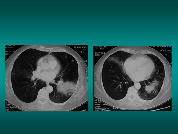
Two exemples of ARDS

Case 1 2008. 01. 29 Woman, 71/2 months pregnant. Fever and dyspnea with rapid deterioration in few days. No improvement despite amoxicilline then erythromycin

2008. 01. 30 2008. 01. 31 Worsening at D 2 then D 3 : lesionnal oedema probably with a viral origin

7 y. , fever, dyspnea and diffuse pain with quick onset. No improvmen antibiotherapy - D 1 : admission in intensive care unit. Tamiflu treatme Bronchial and nasopharyngeal sample for H 5 N 1 virus PCR Case 2

D 2 : no improvment => Non invasive ventilation - PCR + for H 5 N 1 Typical ARDS

D 4: mecanical ventilation with intra tracheal tube; Note chest tube for left pneumothorax drainage Progressive improvement D 8 to D 25 D 4 D 8

ARDS lesionnal edema during a H 5 N 1 flu in a young woman Slow improvement after management in ICU D 12 D 25

Alveolar syndrome Easy to diagnose : Blurred opacity, heterogeneous, ill-defined, except in contact with a fissure (systematized) non retractile with air bronchogram Must especially evoke : Localized : Bacterial pneumonia, TB pneumonia Diffuse : acute pulmonary edema, infection (bacteria, TB, virus, fungus)
- Slides: 45