RADIOLOGICAL PATHOLOGY OF ABDOMENUROGENITAL SYSTEM INTRODUCTION Radiograph external

RADIOLOGICAL PATHOLOGY OF ABDOMEN-UROGENITAL SYSTEM

INTRODUCTION Radiograph – external anatomy of the kidneys Radiographic contrast- visualization It allows us to visualize the kidneys, check for stones in the urinary system, look for calcification that might go along with kidney disease.
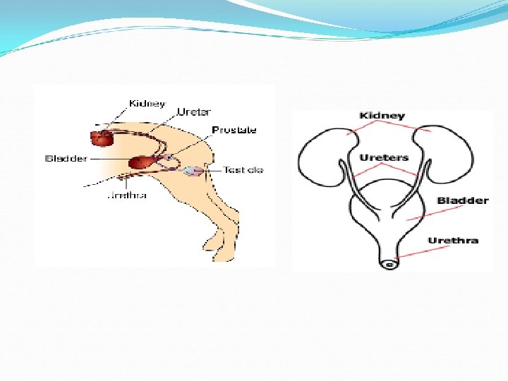

EXCRETORY UROGRAPHY Useful for defining anatomic structure & qualitatively assesing the functions of the kidneys. To demonstrate changes in the shape and structure of kidney substance due to neoplastic or obstructive changes. Size & position of the ureters eg: ureteral ectopia………
 gives")
Excretory Urogram This special test, also know as an IVP (intravenous pyelogram) gives us significant information about the renal system. It has to be used carefully if ARF or CRF is suspected because it can exacerbate the problem. A radiopaque dye is injected into the bloodstream and radiographs are taken of the dye as it passes through the kidneys, ureters, and bladder.

Techniques Two techniques for this investigation: A low volume rapid injection tech…-kidneys A high volume drip infusion tech…-ureters Water soluble contrast medium. (300 -400 mg iodine per ml) Total dose: 600 -800 mg iodine per kg b. wt – ten secs. Total dose: 1200 mg iodine per kg b. wt -10 -15 mins.

Cystography: small systic calculi, neoplasia, lesions in urinary bldder, displacement of urinary bladder… Pneumo cystogram: the bladder is inflated with air untill moderately distened. This is best judged by the gentle abdominal palpation. In some cases it is difficult to maintain adequate filling and it may be necessory to inject air just prior to the exposure if there is leakage around the catheter.

Positive contrast cystogram: water soluble contrast medium to distend the bladder This tech. provides better mucosal detail than the pneumo cystogram but small lesions or calculi may be lost in the contrast pool. Double contrast cystography: best method for detecting radiolucent calculi.

Positive contrast cystogram : UB is displaced cranial from the pubis by a large prostatic mass

Double contrast cystogram

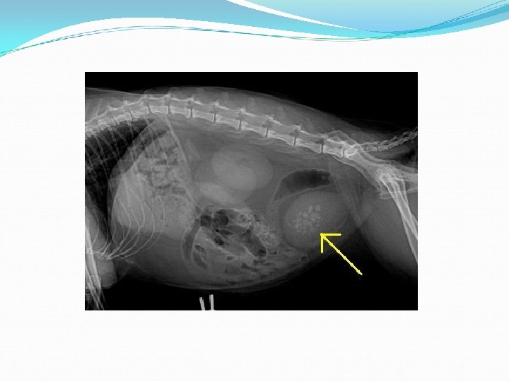
Radiograph showing a urinary bladder full of stones (actually, these are oxalate stones, but it would look the same if they were uric acid stones.

The black dots show stones that are not visible on a normal radiograph. This is a close up of a urinary bladder with contrast material inside. The contrast material is white. the surrounding air is black, and the black dots inside the contrast material are the uric acid stones.

Renal lymphosarcoma

Transitional Cell Carcinoma The urinary bladder is lined by “transitional cells. ” They must protect the body from the caustic urine inside the bladder but also must maintain this barrier when the bladder stretches and distends with larger volumes of urine. A Transitional Cell Carcinoma is a tumor of the Transitional Cell lining of the urinary bladder.
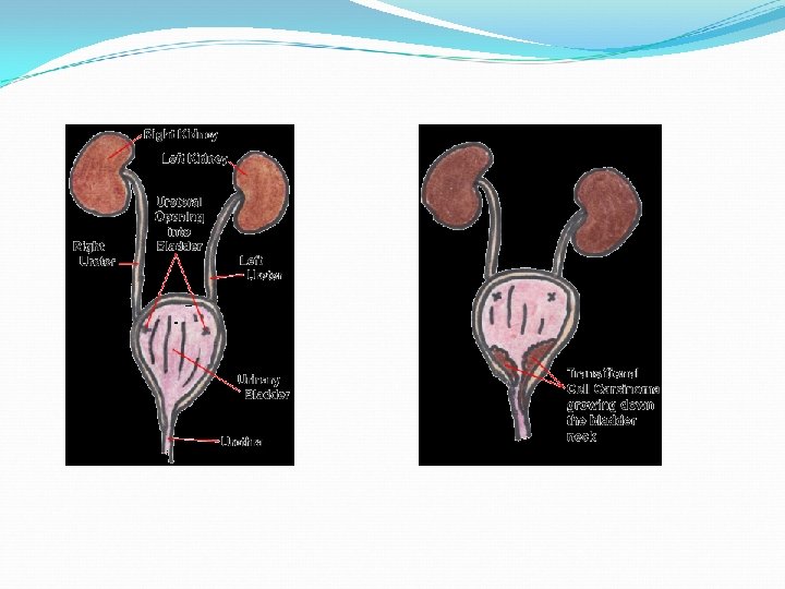

. Radiograph of a bladder injected with radiographic dye. The top right of the bladder oval is obscured, because of a “filling defect: ” A transitional cell carcinoma is taking up that space in the bladder, preventing the dye from “filling up” the entire space
 are hard, rock-like")
BLADDER STONES Bladder stones (also known as uroliths or cystic calculi) are hard, rock-like lumps of mineral that form in the urinary tract. Dogs and cats are different and usually form stones in the bladder. Because it is a bigger space, these stones tend to be much larger and usually cannot be passed, but have to be removed surgically or dissolved with special diets.
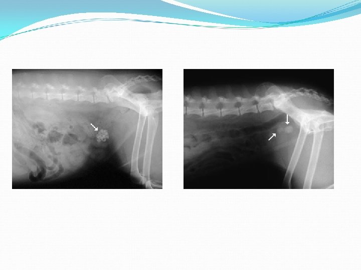

Renal calculi: Urolithiosis : the presence of calculi in the urinary system. Uratic calculi is common in dalmatian dogs. Oxalate calculi –hard, rough & spine surface – irritation, inflammation, hemorrhage.

RENAL CALCULI

Radiograph of an os penis Small black dots are tiny uric acdis stones surrounded by urinary dye. These stones are obstructing urine flow

Hydronephrosis Dilatation of renal pelvis due to obstruction to free flow of urine. It may be uni lateral or bilateral. The obstruction produces stasis of urine which with its back pressure cause atropy of renal parenchyma

Hydronephrosis, intravenous pyelogram, ventrodorsal, lateral projection

This is a radiograph of a cat with a moderately distended bladder from urinary obstruction.

DYSTOCIA: ventrodorsal view of a small-breed bitch. note the transverse presentation of the enlarged, viable fetus.

Bitch in mid-term pregnancy with an inguinal mass the tubular soft tissue opacity representing the uterus extends into the subcutaneous peri inguinal tissues

REFERENCE: http: //www. google. co. in/imgres? imgurl http: //www. marvistavet. com/assets/images/contrast_ radiography_bladder. jpg&imgrefurl http: //www. ncbi. nlm. nih. gov/pubmed/9335093 www. ukvet. co. uk/ukvet/articles/imaging_the%20 dys uric%20 dog. pdf www. vetstreamcanis. com/ACI/October 08/VMD 1/teq 00686. asp Textbook of VETERINARY DIAGNOSTIC RADIOLOGY by Donald E. Thrall, DVM, Ph. D
- Slides: 31