Radiological Pathology of Abdomen Part I GI Tract

Radiological Pathology of Abdomen Part I GI Tract

Normal Positions of GI Tract Small Animals

CATS

Stomach • Type of food and amount of food • Food retention > 12 hrs is abnormal • Position of stomach varies in different breeds of dogs and cats • In cats and Most of dogs – The stomach axis is parallel to the ribs
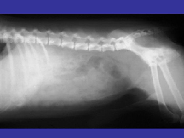


Useful interpretive criteria: • • Liver size influences the “fundic-pyloric” axis Normally parallel to last 2 -3 ribs on lateral view Normally perpendicular to spine on VD/DV views Large liver or right-sided liver mass displaces pylorus caudally and to the left remember F-P axis effect • Small liver allows pylorus to drift cranially remember F-P axis effect
 vs. VD (spinal")
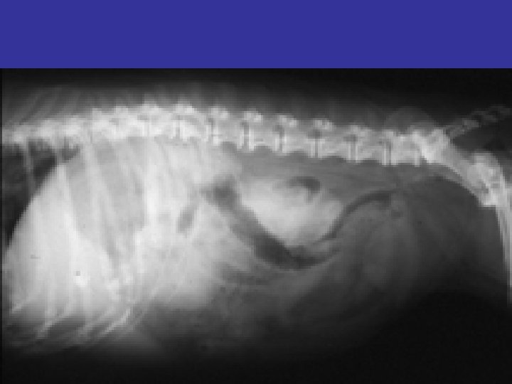
• Stomach: • Appearance changes depending on DV (Sternal recumbency) vs. VD (spinal recumbency) views (with the X-ray beam is centered on the last rib) • On DV views, the gas is primarily in the cardiac region with less in the pyloric region; fluid is in the body region • On VD views, the fluid is primarily in the cardiac region with less in the pyloric region; air is in the body region • Appearance may vary depending on the relative amounts of fluid and gas

DV VD

Right Lateral Recumbency Left Lateral Recumbency

VARIATIONS • Displacement of the organ – Cranial • • Reduced Liver Size Diaphragmatic Rupture Peritoneo-Pericardial DH Hiatal Hernia Mesentric masses Colonic Masses Pancreatic Masses – Caudal • Enlarged Liver • Thoracic Expansion

• Displaced towards the Right – Splenic Enlargement – Left side of Liver Enlarged • Right side displacement – Enlarged Right Liver – Pancreatic Enalrgement

• Variations in Size – GDV – Tumors f stomach • Gastrinoma – Chronic gastritis

Intestines • Ideally the animal should be fasted for 12 hrs before survey radiographs • For Colon an enema can be given • SI fills most of the abdomen – Caudal to stomach and Cranial to Urinary Bladder
- Slides: 19