RADIOLOGICAL EXAMINATION ASSISTED CLINICAL DIAGNOSIS OF CANCER PLAIN


 Gamma camera images using")


")
")


 show metastases in the liver shown in red circles")


 situated within and outside the muscle")


 view of right breast demonstrates the same")


 show")


 and an electric generator (LINAC)")


 Description n Immobilization n Image acquisition n Delineation of volumes (GTV,")


- Slides: 87
RADIOLOGICAL EXAMINATION ASSISTED CLINICAL DIAGNOSIS OF CANCER (PLAIN PHOTO, CONTRAST PHOTO, ULTRASONOGRAPHY, MAMMOGRAPHY CT, MRI AND NUCLEAR MEDICINE) Disampaikan leh: Wigati Dhamiyati Radiology Department Faculty of medicine Gadjah Mada University
INTRODUCTION Plain photo, contrast photo, ultrasound, CT, MRI and nuclear medicine are modalities on radiology imaging. n Mammography is a special examination for the breast using x-rays, usually combined with ultrasound. n All of these examination depend of the interest organ have an important role to make an accurate diagnosis of neoplasm n
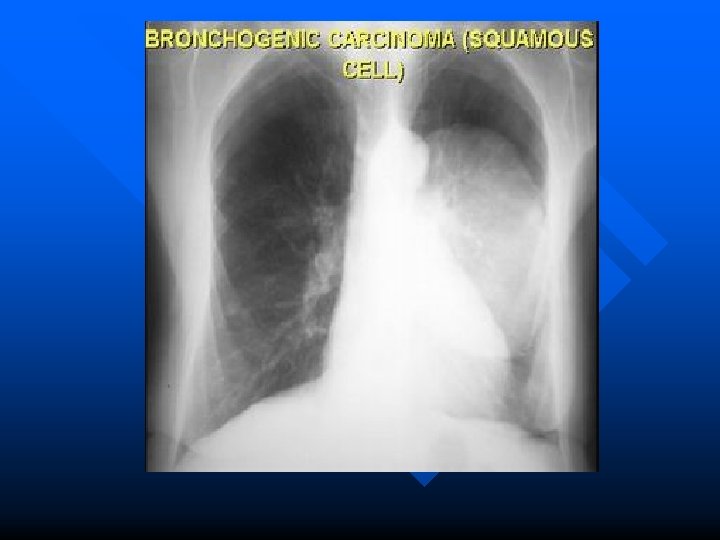
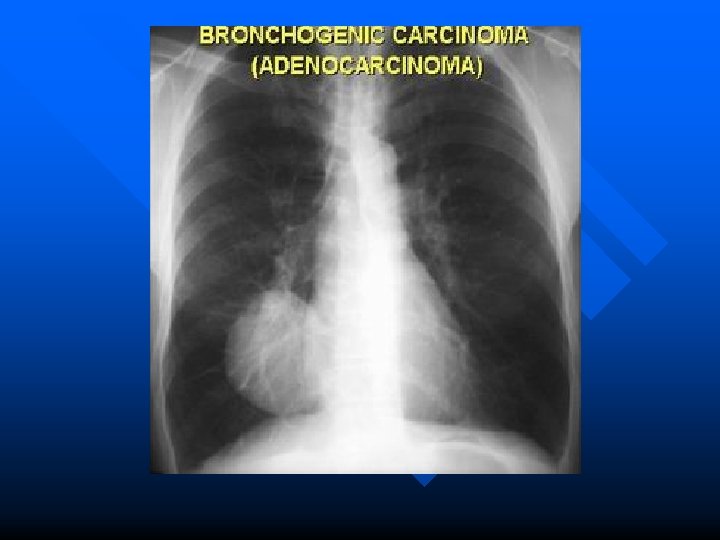
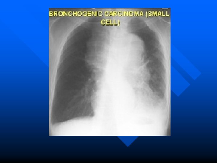
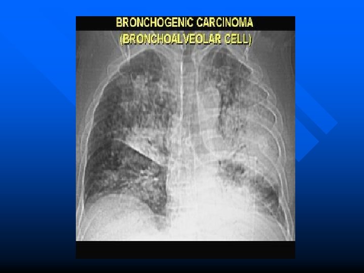
Tumors of the lung in plain chest film
A solitary lung lesion n The questions that have to be answered are : is it a primary or secondary malignant lesions or is it benign?
Radiographic signs a. b. c. The character of the edge Internal calcification Growth of a lesion
CT Scan
PANCOST TUMOR Pancoast tumour , Chest X-ray shows asymmetrical right apical pleural thickening
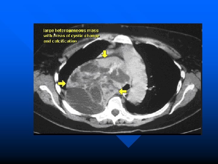
Mediastinal Mass
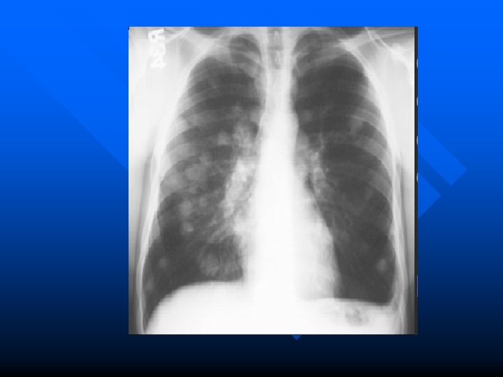
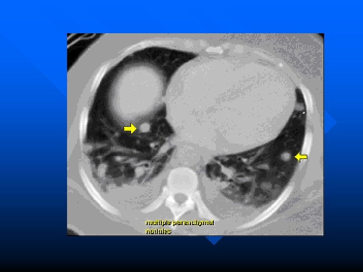
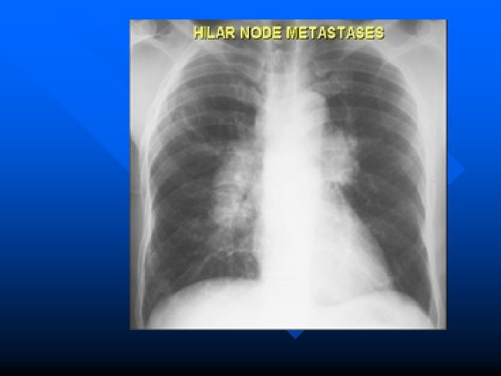
Metastatic tumors Most pulmonary metastases genitourinary and gastrointestinal system. n Hematogenous metastasis Discrete and circumscribed Miliary or snow strom appearance indicated rapid growth. n Lymphatic permeation Mediastinum and its nodes are the first line with this type of spread. n Massive pleural effusion. n
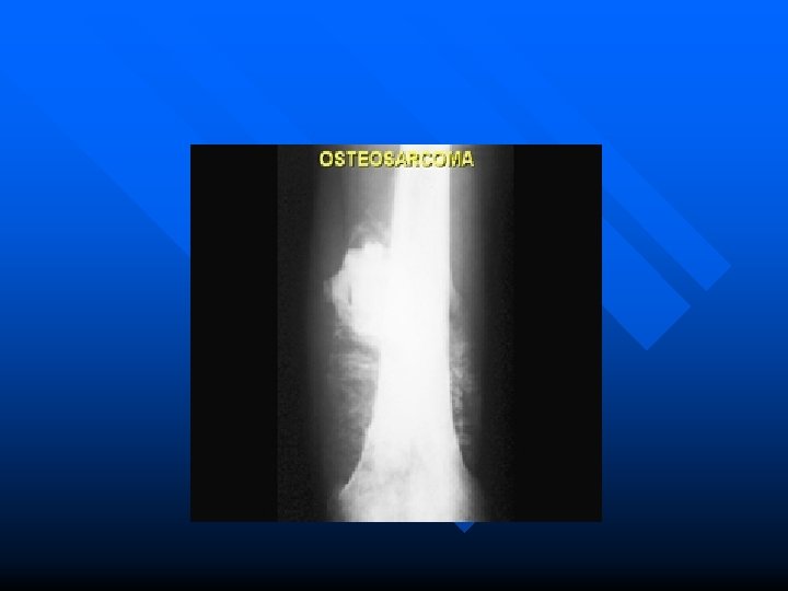
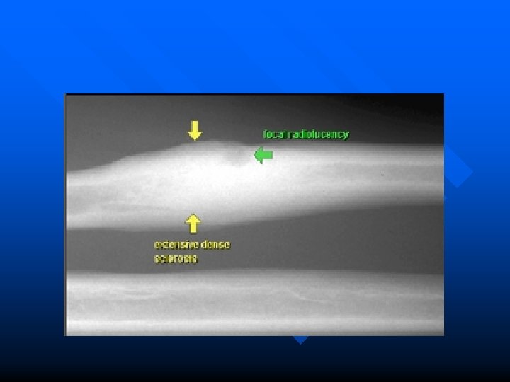
Tumors of the bone in plain photo Three important question requere to answered : 1. Neoplastic or infective 2. Benign or malignannt 3. Primary or secondary neoplasm. n
General principles of radiological diagnosis of bone tumors : n consider the age of the patient and the clinical hystory. a. Lesion soliter or multiple Type of bone involved Position of the lesion within the bone : cortex, disphysis, metaphysis Plain film features : radioluscent focus, radioopaq, ground glass Margins of the lesion look like b. c. d. e.
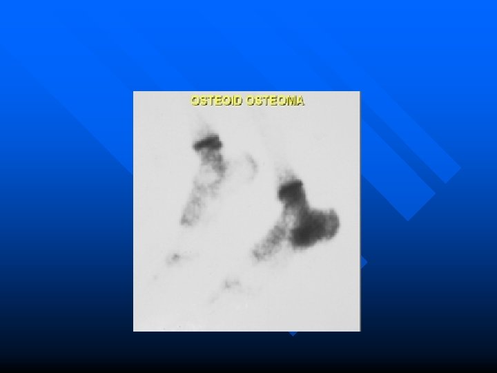
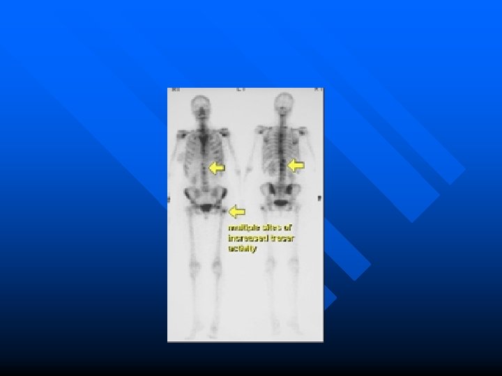
Secondary neoplasm. n n n Bone Scintigraphy (Nuclear Medine examination) Gamma camera images using Tc 99 m labelled diphosphonate compounds After injection 3 hours later the skeleton is imaged when the radiopharmaceutical has localized in bone. Scintigraphy may used to asses the primary presenting lesion and also to detect whether it is monoostotic or accompanied by other skeletal lesions such as metastasis. t
GASTROINTESTINAL AND UROGENITAL TRACT TUMOR CONTRAST EXAMINATION n ULTRASONOGRAPHY n CT n MRI n
COLON TUMOR BENIGN POLIP n MALIGNANT TUMOR n
SESSILE POLYP
PEDUNCULATED POLIP
VILLOUS TYPE
ANNULARE TYPE
Carsinoma colon
UTERINE FIBROID (leiomyoma)
UTERINE FIBROID (leiomyoma)
URINARY BLADDER TUMOR
Renal tumor CT Scan
Both pictures (different cases) show metastases in the liver shown in red circles
MRI n .
LIVER TUMOR
LIVER METASIS
PANCREATIC TUMOR
BRAIN TUMOR CT n MRI n
BRAIN TUMOR CT
GLIOBLASTOMA MULTIFORME AND MENINGIOMA
BRAIN TUMOR MRI
ORBITAL TUMOUR A B A. Haemangiomas. Multilobate mass right which does not fill the tip of the orbit and displays distinct enhancement of 37 HU after administration of contrast medium (In). B. Glioma of the optic nerve. Spindle-shaped enlargement retrobulbar in the region of the optic nerve with dilatation of the tip of the orbit including the optic canal (Su); Meningioma of the optic nerve sheath with calcification in the region of the optic sheath. No enlargement of the optic nerve is visible in this slice
MALIGNANT LYMPHOMAS A. Medial lesion, medial extraocular muscles and almost exclusively extraconal location determined in the horizontal slice. B. Intraconal malignant lymphoma with enlargement of the muscle cone extending into the tip of orbit. Homogenous tissue density lesion.
MALIGNANT LYMPHOMAS A. Lateral malignant lymphoma (polymorphic immunocytoma) situated within and outside the muscle cone and masking the musculus rectus lateralis B. Plasmocytoma with infiltration of the retrobulbar fatty tissue and destruction of the osseous orbit
MAMMOGRAPHIC SIGNS OF BREAST CA DIRECT SIGN OF FOCALLY GROWING INVASIVE CARCINOMA 1. increase density in comparison with the parenchym. 2. equal in density in comparison with the parenchym with outline : . n Spiculated or irregular n Similar to parenchyma, lobulated, geographically illdefinded (=indeterminate mass) n Round or rarely entirely smooth. 3. Microcalsification 4. Distorted architecture. 5. Asymmetry in comparison with the contralateral side. n
INDIRECT SIGN / secondary sign OF A FOCALLY GROWING INVASIVE CA 1. 2. 3. 4. 5. 6. Nipple retraction Local retraction of the skin or the parenchyma overlying the lession Thickening of cooper’s ligaments in the vicinity of the lesion Local thickening of skin overlying the lesion. Trabecular thickening in the subcutaneous space or in the prepectoral muscle Retraction or fixation on the pectoral muscle Enlarged, multiple, homogenously dense, smoothly or unsharply outlined lymph nodes in the axillary extention.
Malignant tumor
Malignnat calcification
Benign tumor
Benign tumor
SONOGRAPHY n • • • Indications: Exclusion of a simple cysts Detection of malignancy in the presence of uncharacteristic and questionable palpatory findings in mamographycally dense tissue. Sonographically guided puncture)
Image presentation of the breast Ca • • Hypoechoic, often with varying echogenicity throughout the mass. Poorly to moderately compressible or moveable Irregularly out; ined with or without hyperechoic rim. With or without acoustic shadow.
Cyst
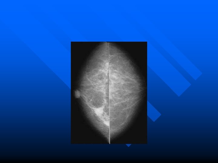
Example Case n n This 61 year old female presented with a dominant mass in the right breast with several other indeterminate lesions seen on mammography. Mammography CC view demonstrates three masses in the right breast, one behind the nipple and two others in its medial portion.
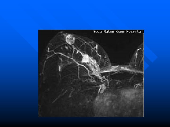
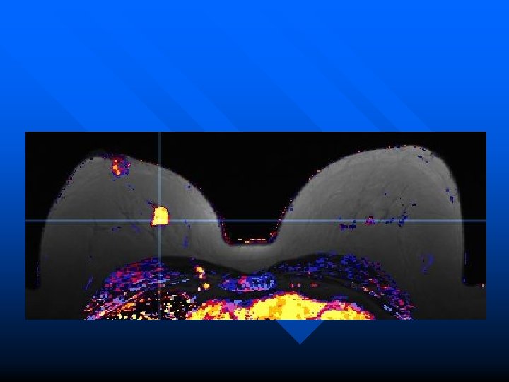
n MRI magnification maximum intensity projection (MIP) view of right breast demonstrates the same three masses with tumor bridging between them and extending toward the chest wall.
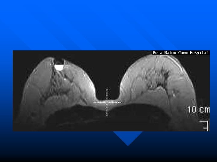
n MRI T 2 axial slice indicates cystic nature of subareolar mass.
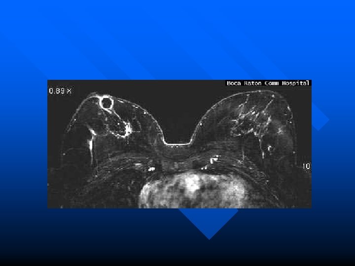
n MRI subtraction and color images show rim enhancement around cystic mass.
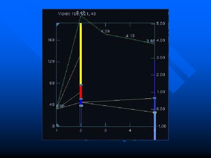
n MRI image of color map of enhancement curves (kinetics of breast tissue) show intense early enhancement and rapid signal loss from the dominant mass (typical malignant characteristics).
n n n Enhancement curve from dominant mass. Final Pathology at mastectomy confirmed that the dominant mass represented infiltrating carcinoma with extension of intraductal carcinoma. The subareolar cystic mass represented encysted papillary carcinoma.
Nuclear medicine Using liquid radioactive material • diagnostic • therapy
Dose measurement
SOURCE PREPARATION INTRA VENOUS INJECTION
SCANNING WITH GAMMA CAMERA SPECT
BONE SCANNING
Radiation Therapy Using SOLID radioactive (for example Co-60; Cs-137) and an electric generator (LINAC)
Radiation therapy Internal Radiation Therapy Brachytherapy External Beam Internal Radiation Therapy Radiation. Therapy Nuclear medicine Cobalt 6 o LINAC Intracavitair, Interstitiil, mould, etc
EXTERNAL BEAM RADIATION THERAPY Cobalt 60 Linear Accelerator
RADIATION THERAPY PROCEDURE PLANNING Cilinical evaluation and staging n Treatment intent n Choice of treatment n
TREATMENT PLANNING (RT) Description n Immobilization n Image acquisition n Delineation of volumes (GTV, CTV, PTV) n Choice of technique and beam modification n Computation of dose distribution n
Fixation with plastic mask is essential for Linac Radiotherapy
SIMULATION PHOTO
CT DOSIMETRI
Treatment Planning System
Cobalt 60 teletherapy Radiation dose 70 Gy in 35 fraction
Radiotherapy with LINAC Synchrony ™ X-ray sources camera Treatment couch Linear accelerator Manipulator Image detectors
Brachitherapy: interstitiil, intracaviter
Thank you