Radiological Category Thoracic chest Principal Modality 1 General
: General Radiography Principal Modality (2): CT Case")


- Slides: 9
Radiological Category: Thoracic chest Principal Modality (1): General Radiography Principal Modality (2): CT Case Report #0104 Submitted by: Beth Chasen, M. D. Faculty reviewer: Marvin H. Chasen, M. D Date accepted: 15 March 2004
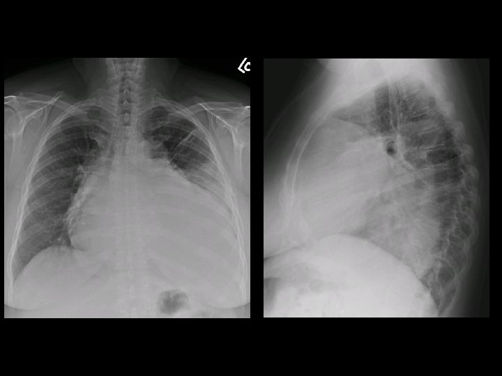
Case History 53 year old female with hypertension, diabetes, and six month history of shortness of breath.
Test Your Diagnosis Which one of the following is your choice for the appropriate diagnosis? After your selection, go to next page. • Cardiomyopathy with pleural effusion • Cardiac amyloidosis • Mediastinal sarcoma • Pericardial and pleural effusions • Cardiac rhabdomyosarcoma
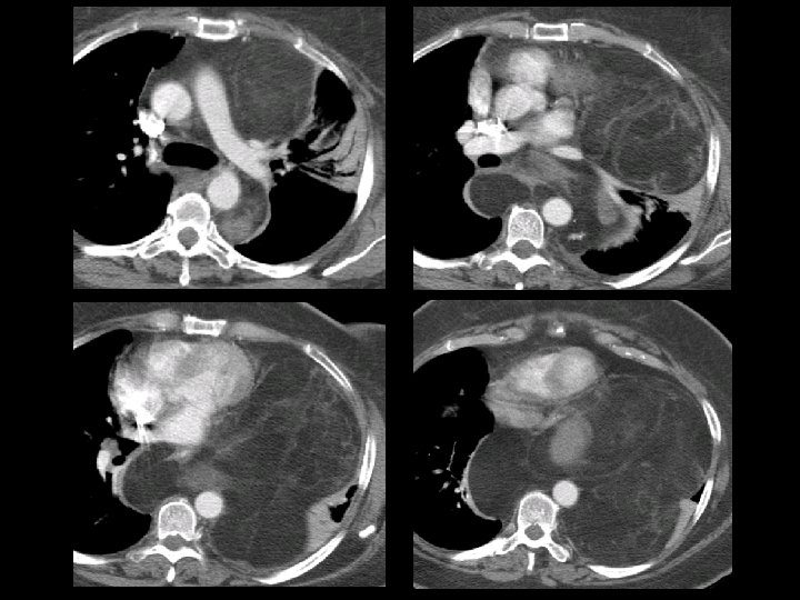
Findings and Differentials Findings: Large, multilobular mediastinal mass containing soft tissue and fatlike components. Mass effect upon the heart is noted with no evidence of cardiac origin. No pericardial or pleural effusion is present. Differentials: • Mediastinal liposarcoma • Mediastinal lipomatosis • Mediastinal lipomas
Discussion When presented with the PA radiograph alone, one could certainly suspect a cardiac process. However, the lateral radiograph shows a lucent plane behind the heart that suggests definition of a posterior cardiac border. In addition, multiple lobular masses are posterior and superior to the expected cardiac contour. The CT findings exclude a cardiac origin. Mediastinal lipomatosis is a benign condition involving excess deposition of fat within the mediastinum. It may be associated with conditions like Cushing’s syndrome, steroid use, or even obesity. This fat deposition surrounds the mediastinal structures uniformly with no appreciable mass effect. CT should show a homogenous fat density to make the diagnosis of mediastinal lipomatosis. Mediastinal lipomas also have essentially uniform fatty density and are welldefined lesions. Lipomas may occur in any location but are most often within the anterior mediastinum. This case is an example of mediastinal liposarcoma. The CT findings of mass effect upon mediastinal structures and of soft tissue elements within the predominantly fatty matrix are essentially pathognomonic.
Discussion Reference J. London, E. Kim, S. Wallace et al. : MR Imaging of Liposarcomas: Correlation of MR Features and Histology. J Comput Assist Tomogr 1989; 13: 832 -835
Diagnosis Well-differentiated mediastinal liposarcoma