
Radiological Category Gastrointestinal Principal Modality 1 MRI Principal

- Slides: 26
Radiological Category: Gastrointestinal Principal Modality (1): MRI Principal Modality (2): None Case Report #0435 Submitted by: Jennifer C. Lin, M. D. Faculty reviewer: Anurada Rao, M. D. Date accepted: 07 January 2008
Case History 57 year-old female with chronic epigastric pain
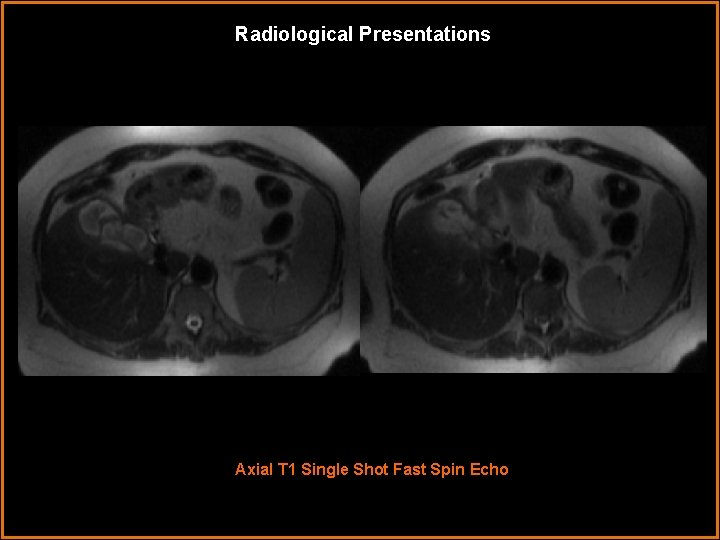
Radiological Presentations Axial T 1 Single Shot Fast Spin Echo
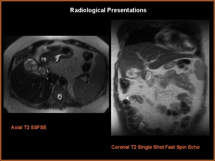
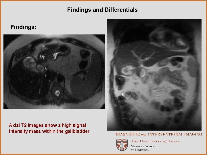
Radiological Presentations Axial T 2 SSFSE Coronal T 2 Single Shot Fast Spin Echo
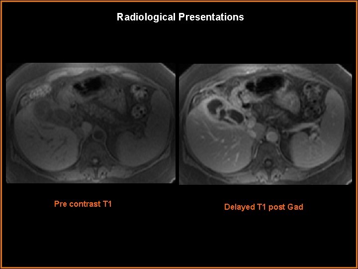
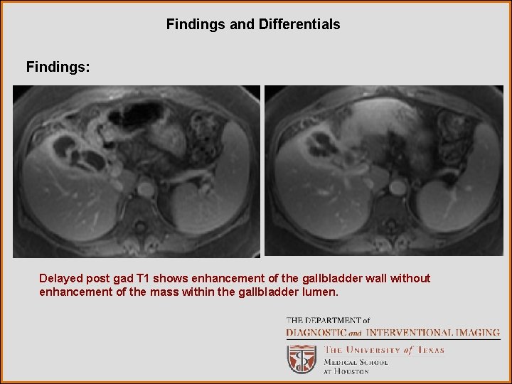
Radiological Presentations Pre contrast T 1 Delayed T 1 post Gad
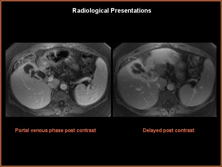
Radiological Presentations Portal venous phase post contrast Delayed post contrast
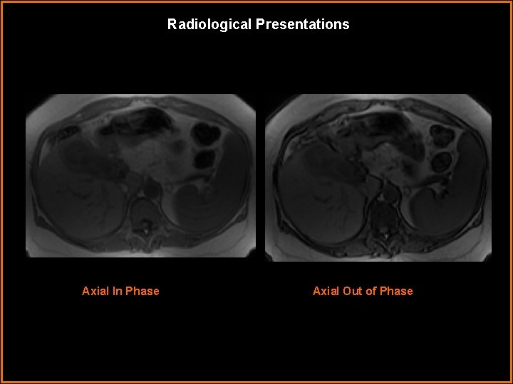
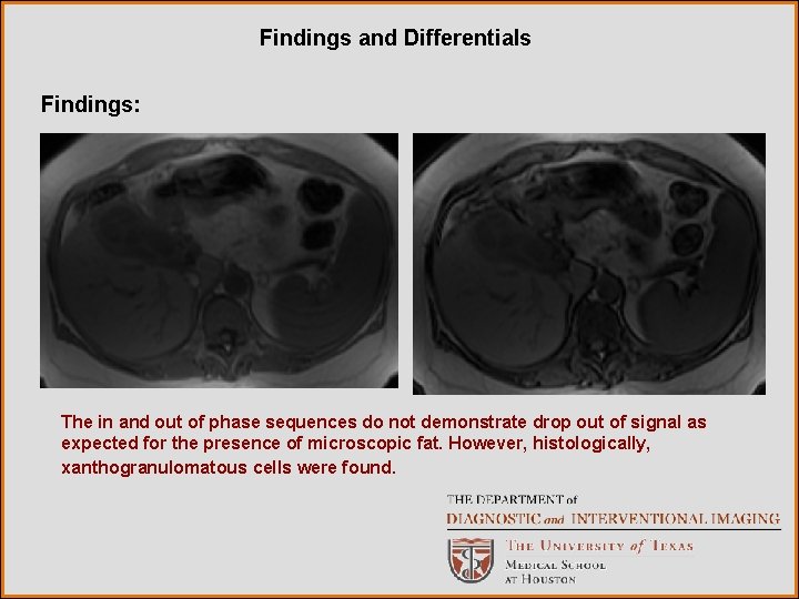
Radiological Presentations Axial In Phase Axial Out of Phase
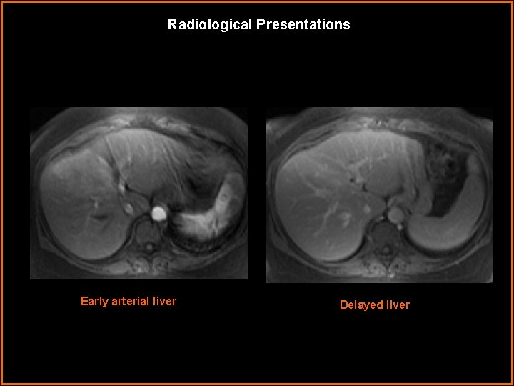
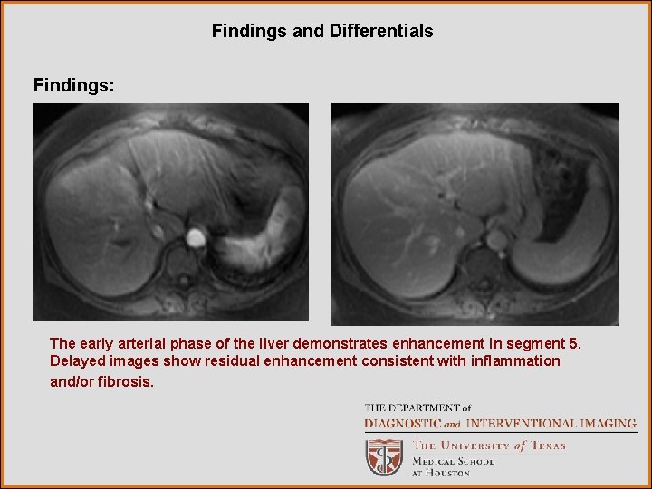
Radiological Presentations Early arterial liver Delayed liver
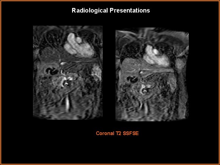
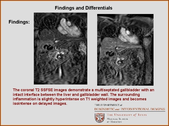
Radiological Presentations Coronal T 2 SSFSE
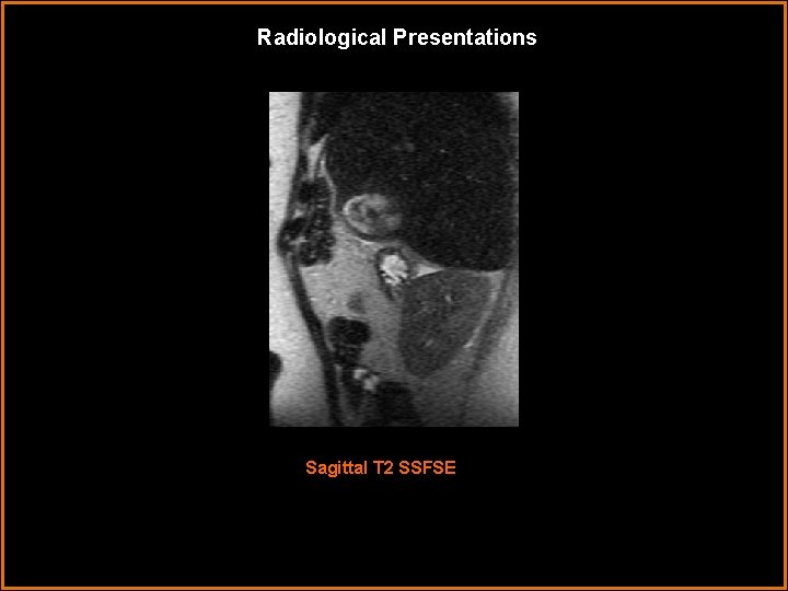
Radiological Presentations Sagittal T 2 SSFSE
Test Your Diagnosis Which one of the following is your choice for the appropriate diagnosis? After your selection, go to next page. • Gallbladder carcinoma • Biliary ductal dilitation • Acute cholecystitis • Xanthogranulomatous cholecystitis
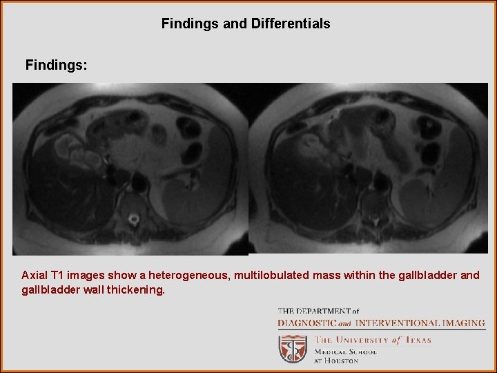
Findings and Differentials Findings: Axial T 1 images show a heterogeneous, multilobulated mass within the gallbladder and gallbladder wall thickening.
Findings and Differentials Findings: Axial T 2 images show a high signal intensity mass within the gallbladder.
Findings and Differentials Findings: Delayed post gad T 1 shows enhancement of the gallbladder wall without enhancement of the mass within the gallbladder lumen.
Findings and Differentials Findings: The in and out of phase sequences do not demonstrate drop out of signal as expected for the presence of microscopic fat. However, histologically, xanthogranulomatous cells were found.
Findings and Differentials Findings: The early arterial phase of the liver demonstrates enhancement in segment 5. Delayed images show residual enhancement consistent with inflammation and/or fibrosis.
Findings and Differentials Findings: The coronal T 2 SSFSE images demonstrate a multiseptated gallbladder with an intact interface between the liver and gallbladder wall. The surrounding inflammation is slightly hyperintense on T 1 weighted images and becomes isointense on delayed images.
Findings and Differentials: • Primary Gallbladder carcinoma • Metastatic disease to the gallbladder • Gallbladder adenoma • Biliary cystadenoma • Adenomyomatous hyperplasia • Xanthogranulomatous cholecystits
Surgical Findings: The patient went to surgery and had an open cholecystectomy. The gallbladder closely adhered to the liver parenchyma without invading it. The gallbladder wall was very friable and needed to be removed in pieces.
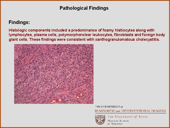
Pathological Findings: Histologic components included a predominance of foamy histiocytes along with lymphocytes, plasma cells, polymorphonclear leukocytes, fibroblasts and foreign body giant cells. These findings were consistent with xanthogranulomatous cholecystitis.
Discussion Almost all malignancies and benign gallbladder lesions occur more frequently in women than men. Gallbladder carcinoma may appear as a mass replacing the normal gallbladder, diffuse or focal thickening of the gallbladder wall, or a polypoid mass within the gallbladder lumen. Invasion of the adjacent liver, gallstones, and/or biliary obstruction are typically present at diagnosis. Additionally, a porcelain gallbladder has been associated with gallbladder carcinoma. Gallbladder adenomas are rare and are associated with Familial Adenomatous Polyposis and Peutz-Jeghers syndrome. They are frequently seen with cholelithiasis and may eventually progress to carcinoma. Radiographically, adenomas are seen as intraluminal polypoid masses. The gallbladder wall should not be thickened. If gallbladder wall thickening is observed, carcinoma should be higher on the differential.
Discussion Biliary cystadenomas are benign unilocular or multilocular cystic neoplasms that may occur in the liver, extrahepatic biliary tree, or gallbladder. Although biliary cystadenomas are benign, they recur after resection, and have the potential to develop into biliary cystadenocarcinoma. On imaging, biliary cystadenomas do not have a fatty component. Typically the fluid of is of varying attenuation on CT and varying intensity on T 1 and T 2 MRI. Adenomyomatous hyperplasia is a common condition characterized by diffuse gallbladder wall thickening with intramural diverticula appearing as cystic spaces within the wall. Radiographically, it presents as reverberation artifact within a thickened gallbladder wall on ultrasound or as Rokitansky-Aschoff sinuses on MRI.
Discussion Xanthogranulomatous cholecystitis is a rare form of chronic cholecystitis that may imitate the appearance of a malignancy. The inflammatory process is believed to be due in part to extravasation of bile into the gallbladder wall from mucosal ulceration or rupture of Rokitansky-Aschoff sinuses due to increased intraluminal pressure from gallblader or cystic duct obstruction. Patients present with clinical signs of cholecystitis including right upper quadrant pain, vomiting, leukocytosis, or positive Murphy sign. It is most common in females between 60 -70 years. Complications include abscess formation, perforation, fistulous tracts to the duodenum or skin, extension of the inflammatory process to the liver, colon, or surrounding soft tissues. Although xanthogranulomatous cholecystitis is benign, it is associated with gallbladder malignancies. The imaging characteristics of the two are very similar, and preoperative differentiation is extremely difficult if not impossible. Therefore most patients will undergo operative cholecystectomy for definitive diagnosis.
Discussion Characteristics more typical of xanthogranulomatous cholecystitis include : 1) A maintained gallbladder coutour 2) No biliary obstruction 3) May show signal drop out with in- and out- of phase MRI 4) Increased enhancement in the portal phase suggest chronic inflammation 5) Pericholecystic enhancement in segment 5 of the liver Gallstones may be seen with both xanthogranulomatous cholecystitis and gallbladder carcinoma. Sonographically, xanthogranulomatous cholecystitis may appear as hypoechoic nodules or bands in a thickened gallbladder wall. On MRI, xanthogranulomatous cholecystitis will appear as an enhancing gallbladder wall without evidence of an intraluminal gallbladder mass. Surrounding inflammation is often seen.
Diagnosis Xanthogranulomatous cholecystitis
References: Levy AD, Murakata LA, Abbott RM, Rohrmann CA. From the Archives of the AFIP: Benign Tumors and Tumorlike Lesions of the Gallbladder and Extrahepatic Bile Ducts: Radiologic-Pathologic Correlation. Radiographics 2002; 22: 387 -413. Levy AD, Murakata LA, Rohrmann CA. Gallbladder Carcinoma: Radiologic-Pathologic Correlation. Radiographics 2001; 21: 295 -314.