
Radiographic Assessment of the Pediatric Patient Special considerations













- Slides: 29
“Radiographic Assessment of the Pediatric Patient”
Special considerations 1. • • • Risk assessment Evidence of caries/hx Trauma Anomalies Fluoride status Diet
AAPD guidelines for radiographs • Based on Age and risk assessment
Child preparation and management • • • Euphemisms Role models Contour film Gag reflex – distraction Parental help Bad taste
Film Sizes • Sizes 0, 1, 2, occlusal/lateral
Radiographic Tools • Snap-a-ray • Bite wings, periapicals
Radiographic techniques 1. 2. 3. 4. 5. 6. Bite wings Periapicals (not p. a. ’s) Max/mand occlusals Extraoral/lateral film Soft tissue x-ray Panoramic radiographs
Bite Tabs
Bite wing x-ray • Mesial surface of canine to distal surface of 1 st permanent molar
Bite wing x-ray • Incipient carious lesion. • Overlapping – common error
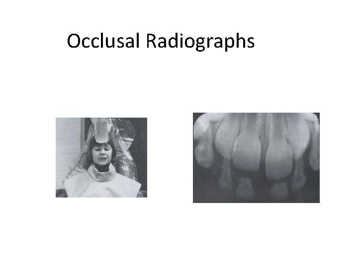
Occlusal Radiographs
Occlusal Radiographs • Posterior max. occlusal radiograph
Extra Oral film • Lateral Film
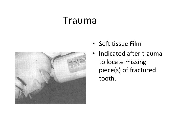
Trauma • Soft tissue Film • Indicated after trauma to locate missing piece(s) of fractured tooth.
Panaramic radiograph
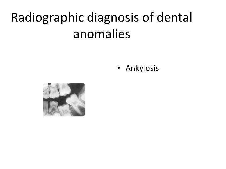
Radiographic diagnosis of dental anomalies • Ankylosis
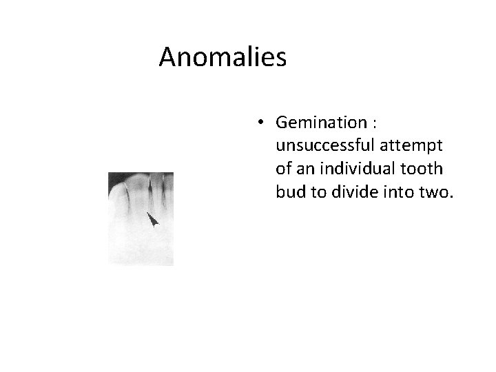
Anomalies • Gemination : unsuccessful attempt of an individual tooth bud to divide into two.
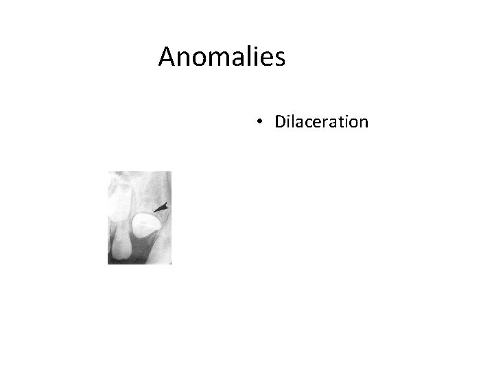
Anomalies • Dilaceration
Anomalies • Peg lateral • Supernumary primary lateral
Anomalies a) Fusion: dentinal union of two teeth. b) Supernumary tooth c) Missing lateral
Anomalies • Concrescence: fusion with a cemental union.
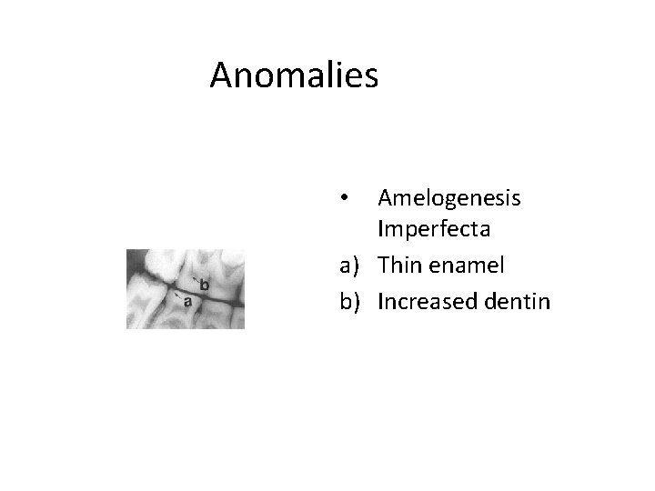
Anomalies Amelogenesis Imperfecta a) Thin enamel b) Increased dentin •
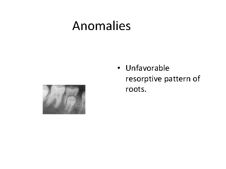
Anomalies • Unfavorable resorptive pattern of roots.
Pathology • Retained primary root tips.
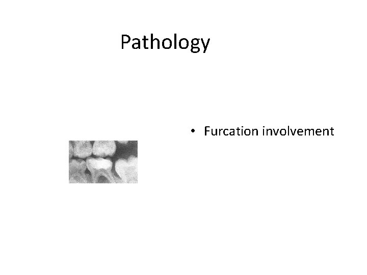
Pathology • Furcation involvement
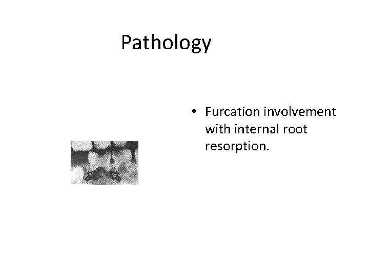
Pathology • Furcation involvement with internal root resorption.
Pathology • Internal resorption with furcation involvement.
Artifacts/optical illusions 1. Cervical burnout 2. Mach band phenomenon Ø It may take 30%-70% demineralisation to occur before it can be evidenced radiographically. Ø Radiographs are 2 D views of 3 D objects.
• THANK YOU!